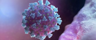
Epstein-Barr virus (IgG antibodies to capsid antigen)
IgG antibodies to the infectious mononucleosis virus (Epstein Barr Virus, EBV) are specific antiviral immunoglobulin proteins produced by the immune system in response to infection with the infectious mononucleosis virus and indicating a current or past infection. Epstein–Barr virus is a widespread virus of the Herpesviridae family that primarily infects B lymphocytes, as well as T lymphocytes and epithelial cells. It is transmitted by airborne droplets. The peak incidence occurs at 15-25 years of age. The first contact of a person with the virus occurs, as a rule, in childhood and leads to the development of a latent asymptomatic or low-symptomatic infection. In adults, the Epstein-Barr virus causes infectious mononucleosis, which in most patients is accompanied by fever, intoxication, enlarged lymph nodes, palatine and pharyngeal tonsils. The liver and spleen often become enlarged, and petechiae appear on the mucous membrane of the upper palate. Infectious mononucleosis can be complicated by splenic rupture, as well as hepatitis, pancreatitis, pneumonia, hemolytic anemia, thrombocytopenia, aplastic anemia, myocarditis, Guillain-Barre syndrome, encephalitis, meningitis. The virus persists in small quantities in memory B cells. About 90% of adults are virus carriers. The persistence of the virus in B lymphocytes and epithelial cells continues throughout life, so that when immunity is reduced (for example, with HIV or immunosuppressive therapy after organ transplantation), reactivation of the infection can occur, which contributes to the development of lymphoproliferative diseases (including Burkitt's lymphoma), nasopharyngeal carcinoma or (most often) infectious mononucleosis. In response to infection, the immune system produces various specific antiviral antibodies. In the acute stage of infection, IgM to the capsid protein (VCA) of the virus is the first to be detected in the blood, which reaches its maximum concentration in the blood plasma at the 3rd week of the disease and disappears by the 4-6th week. Later, IgG to the capsid protein appears, reaching a maximum at 2-4 weeks of the disease, then their concentration decreases, but they still persist for life. When the infection is reactivated, the titers of these antibodies usually increase. Antibodies to early antigens are detected at the acute stage of infection and disappear 3-6 months after the onset of the disease, but in 20% of infected people they can be detected for several years. Antibodies to the viral nuclear antigen (EBNA) are usually not detected at the acute stage of infection, appear in the blood no earlier than the 6-8th week of the disease (usually 2-4 months after the onset of the first symptoms) and persist throughout life. Thus, an antibody test allows not only to detect an infection caused by the Epstein-Barr virus, but also to determine its stage. Research used:
- To confirm current or past infectious mononucleosis.
- To assess susceptibility to infection caused by the Epstein-Barr virus (infectious mononucleosis).
The study is assigned:
- In cases where existing clinical (fatigue, fever, sore throat, enlarged perimaxillary and cervical lymph nodes, enlarged liver and/or spleen) and laboratory (atypical lymphocytes in peripheral blood) signs indicate current or past infectious mononucleosis.
- For flu symptoms in pregnant women (along with tests for cytomegalovirus infection, toxoplasmosis, etc.).
- If the patient (even without symptoms of infection) was in close contact with a patient with infectious mononucleosis - to assess the strength of the immune system and susceptibility to infection.
Reasons for the positive result:
- the presence of active immunity due to a previous infection (along with the detection of antibodies to nuclear antigen (EBNA) and the absence of IgM to the capsid antigen (VCA) of the Epstein-Barr virus);
- current or recent infectious mononucleosis (in combination with detection of IgM to capsid antigen (VCA) and antibodies to early antigens (EA-D) of the Epstein-Barr virus);
- Epstein–Barr virus reactivation.
Reasons for negative results:
- absence of infection caused by the Epstein-Barr virus (IgM to the capsid antigen (VCA) of the Epstein-Barr virus is not detected); if there is a suspicion of infection, it is advisable to repeat the IgG determination after 2-4 weeks;
- early stages of infectious mononucleosis (provided that an increase in the level of IgM to the capsid antigen (VCA) of the Epstein-Barr virus is detected) - repeat the study over time after 14 days;
- low levels of Epstein-Barr virus in the blood;
- absence of an immune response or a weak immune response to the Epstein-Barr virus due to disorders in the immune system (IgM to the capsid antigen (VCA) of the Epstein-Barr virus is not detected).
An increase in antibody titer over time (in paired sera) rather indicates an acute infection or reactivation of an infection, while a decrease indicates a recently resolved infection. The amount of antibodies in the blood does not reflect the severity or duration of the infection. In some cases, high levels of IgG to the capsid protein (VCA) of the Epstein-Barr virus can persist throughout life.
Epstein Barr Virus capsid protein (VCA), IgG
IgG antibodies to the infectious mononucleosis virus (Epstein Barr Virus, EBV) are specific antiviral immunoglobulin proteins produced by the immune system in response to infection with the infectious mononucleosis virus and indicating a current or past infection.
Synonyms Russian
IgG class antibodies to the capsid protein of the Epstein Barr Virus (EBV), class G immunoglobulins to the capsid protein of the Epstein Barr virus.
English synonyms
Anti-Epstein-Barr viral capsid antigens IgG, Epstein Barr Virus (EBV), VCA IgG, Anti-EBV (VCA) IgG, EBV-IgG anti-VCA.
Research method
Chemiluminescent immunoassay.
What biomaterial can be used for research?
Venous blood.
How to properly prepare for research?
Do not smoke for 30 minutes before the test.
General information about the study
Epstein–Barr virus is a widespread virus of the Herpesviridae family that primarily infects B lymphocytes, as well as T lymphocytes and epithelial cells. It is transmitted by airborne droplets. The peak incidence occurs at 15-25 years of age.
The first contact of a person with the virus occurs, as a rule, in childhood and leads to the development of a latent asymptomatic or low-symptomatic infection. In adults, the Epstein-Barr virus causes infectious mononucleosis, which in most patients is accompanied by fever, intoxication, enlarged lymph nodes, palatine and pharyngeal tonsils. The liver and spleen often become enlarged, and petechiae appear on the mucous membrane of the upper palate. Infectious mononucleosis can be complicated by splenic rupture, as well as hepatitis, pancreatitis, pneumonia, hemolytic anemia, thrombocytopenia, aplastic anemia, myocarditis, Guillain-Barre syndrome, encephalitis, meningitis.
The virus persists in small quantities in memory B cells. About 90% of adults are virus carriers. The persistence of the virus in B lymphocytes and epithelial cells continues throughout life, so that when immunity is reduced (for example, with HIV or immunosuppressive therapy after organ transplantation), reactivation of the infection can occur, which contributes to the development of lymphoproliferative diseases (including Burkitt's lymphoma), nasopharyngeal carcinoma or (most often) infectious mononucleosis.
In response to infection, the immune system produces various specific antiviral antibodies. In the acute stage of infection, IgM to the capsid protein (VCA) of the virus is the first to be detected in the blood, which reaches its maximum concentration in the blood plasma at the 3rd week of the disease and disappears by the 4-6th week. Later, IgG to the capsid protein appears, reaching a maximum at 2-4 weeks of the disease, then their concentration decreases, but they still persist for life. When the infection is reactivated, the titers of these antibodies usually increase. Antibodies to early antigens are detected at the acute stage of infection and disappear 3-6 months after the onset of the disease, but in 20% of infected people they can be detected for several years. Antibodies to the viral nuclear antigen (EBNA) are usually not detected at the acute stage of infection, appear in the blood no earlier than the 6-8th week of the disease (usually 2-4 months after the onset of the first symptoms) and persist throughout life.
Thus, an antibody test allows not only to detect an infection caused by the Epstein-Barr virus, but also to determine its stage.
What is the research used for?
- To confirm current or past infectious mononucleosis.
- To assess susceptibility to infection caused by the Epstein-Barr virus (infectious mononucleosis).
When is the study scheduled?
- In cases where existing clinical (fatigue, fever, sore throat, enlarged perimaxillary and cervical lymph nodes, enlarged liver and/or spleen) and laboratory (atypical lymphocytes in peripheral blood) signs indicate current or past infectious mononucleosis.
- For flu symptoms in pregnant women (along with tests for cytomegalovirus infection, toxoplasmosis, etc.).
- If the patient (even without symptoms of infection) was in close contact with a patient with infectious mononucleosis - to assess the strength of the immune system and susceptibility to infection.
What do the results mean?
Reference values
Result: negative.
Signal/cutoff ratio: 0 - 0.9.
Reasons for the positive result:
- the presence of active immunity due to a previous infection (along with the detection of antibodies to nuclear antigen (EBNA) and the absence of IgM to the capsid antigen (VCA) of the Epstein-Barr virus);
- current or recent infectious mononucleosis (in combination with detection of IgM to capsid antigen (VCA) and antibodies to early antigens (EA-D) of the Epstein-Barr virus);
- Epstein–Barr virus reactivation.
Reasons for negative results:
- absence of infection caused by the Epstein-Barr virus (IgM to the capsid antigen (VCA) of the Epstein-Barr virus is not detected); if there is a suspicion of infection, it is advisable to repeat the IgG determination after 2-4 weeks;
- early stages of infectious mononucleosis (provided that an increase in the level of IgM to the capsid antigen (VCA) of the Epstein-Barr virus is detected) - repeat the study over time after 14 days;
- low levels of Epstein-Barr virus in the blood;
- absence of an immune response or a weak immune response to the Epstein-Barr virus due to disorders in the immune system (IgM to the capsid antigen (VCA) of the Epstein-Barr virus is not detected).
An increase in antibody titer over time (in paired sera) rather indicates an acute infection or reactivation of an infection, while a decrease indicates a recently resolved infection. The amount of antibodies in the blood does not reflect the severity or duration of the infection. In some cases, high levels of IgG to the capsid protein (VCA) of the Epstein-Barr virus can persist throughout life.
Also recommended
- Epstein Barr Virus capsid protein (VCA), IgM
- Epstein Barr Virus, DNA [real-time PCR]
- Epstein Barr Virus early antigens (EA), IgG
- Epstein Barr Virus nuclear antigen (EBNA), IgG (quantitative)
- Complete blood count (without leukocyte formula and ESR)
- Alanine aminotransferase (ALT)
- Aspartate aminotransferase (AST)
- HIV 1, 2 Ag/Ab Combo (determination of antibodies to HIV types 1 and 2 and p24 antigen)
Who orders the study?
Infectious disease specialist, pediatrician, ENT, hematologist, therapist, general practitioner.
Literature
- Cohen JI Epstein-Barr virus infection / JI Cohen // N. Engl. J. Med. – 2000. – Vol. 343, N 7. – P. 481–492.
- Hess RD Routine Epstein-Barr virus diagnostics from the laboratory perspective: still challenging after 35 years / RD Hess // J. Clin. Microbiol. – 2004. – Vol. 42, N 8. – P. 3381–3387.
- Isakov V.A. Herpes: pathogenesis and laboratory diagnosis: a guide for doctors / V.A. Isakov, V.V. Borisova, D.V. Isakov. – St. Petersburg: Lan Publishing House, 1999. – 192 p.
- Johannsen EC, Schooley RT, Kaye KM Epstein-Barr Virus (Infectious Mononucleosis). In: Principles and practice of infectious disease / GL Mandell, Bennett JE, Dolin R (Eds) ; 6th ed. – Churchill Livingstone, Philadelphia, PA 2005. – 2701 p.
- Tselix A. Epstein-Barr virus / A. Tselix, Jenson H.V. – NY: Taylor & Fransis, 2006. – 410 p.
Research method - Chemiluminescent immunoassay
Material for research - Blood serum
Infectious mononucleosis is caused by the Epstein-Barr virus (family of herpes viruses), the disease is transmitted primarily by airborne droplets. Epstein-Barr virus (EBV) was first isolated in 1964 from a patient with Burkitt's lymphoma. EBV belongs to the Herpesviridae family. The size of the virus is 180-200 nm. The virus consists of double-stranded DNA and has four main antigens: early antigen – EA (early antigen), which appears in the cytoplasm and nucleus before the synthesis of viral particles; capsid antigen – VCA (viral capcide antigen), contained in the nucleocapsid of the virus; membrane antigen - MA (mempane antigen) and nuclear antigen - EBNA (Epstain-Barr Nuclea antigen), consisting of a complex of polypeptides. There are A and B strains of the virus. They are found in different geographical areas, but significant differences between the strains themselves and the nature of the disease they cause have not yet been identified.
After the primary infection, the virus remains in the B lymphocytes and epithelium of the nasal and pharyngeal mucosa during the incubation period. The virus is most often transmitted to children through saliva, and their infection is often asymptomatic or subclinical. The second peak of infection is observed in young people aged 14 to 20 years. In 2/3 of cases, infectious mononucleosis may develop (Pfeiffer's glandular fever, “kissing disease”). Clinical symptoms of the disease are loss of appetite, fatigue, fever, rash, pharyngitis, tonsillitis, lymphangitis, leukocytosis, headaches, rheumatic pain and liver dysfunction. In some cases, serious complications may occur, such as hemolytic anemia, pneumonia, neurological or cardiac disorders. EBV persists throughout life in B lymphocytes and individual epithelial cells. More than 90% of adults are seropositive and carry EBV virus.
In the routine practice of doctors who consult adult patients, the chronic form of Epstein-Barr viral infection is mainly encountered, which develops on average in 20% of individuals after the acute phase. To diagnose Epstein-Barr virus infection and infectious mononucleosis, enzyme immunoassay is used to determine antibodies to Epstein-Barr virus antigens, which allows for reliable laboratory diagnosis of the infection and determines the period of the infectious process. Additional studies include a complete blood count, leukocyte count (microscopy) and ESR. With infectious mononucleosis, leukocytosis, lymphocytosis, the appearance of atypical mononuclear cells and accelerated ESR are possible. IgG antibodies to the viral core antigen (EBV NA IgG) appear 4–6 months after the initial infection. Then their titer decreases and persists throughout life. These antibodies can be detected in more than 90% of adults and more than 50% of adolescents. IgG antibodies to nuclear antigen can be detected after acute infection in the later stages, during latent infection, as well as during reactivation of the virus and chronic infection. The determination of IgG antibodies to the nuclear antigen must be combined with the determination of IgM antibodies to the capsid protein.
Interpretation of test results is for informational purposes only, is not a diagnosis and does not replace medical advice. Reference values may differ from those indicated depending on the equipment used, the actual values will be indicated on the results form.
A positive result indicates past EBV infection.
A negative test result does not always rule out recent or past infection, unless the test results for IgM and IgG antibodies to the capsid antigen are also negative. In patients with acute infection, an increase in the titer of IgG antibodies to nuclear antigen usually indicates the progression of the early stage of recovery. If, despite a negative result, there remains a suspicion of the possibility of infection, it is necessary to conduct a repeat study after 1-2 weeks.
An equivocal result is usually an indicator of previous exposure to EBV if testing for IgM antibodies to capsid antigen is negative, or an indicator of acute infection if IgM antibodies to capsid antigen are still detected in the blood. Additional serological studies are necessary for clinical interpretation of the results.
Unit of measurement: Unit
Reference values:
- <0.8 – negative result
- 0.8 – 1.0 – the result is doubtful
- ≥ 1.0 – positive result