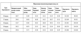
| Name of service | Price, rub |
| Ultrasound of the fetus from 10 to 21 weeks | 1800 |
| Ultrasound detection of pregnancy in the 1st trimester (up to 10 weeks) | 1500 |
| Ultrasound monitoring of a growing follicle (1 study) | 1000 |
| Fetal ultrasound 1st trimester to 12th week (SCREENING) | 2000 |
| Fetal ultrasound 2nd trimester after 12 weeks (SCREENING) | 2500 |
| Fetal ultrasound 3rd trimester after 21 weeks | 2200 |
| Ultrasound determination of fetal sex | 1200 |
| Ultrasound of the fetus up to 21 weeks (multiple pregnancy) | 2200 |
| Ultrasound of the fetus from 21 weeks (multiple pregnancy) | 3500 |
| Vascular Doppler | 1200 |
| Ultrasound monitoring of a growing follicle (folliculometry) | 900 |
During pregnancy, a woman undergoes several routine ultrasound scans.
At 7–8 weeks, such a study is carried out to confirm the presence of pregnancy, determine its duration and see the attachment of the embryo. In the first trimester, at a period of 10 to 14 weeks, the first mandatory screening is carried out, at a period of 20 to 24 weeks - a second screening, and a third - at a period of 31 to 34 weeks.
Our clinic constantly hosts promotions
Body changes and new sensations for women
During pregnancy, premature opening of the cervix should not be allowed. To maintain pregnancy, support the cervix and relieve excessive pressure on the rectum, as well as the bladder, at 21 weeks (sometimes later), for medical reasons, a woman is given an obstetric pessary (special ring). There is no need to worry: this virtually painless procedure takes only a few minutes. Apart from periodic examination of the cervix by a gynecologist and smear tests, no special actions are required when wearing a pessary during pregnancy. It has smooth edges, so it does not injure the uterus and vagina. The pessary is removed just before childbirth, at 36–38 weeks. It is as easy to remove as it is to be installed.
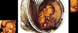
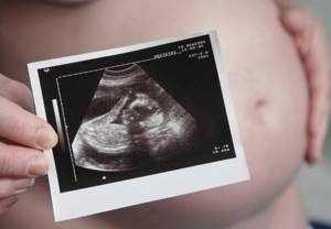
Why do you need to do an ultrasound before 21 weeks?
Ultrasound of the fetus up to 21 weeks of gestation has several goals:
- Identify fetal developmental defects. Some of them (for example, Down syndrome) could have been identified or suspected already at the first screening. A second ultrasound will give a more accurate picture.
- Determine the size of the fetus in order to assess the sufficiency of its nutrition and compliance with gestational age.
- Assess the condition of the placenta, uterus, and cervix.
- Assess the speed of blood flow in the uterine and umbilical arteries (for this, a Doppler examination is performed during an ultrasound).
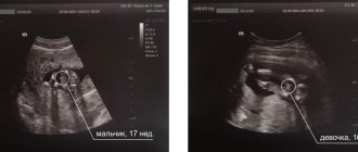
- Determine the sex of the unborn child, since at a period of 18 weeks, his genitals are already almost completely formed.
Ultrasound of the fetus up to 21 weeks does not require special preparation from the woman. The examination is carried out in a transabdominal manner, it is absolutely painless and safe.
It is very important that each screening is carried out on time. This makes it possible to promptly identify pathologies (such as oligohydramnios, isthmic-cervical insufficiency, hemodynamic disorders, etc.) and prescribe effective treatment.
It is equally important that the ultrasound is performed by an experienced diagnostician using modern equipment that will give a clear image from different angles.
Tests and ultrasound
We remind you that 21 weeks is the deadline when the expectant mother can undergo a second planned ultrasound scan during pregnancy. With the help of an ultrasound at 21 weeks, a woman will not only receive an answer to the question of how her baby is developing, but will also find out who she can expect in the near future: a boy or a girl. Sometimes the received data upsets the mother. This is not about the gender of the fetus (in some cases, the baby may not show at all what curious parents expect from him), but about possible deviations in the child’s development. Stay calm and remember: the screening result is not the ultimate truth. If any anomalies are detected, the doctor will suggest that the pregnant woman take additional tests and undergo a series of clarifying examinations.
How the research works
For the ultrasound, the woman is asked to lie on her back. A special water-based gel is applied to the abdomen, which improves the conductivity of ultrasonic waves. The direction of movement of the sensor depends on the position of the fetus. Sometimes the doctor has to press hard to see certain organs better.
The child may sleep and not be active during the study. Therefore, they can try to stir him up a little with light pushes.
The baby's father may be present at the ultrasound.
If technical capabilities allow, the doctor will make a video recording of the procedure.
Nutrition at 21 weeks of pregnancy
The expectant mother's body still needs a balanced amount of vitamins, proteins, carbohydrates and mineral salts. Choose dishes with the least fat content, but eating only low-fat foods is also not recommended. To prevent your child from being born with allergies, do not overuse exotic fruits, citrus fruits and strawberries. Avoid foods that contain a large amount of cholesterol and therefore impede the functioning of the liver (sausages, sausages, butter, fatty meats, lard and baked goods). The future mother's diet must include the following products: meat (preferably dietary), fish, cereals, cottage cheese, vegetables, herbs.
Good to know
Norms of weight gain during pregnancy
When is an ultrasound of the fetal heart performed? Fetal heart rate monitoring
Is yoga good for pregnant women?
What exercises can a pregnant woman do in the pool?
Yoga for pregnant women: what exercises you can do at home yourself
Watch out...office! Expectant mother at work
Is it possible to do lymphatic drainage facial massage during pregnancy?
Preparing the body for childbirth
What physical exercises are good for pregnant women?
All texts for pages about mother and baby were kindly provided by RAMA Publishing - these are chapters from the book by Svetlana Klaas “Your Favorite Little Man from Conception to Birth”, reviewer Irina Nikolaevna Kononova, Candidate of Medical Sciences, Associate Professor of the Department of Obstetrics and Gynecology of the Ural State Medical Academy (Ekaterinburg).
Vitamins
Now the expectant mother needs to regularly take calcium supplements prescribed by the doctor. Otherwise, her teeth may “fly”, her nails will begin to break, and her hair will fall out. Calcium is also found in dairy products: kefir, low-fat cottage cheese and milk. A simple, but proven by our grandmothers, way to get the required amount of calcium during pregnancy is to grind the shells of boiled eggs in a mortar to flour and add it to food.
In addition to calcium, do not forget about vitamins A, B, D, C and E - they also play an important role in the 21st week of the “interesting situation”.
Necessary studies and analyzes
The pregnancy calendar provides for three screenings. By week 21, the results of the second study will be known, which includes:
- analysis for the hormone human chorionic gonadotropin (hCG);
- analysis for the hormone free estriol;
- alpha-fetoprotein protein test;
- ultrasound examination of the fetus.
The first three analyzes make up the so-called triple test, which shows the degree of fetal development and the presence or absence of pathologies. The results of the study reveal how likely the risk of chromosomal abnormalities and hereditary diseases is. If it turns out that the test is positive, the doctor will send the pregnant woman for additional examinations. These are invasive methods that are used only as a last resort, as they can cause harm.
The expectant mother should visit a gynecologist at least once a month, having previously undergone urine and blood tests. With their help, the doctor monitors the well-being of the pregnant woman and evaluates the parameters of fetal development based on indicators such as heartbeat, height of the uterine fundus and abdominal volume.
Lifestyle
Start playing music with your child! Now, at 21 weeks of pregnancy, he can already hear and distinguish sounds. It is known that music stimulates the brain and develops an inner sense of beauty. However, expectant mothers should not get carried away with melodies that contain a large number of low frequencies and sharp or high sounds. This may be unpleasant for the baby. Opt for the classics: Mozart, Tchaikovsky, Vivaldi. It has been proven that children learn and in the future prefer exactly the music that they listened to while still in their mother’s belly.
Recommendations for the expectant mother
- Don't miss an opportunity to communicate with your child. He listens carefully to what is happening in the mother’s body and outside the womb. After birth, he will definitely recognize your voice among everyone else.
- Do not lift heavy objects and avoid situations that can cause stress and nervous tension.
- Visit your gynecologist regularly to keep your well-being and your child's development under control. Follow all recommendations.
- It's time to sign up for courses for expectant mothers. The knowledge gained in the classes will be useful in the last months of pregnancy, as well as during and after childbirth.
- Eat a healthy diet and watch your weight.
When swelling occurs, you must take a lying position with your legs raised up. To prevent them, you need to limit your salt intake and also change your body position more often.
Diagnostic window in the 2nd trimester of pregnancy
The second screening period occurs at 18 weeks. 0 days and ends at 20 weeks 6 days of pregnancy . “lucky days” : 20 weeks. 0 days – 20 weeks. 6 days . Not all the subtleties of the architectonics of the main organs can be examined as successfully, especially in pregnant women with increased body weight at 18 weeks of pregnancy. If so-called “markers of chromosomal dysfunction” are detected, it may be necessary to conduct prenatal karyotyping (obtaining fetal blood or amniotic fluid), which may take some time.
The collection of the material itself lasts several minutes, but the preparation of the pregnant woman (examination, obtaining results of blood and urine tests, etc.), transportation of the material to the laboratory, examination and receipt of results can last several days. If serious deviations in the health of the fetus are detected, in order to resolve the issue of further management tactics, the pregnant woman is sent to undergo a prenatal consultation consisting of an ultrasound doctor, a specialist in the field to which the identified disease relates (for example, a surgeon, neurosurgeon, cardiologist, etc.) .
Before the new order was issued, the second screening period ranged from 18-22 weeks, and even earlier, 18-24 weeks of pregnancy. According to WHO recommendations, the fetus becomes viable from the 22nd week of pregnancy, so before this period it is very important to obtain all possible information about its condition and form a prognosis for health and future life. That is why there is now a restriction regulated by law, so that if serious problems with the health of the fetus are detected, all additional diagnostic procedures are carried out in a timely manner and, if necessary, the pregnancy is terminated without violating the Legislation of the Russian Federation2.
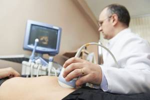
Preparation and performance of ultrasound
Ultrasound and Doppler sonography are performed routinely and urgently; the procedure does not require preparation. A woman should not drink a lot of liquid 2-3 hours before the examination, so as not to experience discomfort due to a full bladder during the procedure.
At the twenty-first week of pregnancy, an ultrasound is performed through the abdominal wall. To do this, the woman lies on the couch on her back. Places legs horizontally or bends at the knees. If necessary, the doctor may ask you to lie on your side.
If you need to measure the length of the cervix, you are worried about discharge, or the pregnant woman is overweight, which prevents the examination, then a transvaginal sensor is used to examine through the vagina.
A woman lies on a couch, her legs are bent at the knees and slightly spread apart. In this way, not only the fetus is examined, but also provisional organs - the placenta, umbilical cord, amniotic fluid.
Doppler ultrasound is performed with a surface sensor on the same principle as a conventional transabdominal (through the abdomen) ultrasound.
The procedure is painless with any method of examination. The examination time is 10–15 minutes.
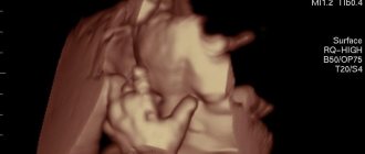
How to perform a fetal ultrasound, watch this video:
Diagnostic window in the 1st trimester of pregnancy
For the first screening study, these days include the period from 12 weeks. 2 days to 12 weeks. 4 days . It is in this interval that the fetus has already grown enough to evaluate the smallest organs (lenses of the eyes, heart), and the probability of ascertaining the most important morphological changes is significantly higher than at 11 weeks 0 days. On the other hand, every day the baby lives increases not only his height and body weight, but also the quality of the image on the ultrasound machine.
According to various authors, the frequency of congenital pathology reaches 5%, and for patients in this group it is especially important to identify the problem at an early stage. In such special cases, it may be necessary to expand the range of diagnostic procedures, including prenatal karyotyping (obtaining samples of fetal tissue or amniotic structures to determine its karyotype). Carrying out these procedures takes time, from preparing the necessary tests for the pregnant woman, ending with directly invasive diagnostics and obtaining results on the health of the fetus.
In some cases, an established fetal disease raises the question of the impossibility of prolonging pregnancy. According to the current legislation of the Russian Federation, the procedure itself for medical termination of pregnancy, by the woman’s decision, can be performed “... no later than the end of the twelfth week of pregnancy,” but “... no earlier than 48 hours from the moment the woman contacts a medical organization for an artificial termination of pregnancy” (clause 3.1, Clause 3-b of Article 56. Federal Law No. 323 dated November 21, 2012)2. In other words, if a patient applies for an ultrasound at 12 weeks 5 days, if a serious pathology is detected, she is no longer referred for an artificial medical abortion, but for termination of pregnancy, which is a more complex and traumatic procedure. Therefore, when asked about the best time to conduct the first screening, almost any practitioner will answer: “up to 12 weeks 4 days .