We tell you how and at what time it is possible to find out the gender of the child by ultrasound, boy or girl
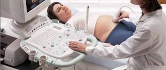
Boy or girl? is a question that sometimes interests future parents the most. You can find a dozen different ways to determine the gender of a child “without leaving home” - folk signs based on a woman’s taste preferences, calculations based on the date of birth of the mother, father, etc. In this case, there is no need to talk about accuracy, and the birth may end in a surprise. It is better to entrust sex determination to specialists, but even they can rarely predict sex with 100% probability, especially in the early stages. The usual way to find out the sex of the child is to perform an ultrasound examination of the fetus. But at what stage of pregnancy will the result be most accurate and unambiguous? It's worth looking into it in more detail.
To find out which period of pregnancy will be the most optimal for determining gender, you need to understand the characteristics of the baby’s development in the womb.
Initially, “sexual characteristics” are laid down by the father. This happens at the moment of conception. That is, when a sperm fertilizes an egg. After all, all children receive an X chromosome from their mother, and it all depends on which father’s sperm wins the “race” and reaches the egg first:
- If with an X chromosome, a girl will be born (genotype XX);
- If with a Y chromosome, you are supposed to expect a boy (XY genotype).
Naturally, none of the parents knows about the results of conception. Therefore, they are waiting for the moment when it will be possible to find out the sex of the child more or less reliably. A mandatory event during pregnancy is to visit an ultrasound specialist. It is at such appointments that you can find out for the first time the expected gender of the child.
Why do you need to determine the gender of your child?
The content of the article
There are several reasons why future parents want to determine the gender of their baby. First of all, it is interest and curiosity that does not allow one to wait until birth in ignorance. In addition, some couples want to prepare the room in advance, buy things and choose a name, and some parents dream of a boy or girl.

The importance of determining the sex of a child is also due to medical indications. When it comes to chromosomal pathologies, such information about gender is vital.
It is safe to perform ultrasound on newborns
An ultrasound scanner is an analogue of the echolocation organ of dolphins. If your child is worried about the upcoming procedure, tell him how dolphins scan the water column for fish by emitting ultrasound. Likewise, the doctor will use the device to scan the child’s body to understand whether everything is in order in his body.
Ultrasound is prescribed:
- Newborn children. Neurosonography or brain examination. It is carried out until the “fontanelle” closes
- Infants – for dislocations, subluxations, ECHO-ECG
- Children under 1 year old
- Children over 1 year of age as directed by a pediatrician
Our clinic has a comfortable, trusting atmosphere. Children feel great when visiting a doctor, which means they are happy to come for a second examination. We use the most modern equipment to obtain the most accurate research results.
Why should you choose diagnostics in our clinic? For most children, having an ultrasound in the hospital can be a real ordeal. It's no secret that kids literally become infected with emotions from each other. While you sit in line, the child will get tired, exhausted and guaranteed to start acting up. This is troublesome for parents and inconvenient for the doctor, because the little patient needs to be calm during the procedure.
Our clinic differs from municipal clinics in its client-oriented approach. This means that the ultrasound doctor will see you at the appointed time, and you can calmly go about your business after the procedure.
- We employ specialists of the highest and first categories
- The doctors are friendly and easily get along with children
- The diagnosis will be comfortable, and the result will be ready quickly
The child will feel great after visiting the doctor, and the treatment is carried out in a timely and effective manner.
What to do on the day of ultrasound
The doctor will definitely tell you in advance what rules you need to follow on the day of the ultrasound.
- Almost half of the studies do not require special preparation, but if we are talking about the internal organs of the abdominal cavity, you will have to bring the child on an empty stomach. It is important to explain to the baby why this is needed so that he does not get nervous.
- Infants are examined no earlier than 3-3.5 hours after the last feeding.
- The kidneys, bladder, and pelvic organs may require a full bladder at the time of the examination. This means that approximately 40 minutes before the procedure, the child needs to be given water.
- Considering that the appointment is strictly by appointment, the child will not have to endure the urge to urinate for a long time; the ultrasound will be done quickly.
Comfortable conditions, high-quality equipment and professionalism of doctors are the components of our success. Based on the diagnostic results, the doctor will prescribe therapy and other required treatments that will help eliminate the problem. Visiting a doctor and undergoing procedures is now comfortable and accessible at a time convenient for you. We will be happy to help you with the examination so that your baby grows up healthy!
Development of the fetus and its sexual characteristics by stage of pregnancy
In the first trimester, it is almost impossible to determine the sex of the child. One can only make an assumption about the gender of the fetus.
At 8 weeks of pregnancy, the size of the fetus is only 12 mm. It already has a small bulge - the genital tubercle, which does not differ in girls and boys until the 11th week, so it is impossible to identify the gender.
By the 11th obstetric week, the fetus grows to 45 mm. There are also no external differences in the genital organs yet. On an ultrasound scan of the embryo, you can see the anus, the recess of the urethra, the genital tubercle and the genital folds, surrounded externally by the labioscrotal tubercles.
The length of the embryo at week 13 is 64 mm. At this stage, in boys, under the influence of testosterone, the penis begins to form from the genital tubercle, and the body of the organ begins to form from the genital folds. There is an increased growth of the labial-scrotal tubercles, which, merging, turn into the scrotum. By this time, the foreskin is already fully formed.
The girl’s body contains a small amount of testosterone, so the external appearance of her genitals after 8 weeks remains virtually unchanged. The clitoris is formed from the genital tubercle, the labia minora from the urogenital folds, and the labia majora from the labioscrotal tubercles. The open urogenital groove forms the entrance to the vagina. The location of the external opening of the urethra is determined at the 14th week of pregnancy.
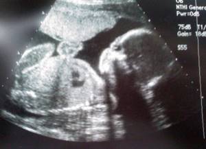
By the 20th week, the genital organs of both sexes are almost completely formed and can be identified - in other words, on an ultrasound you can already see who will be a boy or a girl.
How to prepare for an ultrasound and what features does the procedure have?
At week 20, an ultrasound is performed using the external method. The doctor can examine the fetus through the abdominal wall. A vaginal sensor is rarely used for such a long period of time: it is usually used due to the obesity of the pregnant woman, as well as when there is a suspicion of a threat of premature birth and other pathologies.
During the second screening, the procedure is not performed in an examination chair. No special preparation is required for the examination. There is no need to drink large amounts of liquid or limit yourself in food. The uterus is large, and the intestines are located far away, so the doctor’s view is open.
When can you most accurately determine the sex of a child?
The optimal time to determine the baby's gender is the second or third trimester. Starting from the 20th week, a specialist can most accurately determine the result based on the already practically formed genitals of the child. On the ultrasound screen, only the scrotum is visible in boys; often the penis itself is not visible. But in girls, the labia majora are very clearly visible.
It happens that during the examination the child is not mobile enough or turns around in such a way that it is quite problematic to examine the genitals even at the third planned ultrasound. Sometimes a boy “hides” his penis by tightly squeezing his legs, and may mistakenly “call himself” a girl. Or, conversely, a girl can be identified as a boy by confusing the umbilical cord with a penis. Therefore, to ensure the reliability of the result, the ultrasound procedure is repeated.
An experienced professional sonographer can determine the sex of the child using other indicators. So, boys have a thicker umbilical cord, much more amniotic fluid, and the weight of the fetus is significant. In girls, the opposite is true - a thin and shorter umbilical cord, less weight and volume of amniotic fluid.
On what factors does the accuracy of the research results depend?
Determining the sex of the fetus using ultrasound is an informative and safe method, but is not 100% accurate. Factors influencing the result:
- gestational age;
- position of the fetus inside the uterus;
- volume of amniotic fluid;
- abdominal wall thickness;
- resolution of ultrasound equipment;
- doctor's experience and qualifications.

Methods for determining sex by ultrasound in the early stages
The generally accepted way to determine a child’s sexual characteristics is ultrasound. This informative, painless and safe method has no contraindications and allows you to determine the sex of the baby with an accuracy of 80 - 99%. The reliability of the result will be influenced by the doctor’s experience, the quality of the equipment and the stage of pregnancy at which the examination takes place.
The gender of the child is determined at the moment of conception. It is impossible to change it, no matter how the media instills the idea of genetic engineering. You should not believe in “folk methods”, which are refuted by traditional medicine, and some are even dangerous (for example, drinking garlic juice every day).
It is possible to make an assumption about gender starting from the 12th week, but subject to high-quality equipment and an experienced, qualified doctor. Otherwise, the answer may be wrong.
There are several indirect signs by which future parents can find out the result:
- The angle formed by the back of the child and his genital tubercle
. An angle of less than 30° indicates the development of a girl. If the angle is greater, the woman is carrying a boy. - Localization of the placenta
. The likelihood of having a boy increases significantly if the placenta is located in the uterus on the right side. If the “children’s place” is on the left, you can quite expect a girl. This relationship was established by the Canadian uzologist Dr. Ramsay, after whom this method of sex determination is named. Surprisingly, many note the reliability of the method; - Skull shape
. A square skull and jaw indicate the birth of a boy, round ones indicate a girl.
Gender of the child: diagnostic errors
The shorter the period for which the study is conducted, the greater the likelihood of receiving an erroneous answer. But even at a later date, an experienced professional will not be able to determine the gender 100%. Errors may occur for the following reasons:
- Inexperience or unqualification of the doctor.
- Low-quality or outdated equipment - blurred images and shallow penetration depth of ultrasound beams distort the reliability of the result. The high resolution of the equipment and the presence of Doppler reduce the likelihood of error.
- Conducting the study too early. All women, as a rule, are very curious, so they want to find out the gender of their unborn baby as soon as possible. But it is worth remembering that in the early stages the fetal genital organs are practically the same or not formed at all, and ultrasound in the first trimester has a different goal - identifying possible pathologies.
- Multiple pregnancy - babies can cover each other.
- A belated ultrasound in late pregnancy - the rather large size of the fetus does not allow it to move in the uterus; it sits so compactly in it that it unintentionally hides the genitals. This prevents the doctor from viewing them on the monitor.
- During intrauterine development, a girl's labia may swell. This is not a pathology, but, resembling the scrotum in appearance, it can distort the result.
Boys can “hide” their genitals and appear to be girls. Also, girls' umbilical cord or exposed finger can be assessed as a penis.
Common features of fetuses of both sexes
Until a certain period, female and male embryos have the same appearance. The reason for this similarity is that the genital organs of boys and girls are formed from the same structures (Müllerian and Wolffian ducts).
At an early stage of differentiation, the reproductive organs of fetuses of both sexes are very similar and can only be distinguished from each other using high-resolution equipment, such as 4D.
As for the remaining parts of the body of babies developing in the womb, they are structured the same regardless of the child’s gender. Observations have shown that girls grow more slowly, and boys are more sensitive to nutritional deficiencies and other unfavorable factors.
Determining the sex of a child using 3D and 4D ultrasound
Three-dimensional and four-dimensional ultrasound is the most reliable way to determine the sex of a child using ultrasound. This type of research allows you to examine individual parts of the child’s body and even which parent he looks like. The image of the fetus is so realistic that future parents can get an answer even without the help of a doctor. In this case, the error is practically eliminated.
The optimal period for 3D and 4D ultrasound is 25–30 weeks, when it is possible to obtain the most reliable result. The ultrasound power is the same as with two-dimensional ultrasound. It is absolutely safe and does not harm either the child or the expectant mother.
In addition, it differs from 2D ultrasound in its greater capabilities, since it provides additional information that contributes to the successful and timely diagnosis of many abnormalities.
Where can I get an ultrasound during pregnancy?

Waiting for the birth of a new family member is always exciting. Parents want to know in advance who will be born to them. This will allow you to choose a name, an outfit for discharge, and buy children's furniture in accordance with the design and color. In order not to wonder whether ultrasounds are mistaken about the sex of the child, you need to take your choice of clinic seriously. You can’t always trust colorful advertising signs, because in them medical centers position themselves only from the best side. At the same time, the reputation, qualifications of specialists, and available equipment are not announced.
You can find a complete list of highly rated clinics on our website. The card of each medical center contains detailed information about the address, rating, reviews, equipment used and the cost of the study. Each visitor can find out the clinic’s rating and read reviews from patients who have previously been examined.
On the website you can sign up for an ultrasound scan at any time in order to find out the sex of the child. The advantage of the service is the provision of a discount on ultrasound diagnostics to each registered patient. For detailed information just call us.
Methods for determining the sex of a child without an ultrasound
Some future parents use other methods to determine the gender of their baby: some use “grandmother’s” methods, others use different medical methods. It is worth remembering that many of them are “dubious” and unreliable, and some are even dangerous.
Invasive methods
- Villus biopsy of the placenta embryo
- chorion. This method gives a 100% result, but is quite dangerous - it can cause miscarriage or infection. Therefore, it is used only as a last resort - for genetic diseases of the fetus. The method itself is quite unpleasant. To take biopsy material, long special forceps are inserted into the amniotic sac. - Puncture of the amniotic sac (amniocentesis)
. The method is also uncomfortable and painful. It involves puncturing the amniotic sac and taking a small amount of amniotic fluid for examination. The duration of the procedure is 16–18 weeks. Amniocentesis also poses a risk to mother and baby, risking miscarriage or infection.
Non-invasive methods for calculating the gender of a child
- By date of ovulation and conception
. If sexual intercourse leading to fertilization occurred 3 to 5 days before ovulation, there is a high chance of having a girl. If 1-2 days in advance or directly on the day the egg is released, it’s a boy. The online ovulation calculator is here: FOLLICULOMETRY. - Based on the combination of parental blood groups through the AB0 system
. Special tables are used for calculations, but the method itself is refuted by traditional medicine. - By “renewing” the blood using age division
. It is necessary to divide the age of the mother by 3, and the age of the father by 4. Whichever parent has the lower value will have the child of that gender. - By heart rate
. Using this method, gender is determined at 12-14 weeks. The threshold value for boys is 140 beats/min. Exceeding this indicator indicates the girl’s development. - By the appearance of a pregnant woman
. “Loss of beauty” of the expectant mother during pregnancy, a round belly, darkening of the nipples, increased irritability and moodiness indicate the birth of a girl. - A sharp belly and a great mood
are “masculine” signs. - At the first movement
. If a woman feels the first movement of the fetus on the left, it means the birth of a girl, and on the right, a boy.
Thus, ultrasound is the most reliable, and, most importantly, safe method for determining the sex of the unborn child. Yes, there is a possibility of error, but this is not a reason to endanger yourself and your child out of curiosity using invasive calculation methods.
Non-invasive methods are quite questionable and not scientifically confirmed, so relying on them or not is an individual choice of each future parent. Doctors do not recommend trusting them, considering them “grandmother’s” superstitions and nothing more.
The discovery of the presence of extracellular fetal DNA in the mother's blood was a breakthrough in prenatal research. All dead fetal cells enter the mother's bloodstream through the placenta. It is through its walls that the exchange of various nutrients, oxygen and waste products of the fetus occurs.
Thus, fetal DNA begins to accumulate in the mother’s blood from the first month of pregnancy. A sufficient amount of fetal fraction for non-invasive testing is achieved no earlier than 9 weeks of pregnancy. On average, the content of the fetal fraction in pregnant women at this stage is 4-5%.