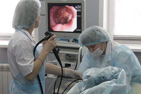
Colonoscopy
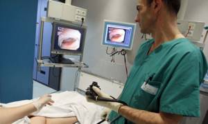
Colonoscopy is a method of diagnosing the intestines, during which the condition of the mucous membrane of the large and small intestines is examined and assessed using an endoscope.
Unlike endoscopy, the device is inserted through the patient's anus. Before the procedure, it is necessary to prepare several days in advance. You can read about this here. An examination of the intestines can be performed using colonoscopy under anesthesia (sedation, anesthesia). The procedure does not cause pain, discomfort and does not take much time. During the examination of the intestine, biological material may be taken (biopsy) for histological examination. This procedure can remove polyps and small tumors without surgery. At KDS Clinic you can undergo an intestinal examination using fibrocolonoscopy (FCS). Proctologists will carry out highly accurate diagnostics and determine the causes of the disease. In Moscow medical clinics this procedure is more expensive, we are pleased to offer you lower prices for diagnostics.
Symptoms to watch out for
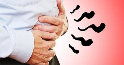
In what cases should you consult a doctor and undergo a comprehensive examination of the intestines? According to medical statistics, patients notice nonspecific symptoms, including:
- flatulence;
- pain in the abdomen and anus;
- gastrointestinal disorders, constipation and diarrhea;
- prolonged nausea and vomiting;
- lack of appetite;
- belching after eating;
- bloating;
- blood impurities in stool;
It is recommended to undergo an intestinal examination if there is constant malaise, weakness and drowsiness, weight loss or rapid obesity with adequate nutrition. Diagnosis is mandatory if the patient has undergone surgery to remove a tumor in the intestine. Annual preventive diagnostics are carried out for people over forty years of age. At this age, age-related changes occur in the body and intestinal pathologies develop.
Patients who suffer from colorectal cancer of the colon and rectum attend bowel screening on a regular basis. This disease causes death in most cases if there is no timely treatment and medical supervision.
In 95% of cases, when pathology occurs, it is possible to stop the development of the disease and be cured completely. To do this, you should begin treatment, remove intestinal polyps, thereby stopping the growth of tumors. As a rule, polyps develop into malignant tumors in a short period of time. It is recommended to visit a proctologist and gastroenterologist not only for older people, but also for young people. In recent years, the statistics of inflammatory processes in adolescents and young people of working age has increased.
If there are no specific symptoms, the patient has no idea about the development of the disease and, accordingly, does not seek help from a qualified specialist doctor. The early stage quickly turns into an advanced stage. Determining the presence of the disease is only possible through laboratory tests and diagnostic tests. That is why preventive studies must be carried out without fail even in the absence of symptoms.
Irrigoscopy
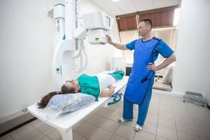
An examination of the intestines is performed using x-rays.
Before the procedure, the patient must cleanse the intestines using laxatives, enemas, and refrain from eating. A few hours before the examination, the patient takes a liquid with a contrast agent. The drug fills parts of the intestines. Using x-rays, the doctor evaluates the intestinal structure, lumen and contours. Examination of the relief of the intestinal mucosa allows us to identify the presence of fistulas, tumors, ulcers, scars and other neoplasms. The method is painless and has a low degree of radiation exposure.
Irrigoscopy is used in the following cases:
- for diagnosing intestinal obstruction
- purulent discharge comes out of the intestines
- presence of bleeding
- severe pain in the anus and colon area
- if it is not possible to use a colonoscopy
Prices
| Name | Price |
| Anoscopy | 1500.00 rub. |
| Examination of the rectum with rectal mirrors | RUR 800.00 |
| Sigmoidoscopy | RUB 2500.00 |
| Endoscopic examination of ENT organs (diagnostic) | 2000.00 rub. |
* Please note that the website aksis-med.ru is not a public offer. All information presented on the website about our prices and services is for informational purposes only. To clarify current information, you can call +7. Show all prices
Intestinal examination with capsule
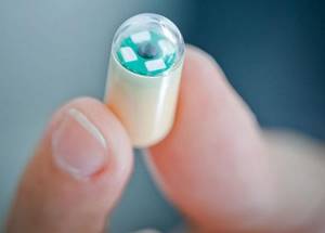
The procedure is performed using an enterocapsule equipped with a video camera. The technique is considered minimally invasive and is performed on an empty stomach. Examination of the intestines with a capsule can detect the presence of cancer, hidden bleeding, congenital pathologies and tumors. A recording device is attached to the patient's body, after which he swallows the capsule. The camera moves around the stomach and intestines, transmitting information to the computer. After the procedure, the capsule leaves the body naturally.
Small intestine examination
The small intestine can be examined in several ways, including ultrasound, fibrocolonoscopy, endoscopy and radiography. During diagnosis, the presence of pathologies will be checked in the jejunum, ileum and duodenum.
One of the most effective and modern research methods is double-balloon enteroscopy. This procedure will help not only to examine, but also to carry out such manipulations as:
- excision of polyps
- tumor detection
- blocking bleeding in the small intestine
- removal of foreign objects
You can undergo such an intestinal examination only after consulting a gastroenterologist. The medical clinic is attended by the best specialists in Zelenograd and Moscow, who will quickly help diagnose, examine and prescribe treatment for intestinal diseases.
Colon examination
To diagnose colon diseases, a comprehensive examination is performed. The patient needs to donate blood and feces for analysis and undergo an endoscopic examination procedure. Basic methods for examining the rectum:
- fibrocolonoscopy;
- general blood analysis;
- stool analysis for dysbacteriosis;
- digital rectal examination.
The diagnostician identifies the presence of diseases localized in the anal area (hemorrhoids, fissures, tumors). A rectoscope is used for deep examination.
Methods for diagnosing intestinal diseases
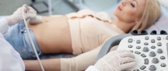
Today, the all-Russian medical examination program includes the analysis of stool for occult blood for people over forty years of age. Laboratory testing reveals signs of the presence of malignant tumors in the body. After receiving the test results, the attending physician decides to conduct further diagnostic studies. Abdominal ultrasound and computed tomography are often performed.
The following types of research are used in diagnostic centers, private and public clinics:
- laboratory methods;
- instrumental methods;
The diagnostic method used depends on the patient’s condition, the presence of concomitant diseases, and complications.
Laboratory methods
After the initial consultation, the patient is required to undergo laboratory tests. These include a general analysis of blood, urine and feces. The results are analyzed by the attending physician, after which the presence of problems and abnormalities in the body is determined. Thus, the following are diagnosed:
- infectious and inflammatory processes in the body;
- parasitic diseases;
- gastrointestinal bleeding;
- malabsorption syndromes;
- presence of neoplasms.
Tests are carried out on an empty stomach in the morning, since it is at this time that the test results will be most accurate. Blood is taken from a vein or finger. If there is a suspicion of intestinal pathology, ESR levels increase, anemia or anemia develops. A biochemical blood test will detect malabsorption of nutrients in the intestine.
A general urine test conducts detailed studies that are associated with impaired absorption of substances and dehydration. We are talking about constant diarrhea and vomiting. To do this, the patient uses a sterile container to collect biological material. Urine collection is carried out in the morning. After this, the materials should be delivered to the diagnostic center as soon as possible. A signal of impaired functioning of the body is called saturated color of urine, turbidity, and increased density.
The coprogram allows you to examine feces and detect the presence of an infectious and inflammatory process.
How to examine the intestines?
Often a person may be bothered by inflammatory processes, impaired passage of food and feces, tumors and many other intestinal problems.
The condition of the initial parts of the digestive tract (esophagus, stomach and duodenum) is best determined using a gastroscope (endoscope 110 cm long).
Next is the small intestine, which is about 6 meters long, starting behind the stomach, from the duodenum, beyond which a standard endoscope does not pass. Therefore, to study it, we recommend x-ray methods, or a video capsule. Unfortunately, the video capsule has poor optical resolution, is uncontrollable, and is quite expensive. The study lasts about a day. But she has the opportunity to see parts of the intestine that are inaccessible to the endoscope.
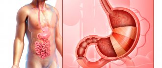
The large intestine has a length of up to 1.5 meters and consists of several sections (cecum, ascending, transverse and descending sections, sigmoid and rectum).
For its study, special endoscopic devices 170 cm long (colonoscopes) have been developed, which have a unique combination of a video system, fiber-optic light guides and fine mechanics, which allows you to control the moving part of the device. There is also a water and air supply system and an instrumental channel that allows you to insert instruments for performing operations and taking tissue for examination under a microscope (histology). This method is today considered the gold standard because of its information content, harmlessness and the possibility of carrying out therapeutic and diagnostic manipulations inside the intestine.
The quality of the examination depends on adequate preparation. The day before, the intestines need to be cleansed with enemas or laxatives. With insufficient preparation, solid contents of the intestine, which cannot be removed by suction during the examination, can clog the optics and channels and will not allow the procedure to be completed in full.
In addition, the presence of adhesions in the abdomen, pressure on the intestine from the outside (tumors, cysts), congenital anomalies (sharp kinks, narrow pelvis, etc.) may prevent the endoscope from being passed to its full length. Then the intestines are examined only along the accessible length.
There are many other research methods, and none of them are absolute. The intestine, like a hollow organ, can be filled with a contrast agent (for example, barium sulfate of a creamy consistency) - drunk, or administered as an enema, and then a series of x-rays taken. This makes it possible to see the location of various parts of the intestine, the rate of their emptying (passage), filling defects (ulcers, polyps, tumors). There are also methods for three-dimensional reconstruction of the intestine using computed tomography. Unfortunately, all these methods have limited diagnostic ability, use ionizing radiation and do not provide the opportunity to obtain material for histological examination.
There are also laboratory tests of blood and stool that can detect enzymatic insufficiency of the liver and pancreas, the presence of tumors (tumor markers), parasites, flora disorders, gluten deficiency, etc.
That is why all available methods do not exclude, but only complement each other . And only a frank conversation with a clinician can help you understand the variety of methods, jointly develop a rational examination plan and prescribe adequate treatment.
The article was prepared by specialists from the endoscopic department of ACMD-MEDOX
Testing for mannitol/lactulose
An effective test for intestinal permeability is the intake of mannitol and lactulose - sugars that are harmless to the body, which are not absorbed by the body and are excreted in the urine after 6 hours. To complete the test, it is enough to eat 5 grams of both substances. The absorption of mannitol by the intestinal wall is more active than the absorption of lactulose, respectively, the absorption of these substances is 14% and 1%.
In the urine of a healthy person with a normal intestinal wall, much more mannitol is always found than lactulose, the normal ratio is 0.03. If the content of lactulose in the urine is increased, then this test result indicates increased permeability of the intestinal wall, since lactulose was able to penetrate through the “leaky intestine” into the patient’s blood, and from there into the urine.
Back to list Previous article Next article
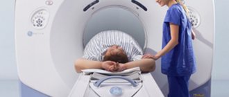
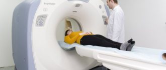
Will an MRI show colon cancer?
Malignant neoplasms of the digestive system occur equally often in men and women. An asymptomatic course in the initial stages makes timely diagnosis difficult, so suspicion of the development of oncology is one of the main indications for intestinal MRI.
Magnetic resonance imaging makes it possible to study the pathologically changed area in detail. If a tumor is present, the doctor evaluates the size, localization of the lesion, and the interaction of the tumor with surrounding structures. The malignant process is characterized by the absence of a clear contour; atypical cells “grow” into healthy tissues.
A cancerous tumor accumulates gadolinium chelates more slowly on contrast-enhanced MRI of the intestine. This occurs due to the pathological tortuosity of its own blood vessels. Magnetic resonance scanning shows formations with a diameter of 3 mm, suggesting the nature of the process. The final diagnosis is made based on the results of laboratory tests.