Every woman involuntarily thinks about the onset of conception if she experiences characteristic symptoms or experiences a delay in menstruation. The fastest way to determine a possible early pregnancy is to do a rapid test. In case of positive or contradictory results, he may push the woman to go to the gynecologist for an ultrasound. But at what day of delay will an ultrasound show pregnancy? And when should you go for testing? You will find answers to these questions below.

Timing of pregnancy determination by ultrasound.

Visualization is difficult: what does this mean?
Pregnancy and the birth of a child are the most touching, exciting, love-filled, and unforgettable stage in the life of any woman. When the expectant mother learns about the birth of a new, small life inside herself, along with a feeling of great happiness, questions arise regarding the timing of conception.
The most important question is: “At what stage can a pregnancy be detected by ultrasound?”
Because, based on this, doctors will determine the state of development of the unborn child and calculate the date of the upcoming birth.
Preparation for ultrasound examination
Before going for a transvaginal ultrasound, a woman should empty her bladder. If we are talking about a transabdominal ultrasound, then it should be complete. To do this, you will have to drink about a liter of water half an hour before the procedure, even if you don’t really want to. In emergency cases, fluid can be administered using a catheter.
If in doubt, a woman should undergo an ultrasound no earlier than the tenth day, when the ultrasound will show pregnancy after a delay. Then the device is able to show the fertilized egg in the uterus. But the examination can give a 100% guarantee only after the twentieth day of delay. During this period, you can already hear the heartbeat of the unborn child. This will be absolute proof that the woman is pregnant and will help rule out a number of pathologies. One of them is anembryony - the presence of a fetal sac without an embryo in it. This pathology is accompanied by all the signs of pregnancy, however, in essence it is not it.
When will the ultrasound show the future baby?

At what time does an ultrasound show conception? - this question worries every woman, in case of expecting a long-awaited conception, a delay in menstruation, or simply because of symptoms indicating pregnancy. But first, you need to know how the fertilization period is determined :
- According to the woman’s calculations, starting from the last day of the delay;
- Based on the results of a gynecological examination;
- But the most effective and more accurate will be ultrasound diagnostics.
Ultrasound diagnostics in the case of a vaginal examination (that is, a condom is put on the sensor of the device and inserted into the vagina), provided that the examination is performed by an experienced doctor using a modern device, allows you to record the fact of successful fertilization for a period of 14 days after conception.
This means that on the ultrasound monitor, a device in the uterus, you can see a fertilized egg measuring 0.4 cm after a week of missed period or a month later according to the obstetric period. If you go for an ultrasound a little later, at 1.5 months, then already in such a short time, the device will record the heartbeat of the unborn child.
At what day of delay will an ultrasound show pregnancy? In the case of an abdominal examination (that is, diagnosis is carried out through the abdominal wall), pregnancy will be visible no earlier than 4 weeks of embryonic age, or at least 14-21 days after the 1st day of delay.
How is pregnancy determined?
The first and most important rule that any woman should learn is that you should not run to an ultrasound at the first missed period. Ultrasound diagnostics is an ineffective method for very early periods of gestation. Therefore, to understand whether a woman needs to make an appointment with a doctor, she needs to do a home rapid test. They are readily available in pharmacies, and the results are quite informative. How does he work?
Fertilization of the egg occurs during ovulation. If this happens, then after a certain period of time the production of the pregnancy hormone, human chorionic gonadotropin (hCG), begins. It is on the presence of a reaction to this hormone that the work of any rapid test is based.
To carry out the test, a paper strip soaked in the reagent must be kept in urine for some time or simply urinated on it. One strip on the test is always a control, the second determines the presence of this hormone (if it appears) or its absence (if the strip does not appear).
In the morning, the concentration of hCG in the urine is maximum, so doctors recommend taking the test in the morning. A test taken during the day or evening may be incorrect. This happens due to the liquid you drink throughout the day. As a result, the concentration of the hormone may decrease. It is also necessary to carefully check the packaging and expiration date of the test during purchase to avoid distortion of the results.
To determine conception as early as possible, it is better to use particularly sensitive tests. With their help, you can establish conception already in the first days of delay. How many of these tests can be performed? They are safe, because the amount depends entirely on desire.
Terms of pregnancy: embryonic and obstetric
The gestational age indicated in the maternal card, filled out when registering at the antenatal clinic, differs slightly from the actual time elapsed from the day of fertilization by about 2 weeks.
But there may be other options, for 1-1.5 weeks, depending on the length of the menstrual cycle. This is considered the norm, because we are talking about different periods: embryonic and obstetric:
- Embryonic - counted from the actual day of conception, which coincides with the date of ovulation. For women, it is not difficult to calculate the day of conception only if they keep an ovulation calendar.
- Obstetric period is the period that has passed since the last menstrual period, after which conception occurred.
It is this period, which is easier for a woman to determine than the embryonic period, that is considered a reference point for scheduling routine examinations and establishing the upcoming date of birth.
Sometimes, women wonder “can an ultrasound not show pregnancy”? - yes, maybe, in the case when the doctor cannot see the fertilized egg, a repeat examination is prescribed due to the probable risk of an ectopic pregnancy.
Advantages of detecting pregnancy in the early stages:
- Timely detection of ectopic pregnancy;
- Establishing the fact of successful conception and its duration;
- Determining the cause of delayed menstruation in the absence of a positive pregnancy test;
- Determination of the number of conceived embryos;
- Recording possible threats to pregnancy.
When to suspect pregnancy
There are several signs when you should take a pregnancy test:
- unusual sensations in the chest and pelvis area, breast swelling;
- drowsiness, lethargy;
- change in appetite or lack thereof;
- a previously unusual aversion to certain odors;
- the appearance of skin pigmentation, allergies, rashes;
- changes in the nervous system (irritability);
- dizziness, nausea.
If a woman is healthy and does not suffer from any menstrual irregularities, then determining her “interesting position” is quite simple - any deviation from the schedule, even for 2-3 days, can serve as a reason for conducting a test.
Qualities and benefits of ultrasound

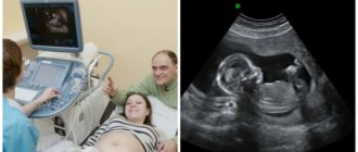
Ultrasound examination is a widespread, painless, very effective method for determining pregnancy and its timing.
The very first ultrasound machines were bulky and unable to produce a clear image on the screen. Therefore, studies often gave inaccurate conclusions. But modern devices are capable of transmitting more detailed information about the state of the human body.
In the modern world, there are various technologies for examining internal organs.
But it is ultrasound that is ideal for determining the timing of conception and the course of pregnancy for the following reasons:
- Determination of conception in the early stages;
- Ultrasonic waves do not harm mother and child;
- Clear visualization of the fetus;
- You can do an ultrasound at any time convenient for the expectant mother;
- No rehabilitation period;
Ultrasound radiation makes it possible to eliminate various types of damage to the skin and mucous membranes. Nowadays, a woman does not have to torment herself with the question “Where to get an ultrasound during pregnancy”?
Because there are a lot of possibilities. An ultrasound scan for pregnancy can be done from private medical institutions to local clinics.
Preparing for examination by a gynecologist during pregnancy

Many women, even before pregnancy, avoid visiting the gynecologist’s office, each with their own reasons for this. But while waiting for the baby to be born, they unwittingly become frequent visitors to various medical institutions. Of course, the first appointment with a gynecologist during pregnancy is an important event for every expectant mother, and in order for the result of the examination to be good, it is necessary to prepare for it.
- First, you should remember the characteristics of your menstrual cycle. When was your period on the last day, how long did it last, did you have any pain, etc.
- Secondly, the doctor will definitely create a personal medical record for you, which will indicate data about your health and the condition of the fetus. The gynecologist will need to know about your past illnesses, infections, bad habits, etc.
- Thirdly, the specialist will ask you about your sex life, the age at which you started having it, the number of partners, whether you have ever taken oral contraceptives, etc.
- Fourthly, the gynecologist is also interested in information about your previous pregnancies (if any), how they ended (childbirth, abortion, miscarriage), how many children you have.
We have listed the main topics that will be discussed at your first appointment with a gynecologist. To help you remember any questions you want to ask your doctor, write them down in a notebook and take it with you. Usually, many expectant mothers start keeping a special diary in which they note all the important information regarding their pregnancy.
We have also prepared for you a small list of recommendations that will help you prepare correctly for a gynecological examination:
- You should not douche before the examination; it will be enough to just take a shower, without using perfumed products, gels and deodorants for intimate hygiene.
- You should not shave your perineum before the appointment, because... this can lead to irritation, which in turn makes inspection difficult.
- Avoid sexual intercourse several days before your visit to the gynecologist.
Try to relax and the examination will be comfortable for you. If something bothers you, be sure to tell your doctor. Choose a time of examination that is convenient for you, the administrator of the A-medic clinic will help you find your way and make an appointment for the appointment day you need.
Scheduled diagnostics

After confirmation of successful conception, the expectant mother is registered with a gynecologist. At the antenatal clinic, she learns that during pregnancy, if the pregnancy progresses normally, she will have three scheduled ultrasounds. This procedure does not have any adverse reactions on the fetus or the expectant mother. There are times when, in order to more carefully monitor the course of pregnancy, the diagnosis is carried out 1–2 times more.
The dates for scheduled examinations are distributed by trimester, because each of them differs in the development and growth of the unborn baby:
During the first trimester (period from 10 to 14 weeks), diagnosis may show chromosomal abnormalities that can cause genetic diseases in the fetus. The features of anatomical development are also determined, and the estimated date of birth is calculated;
During the second trimester (period from 20 to 24 weeks), ultrasound shows the structure of the fetal organs, due to which there is a chance to detect deviations from the normal functioning of the heart, kidneys, gastrointestinal tract, liver, as well as malfunctions in the nervous system. At this stage, very often, the sex of the child is determined, you can see the movements of his arms and legs. The length and estimated weight of the fetus are measured, and the placenta is characterized. In the same trimester, a Doppler ultrasound examination is performed to determine the blood flow in the fetal vessels;
The third trimester (the period from 31 to 36 weeks) is considered the final trimester. At this stage, the diagnosis of organ development disorders that could not be identified earlier is carried out. The position of the fetus is determined, which by this time should have taken a position upside down, and the probable entanglement of the umbilical cord around the neck. The child’s parameters are still measured and the date of the upcoming birth is specified. In the third trimester, the doctor gives an opinion - whether the woman will give birth naturally, or will have to resort to a cesarean section.
Along with routine examination procedures, after 12–13 weeks, biochemical screening of the fetus is carried out. A similar method is used to identify pathologies, as well as for early detection of Down syndrome. Ultrasound of the pelvic organs
What does a pelvic ultrasound show? During a pelvic diagnosis, the doctor examines the uterus, appendages, ovaries, as well as the bladder and rectum. For a pregnant woman, this study is carried out to determine the condition of the reproductive organs and, of course, to examine the fetus, amniotic fluid, and placenta.
A feature of a pelvic examination can be considered the ability to detect pathologies such as:
- Ectopic pregnancy;
- Abnormal development of the uterus during pregnancy;
- The process of inflammation of the ovaries, uterus, vagina;
- Malignant and benign tumors on the organs of the reproductive system.
Screening ultrasound at the Bionika clinic in Krasnoyarsk
Prenatal screening is a complex of laboratory and ultrasound examinations aimed at identifying fetal malformations and complications during pregnancy. Screenings are carried out every trimester and include screening ultrasound, biochemical screening, and medical genetic counseling. Women at risk include:
- aged 35 years and above;
- those who gave birth to children with chromosomal abnormalities and developmental defects;
- with a history of miscarriages;
- those exposed to radiation or living in places with unfavorable environmental conditions;
- took drugs prohibited for pregnant women during the first trimester;
- with a history of regressive pregnancy;
- those who abuse alcohol and drugs;
- with genetic diseases in the family;
- survivors of a severe infectious disease in early pregnancy.
You can undergo complete prenatal screening for expectant mothers at the clinic. In “Bionika” it is possible to perform an ultrasound without queues and hassle, in comfortable conditions and under the supervision of specialists of the highest category.
Ultrasound of the cervix

Ultrasound examination of the cervix during pregnancy is prescribed as an auxiliary precaution during a routine examination by a gynecologist.
A timely examination makes it possible to detect and exclude various types of pathologies that can harm the health of the fetus and mother.
Throughout pregnancy, the uterine canal tends to change. This is considered the norm, since the physiological process of preparation for the upcoming childbirth is in full swing. Therefore, the slightest deviations in this process may be considered a threat to the normal course of pregnancy, which means you should not postpone visiting a specialist for a long time. The sooner the examination occurs, the greater the chances of a positive birth.
Containment of the fetus is the most important function of the cervix in a pregnant woman. Because, during most of the pregnancy, its closed position protects the unborn child and prevents various types of infections from entering. But after 36 weeks, the cervix becomes deformed, becomes shorter, softer and moves towards the center, thereby starting the process of its opening. Afterwards, a single canal is formed connecting the cervix with the external os. Its full opening reaches dimensions of 12 cm.
If you look at it from an anatomical point of view, the cervix is the entrance to the body of the uterus itself. The organ also includes: internal and external pharynx, which is covered with muscle connective tissue. As a result, a cervical canal is formed, smoothly connecting to the vagina. The muscle holding the unborn child in the form of a ring is located in the internal pharynx.
If the muscle loses tone, the cervix loses the ability to contain the child inside itself.
During pregnancy, the length of the cervix plays an important role. It determines the possible risk of premature birth. It happens that it may have a shortened state, which indicates isthmic-cervical insufficiency (premature pathological dilatation of the cervix due to strong internal pressure of the fetus and amniotic fluid).
For this reason, unfortunately, a miscarriage occurs. But in our time of modern technologies, timely treatment helps prevent premature initiation of the birth process and miscarriage.
Ultrasound picture of pregnancy
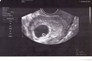
In the first weeks, when women want to visually confirm their pregnancy, it is possible to visualize only the fertilized egg. It is a hypoechoic round formation in the uterine cavity with a diameter of 3-6 mm, depending on what day after the onset of the delay.
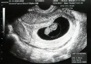
embryo at 7 weeks of pregnancy
If you do a transabdominal examination, then only the fertilized egg is visualized when its size exceeds 7 mm. In the first weeks of pregnancy, the growth of the ovum is quite intense and amounts to approximately 1 mm per day. The size of the fertilized egg during the first ultrasound examination determines the stage of pregnancy.
How is the research done and when is it carried out?
A planned anatomical ultrasound examination is prescribed for the period 14–16 weeks of pregnancy. But if in a woman who has previously given birth, with a previous pregnancy, the labor process began earlier than the appointed time, an ultrasound of the cervix is prescribed approximately at the end of the 12th week. After which, observations are repeated at intervals of two weeks.
The following indicators are considered additional reasons for ultrasound of the cervix:
- Miscarriage that occurred in the first pregnancy at a late stage;
- Conception with the presence of two or more fetuses;
- Various reasons for terminating a previous pregnancy;
- Suspicions of isthmic-cervical insufficiency;
Any previous surgical interventions in the field of gynecology.
Ultrasound of the cervix, as well as diagnostics to detect pregnancy, is done in two ways, abdominal and vaginal. In the first case, the uterus is visible through the anterior wall of the abdominal cavity. The doctor makes a conclusion about the position, condition, parameters of the uterus, and also gives a description of the external pharynx.
Vaginal ultrasound examination is performed by inserting a special sensor into the vagina. This case is more effective than the previous one, since it becomes possible to measure the length of the cervix, the size of the internal pharynx and the cervical canal.
As a rule, special preparation for an ultrasound is not required; only in the early stages of pregnancy, the doctor may ask you to pre-fill the bladder.
Diagnosis through vaginal and abdominal examination is considered absolutely safe for pregnant women. Visualization is difficult: meaning
When during an ultrasound, your doctor, based on the examination results, says that visualization is difficult, this is not a reason to panic. For personal peace of mind, you should understand what this means.
If the pregnancy proceeds without any problems, a routine ultrasound examination is performed during the 24th week. An ultrasound of the pelvic organs in the first trimester will not provide as much important information as in the second trimester.
Therefore, during this period, future parents will hear the long-awaited and joyful news about the gender of their baby, as well as whether there are any abnormalities or pathologies in the development of the fetus.
The size of the unborn baby in the second trimester is quite large, which allows the doctor to clearly examine all its organs. In addition to growth and assessment of the child’s condition, at 24 weeks, the doctor will examine the placenta, which by this time has become denser in structure than in the first trimester, and will also diagnose the amount of amniotic fluid.
Visualization is difficult during pregnancy; in simple words, this is when the fetus is located in such a way that none of the above is simply visible to the doctor. Of course, this does not mean that you should be upset or consider it a violation in the child’s development, because there will be another third planned ultrasound examination ahead, at 34 weeks.
Types of ultrasound diagnostics
There are two modern methods:
- Transvaginal ultrasound. It is used after the third week of pregnancy, that is, on the 7th–10th day of delay. The countdown is from the first day of the last menstruation. This technique is rarely used at earlier stages, since for a qualitative study the size of the embryo should be about 0.5-0.7 cm and before it simply will not be visible. It is more often used as a screening.
- Transabdominal ultrasound. This study is carried out with a device whose sensor is placed on the front wall of the abdomen. In this case, the distance to the uterus is quite large. This nuance allows for accurate diagnosis no earlier than the fifth week and later. Otherwise, the results may be erroneous. Both methods are considered safe and cannot in any way harm the woman’s health or affect the development of the embryo.
Cost of an initial examination by a gynecologist during pregnancy
An appointment with a gynecologist during pregnancy is very important and should not be ignored. In many ways, the baby’s health depends on your well-being. This specialist will accompany you throughout your pregnancy; he will be able to promptly determine whether there is a threat to the fetus or you, and quickly take the necessary measures. And for this assistance to be qualified, you should choose experienced doctors and proven clinics. Multidisciplinary medical is responsible for the quality of services provided! The cost of a consultation with a gynecologist during pregnancy is only 900 rubles, the price of a repeat examination is much less - 750 rubles.