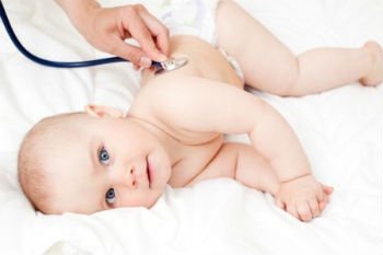
- Primary signs of pneumonia
- Viral pneumonia
- Acute pneumonia
- Atypical pneumonia in children
- Latent pneumonia in children
- Sluggish pneumonia
- Focal pneumonia in children
- Hilar pneumonia
- Community-acquired pneumonia
- Symptoms of inflammation in children
- Prices
- Make an appointment
Primary signs of pneumonia
Like other diseases, pneumonia needs to be treated at the very beginning. This will avoid complications and sometimes save the lives of babies. But there are also difficulties here, since the symptoms of the disease are one-to-one similar to the course of the flu or an acute respiratory infection. In addition, in some children, pneumonia begins sharply, suddenly, while in others, symptoms appear slowly, gradually.
Among the most common signs of pneumonia are the following:
- severe paroxysmal cough;
- sputum discharge;
- rapid breathing (over 40 breaths per minute);
- tachycardia (fast pulse);
- body temperature rises to 38? C and above and lasts longer than three days;
- increased sweating;
- redness of one of the cheeks.
“Parents should know that if a child has suffered from an acute respiratory infection, and a few days later he has a high fever, then this is a sure sign of a complication - an inflammatory process in the lungs.”
Despite the similarity of symptoms with other diseases, a combination of two or more signs should be alarming and cause you to seek emergency medical help and a more in-depth examination. Otherwise, if the presence of pneumonia is confirmed, the disease will manifest itself in more severe forms.
Insidious “silent” pneumonia
In up to half of all cases of pneumonia, in the first stage the disease occurs without classical symptoms, and general symptoms become the main ones. In such situations, a stethoscope is used for diagnosis, and radiography is used as confirmation.
A common feature of the initial stage of pneumonia is a condition called “silent” pneumonia. It is accompanied by the filling of the lungs with inflammatory fluid and blood flow, which makes listening difficult. Experienced specialists, if a disease is suspected, carry out repeated listening after 12-14 hours, since at this time the condition of the organ can change significantly.
Forms of the disease by type of pathogen
Bacterial pneumonia: types and signs
Bacterial pneumonia is an infectious disease caused by various pathogenic microorganisms. Statistics show that most often preschool children and people of retirement age suffer from bacterial pneumonia. Among the factors conducive to development, one can note a decrease in the body’s immune functions.
All pneumonias of this type are divided into nosocomial (hospital), in this case, infection occurs in a medical institution and out-of-hospital (home). The hospital-acquired disease is very severe and difficult to cure. This is due to the resistance of pathogens to antibiotic drugs.
The main causative agents of bacterial pneumonia are:
- hemophilus influenzae;
- mycoplasma;
- pneumococci (Streptococcus pneumoniae);
- staphylococci;
- Pseudomonas aeruginosa;
- chlamydia;
- Klebsiella and many others.
Symptoms and features in one-year-old children:
- In most cases, the lesion is focal, less often - lobar;
- In more than 70% of cases it is complicated by pleurisy;
- Occurs with increasing temperature;
- Accompanied by a cough - initially dry, then wet, with sputum;
- Patients experience impaired breathing;
- The disease progresses in waves.
Features of children over 5 years old:
- The disease begins gradually;
- Among the symptoms there is a cough, paroxysmal, with a small amount of sputum;
- Dry wheezing can be heard;
- Body temperature is subfebrile (from 37 to 38°C) with periodic increases to 40°C;
- Breathing is accompanied by pain in the chest;
- In one case out of five, pneumonia is not auscultated, but is detected using an x-ray.
Pneumococcus:
- Begins acutely;
- The disease is accompanied by chills and weakness;
- The temperature can be very high, or, conversely, drop to critical levels;
- When inhaling, the child feels pain;
- The cough is accompanied by the release of “rusty” sputum;
- There is redness of the cheek on the side of the affected organ;
- When exhaling, the child groans;
- Heart rate increases;
- Patients refuse to eat;
- Blood pressure decreases;
- The skin turns bluish, especially in the area of the nasolabial triangle and fingertips.
Staphylococcus:
- Affects infants;
- Accompanied by frequently recurring chills;
- Often becomes a symptom of sepsis;
- The course of the disease is severe;
- High temperature;
- Severe signs of intoxication;
- Dyspnea;
- Lung tissue is in most cases significantly damaged.
Pseudomonas aeruginosa
- More often occurs in newborns and children under three years of age;
- Develops acutely, the patient quickly reaches a serious condition;
- High temperature with characteristic morning fever peaks;
- Symptoms of intoxication are present;
- Dyspnea;
- Blue coloration of the skin (cyanosis);
- Increased heart rate.
Klebsiella
- Affects newborns and infants;
- Has an acute onset;
- Body temperature can rise to 40°C;
- Chills;
- The cough is severe;
- Separation of viscous sputum with blood clots;
- Often combined with other infections.
Viral pneumonia: pathogens and signs
Pneumonia can be caused by various viruses, most often it is provoked by parainfluenza virus, chickenpox, measles, cytomegalovirus, viruses of categories A and B. In this case, the child’s pulmonary alveoli are filled with purulent exudate (liquid).
This form of pneumonia affects both children under one year old and older ones. Among the main signs of the disease are the following:
- in the initial stage, the cough is dry, “barking”, and with the development of inflammation it turns into a wet one;
- rapid breathing, whistling often appears;
- when coughing, the patient feels pain in the area of the shoulder blades or chest;
- the temperature is elevated;
- cyanosis of the skin of the fingertips and nasolabial triangle;
- decreased appetite;
- chills;
- dyspnea;
- headaches, muscle, joint pain.
Often, pneumonia is “masked” as a common cold and it becomes difficult to recognize. But with pneumonia, unlike a common cold, a decrease in temperature is not observed even 2-3 days after the onset of the disease.
Initially, a viral infection develops in the first 2-3 days, and then, from 3-5 days, an infection of bacterial etiology occurs. Pneumonia becomes viral-bacterial.
Acute pneumonia: features of the course and symptoms
Acute pneumonia (pneumonia acuta) is a disease that has different etiologies and, depending on the pathogen, different symptoms. Most often, this form of the disease occurs in children under 3 years of age. It is characterized by the presence of infiltrates in the lung tissues (accumulations of cellular elements with the addition of blood and lymph particles) and filling of the alveoli with exudate containing neutrophils (leukocytes that protect against infectious pathogens).
Acute pneumonia can be caused by:
- viruses;
- bacteria;
- atypical pathogens.
Pneumonia in its acute form can be either an independent disease or a complication of previously suffered diseases. Depending on the pathogen, symptoms of acute pneumonia may include the following conditions:
- signs of intoxication - fever, lack of appetite, weakness, etc.;
- when listening, changes are detected - noises, wheezing, short breathing, etc.;
- X-ray reveals areas of infiltration in the form of darkening;
- the disease in most cases is accompanied by a cough;
- respiratory failure is observed (shortness of breath, rapid breathing, etc.)
The main predisposing factor for the development of pneumonia is hypothermia.
Atypical pneumonia in children: symptoms and signs
Atypical pneumonia, or otherwise acute respiratory syndrome, is a certain form of pneumonia caused by atypical microbes - chlamydia, mycoplasma, coronaviruses, legionella. The peculiarity of atypical microorganisms is that they can multiply intracellularly. Moreover, unlike other bacteria, they are not able to exist outside the “host” cells.
Symptoms of atypical pneumonia depending on the pathogen:
Mycoplasma
More often diagnosed in children over 5 years of age. Rarely, but still there are cases of mycoplasma pneumonia in newborns.
- The disease begins gradually;
- Among the symptoms there is a cough, paroxysmal, with a small amount of sputum;
- Dry wheezing can be heard;
- Body temperature is subfebrile (from 37 to 38°C) with periodic increases to 40°C;
- Breathing is accompanied by pain in the chest;
- In one case out of five, pneumonia is not auscultated, but is detected using an x-ray.
Chlamydia
Most often affects children over 5 years of age; the first obvious symptom is a dry cough, which then turns into a wet cough.
- Occurs simultaneously with pharyngitis;
- The temperature is often subfebrile, a significant increase is possible, but without chills;
- The disease is accompanied by headaches, muscle pain, and general weakness;
- Auscultation reveals dry rales;
- Often there are no clear signs of damage on x-rays.
Coronavirus
It can affect the body of children of any age, but it is more severe in children under 2 years of age. The incubation period lasts 2-3 days
- Runny nose;
- Pain when swallowing;
- General malaise, muscle weakness;
- Headache;
- Bronchial obstruction (a form of respiratory failure that occurs due to obstruction of the bronchial tree).
Legionella
The disease is recorded in children of all ages, including infants. Average incubation period 3-5 days
- The onset of the disease is acute, with fever, chills, muscle pain and headaches;
- Initially, the cough is dry, which over time develops into a wet cough with mucus discharge;
- Pain appears behind the chest;
- When listening, fine bubbling rales are detected;
- Severe signs of intoxication (loss of appetite, vomiting, weakness, etc.);
- Tachycardia;
- Decreased blood pressure.
The first case of legionellosis was recorded in 1976 in the city of Philadelphia. During the Congress of Legionnaires - participants in military operations in Indochina, more than two hundred participants fell ill. The cause turned out to be contaminated water entering the hotel where the congressmen were staying. 34 cases of infection resulted in death.
What is atypical pneumonia
Atypical pneumonia is an inflammation not of the lung sacs themselves, where the exchange of gases occurs, but of the connective tissue surrounding them (the lung framework).
In addition, atypical pneumonia is called because it is not caused by bacteria, as in the “classical” disease, but by viruses, chlamydia, mycoplasma or even rarer pathogens. Outbreaks of atypical pneumonia, especially viral ones, are often observed. They were in 1960, 2002 - 2003, then in 2015 and at the end of 2021, when the most serious epidemics provoked by coronaviruses occurred.
Latent pneumonia in children: features and symptoms
This form of pneumonia is the most dangerous, as it often occurs without the symptoms characteristic of pneumonia. However, children's lungs are affected and the condition can lead to significant complications. The main reasons for the absence of obvious signs are a significant decrease in immune functions and the body’s addiction to certain drugs.
This type of pneumonia can affect children of any age, but increased danger threatens children under three years of age who cannot talk about their feelings. Parents need to be more attentive to the baby’s condition, since unexpressed signs of the disease are still present:
- drowsiness, weakness, lethargy;
- mood swings, tearfulness;
- pale skin;
- shortness of breath during physical activity;
- lack of appetite;
- pain in various parts of the body;
- increased sweating;
- increased thirst.
Statistics show that about 1,500 children die annually due to undetected latent (silent) pneumonia.
Sluggish pneumonia: symptoms and features
This type of pneumonia, which is classified as a separate category, is in most cases focal and occurs in a mild form without pronounced clinical signs. Pneumonia of this type is a complication of acute respiratory viral infection or influenza virus and has a favorable outcome. It is treated at home; difficulties can only be caused by primary diagnosis, which only includes examining the patient and collecting information.
In case of prolonged colds, the following symptoms should alert you:
- an increase in temperature two to three days after a decline;
- During the recovery period, a sudden deterioration in condition.
If there is a second “wave” of the disease, then this is a signal to seek advice from a doctor. Even without visible signs characteristic of pneumonia, such a phenomenon can be alarming and cause an x-ray to be prescribed.
Do not confuse the two concepts - sluggish and prolonged inflammation; in the second case, the disease begins acutely, and then the symptoms subside significantly.
Focal pneumonia in children: signs of the disease
Focal pneumonia, or bronchopneumonia, is most often a complication of ARVI. It develops within 5-7 days after infection. The disease is characterized by localization of the lesion in a limited area of the lung. This can be a single lesion, the minimum size of which is 10 mm. When infected with chlamydia, the lesions can be multiple. The primary symptoms of bronchopneumonia are very similar to those of a cold - cough and runny nose.
The development of infection leads to damage to the epithelial cover of the bronchi, and then damage to the lung tissue. After 5-7 days, a sharp deterioration in the condition occurs, the symptoms become more pronounced:
- severe cough, it can be both dry and wet;
- shortness of breath (in infants it can be observed even during sucking, in older children - with any, even minor, load;
- the skin becomes pale;
- breathing becomes noisy, with obvious participation of the chest muscles;
- body temperature rises to 38-39? C and can last for several days;
- feverish condition;
- increased heart rate;
- vomiting and nausea;
- enlarged liver;
- pain in the abdomen.
What type of CT scan is needed to detect coronavirus?
To carry out computer screening, different installations are used, from step-by-step to multispiral type (MSCT). They differ in operating speed, image resolution, and the distance between individual slices. The smaller the distance, the more accurate the research will be. Currently, multislice tomography with a step between scans of 1-2 mm is recognized as the “gold standard”. Thanks to the large number of sensors in the device, screening takes several minutes, reducing the total radiation dose by several times. This is important, since the patient may require a repeat or follow-up examination.
Hilar pneumonia: signs and special properties
This form of the disease differs in the location of the infection - it affects tissue at the pulmonary root, so hilar pneumonia is very difficult to diagnose. Different studies - x-ray, inspection and listening - can provide different information about the location of the lesion. If this happens, then most likely the child has basal pneumonia.
The disease is protracted; it may occur with pronounced symptoms of intoxication, but it may also not have them. Often it occurs without breathing problems or wheezing, with only a slight increase in body temperature. The presence or absence of symptoms depends primarily on the type of pathogen.
In infants, the presence of the disease can be determined if the baby often asks for the breast, but sucks sluggishly, the nasolabial triangle takes on a bluish color, and the skin in the intercostal space is retracted when breathing. If signs are present, they manifest themselves in the following conditions:
- severe, paroxysmal dry cough;
- release of large amounts of sputum;
- hard breathing;
- wheezing;
- rhinitis;
- sore throat;
- weakness and chills at the very beginning of the disease.
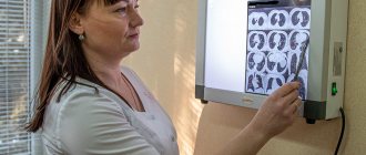
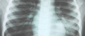
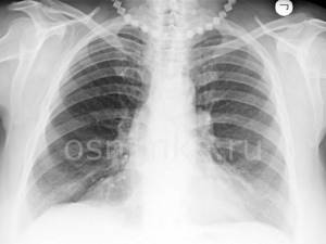
What does pneumonia look like on an x-ray?
What pneumonia looks like on an X-ray depends on the stage of the disease and its nature. However, in any case, signs of pneumonia on x-ray are visible as disturbances in the structure of the lung. Doctors pay attention to the features of the pulmonary pattern, the roots of the lung, the pleura and the location of infiltrative foci. Light areas in the image are interpreted as airless.
Krupoznaya
Croupous pneumonia on X-ray is characterized by increased pulmonary pattern and thickening of the root, slight compaction of the pleura, as well as reduced airiness of the lung. The reduction in airiness directly depends on the stage of the disease. This disease looks like medium-intensity shading. Lobar pneumonia is one of the most dangerous.
Focal
As with lobar pneumonia, with focal pneumonia, X-rays show an increase in the pulmonary pattern and thickening of the root, as well as thickening of the pleura. Such pneumonia is characterized by focal shadows of different sizes with unclear contours. There is also a deformation of the pulmonary pattern, which is very clearly visible on x-ray. It is difficult to identify symptoms of focal pneumonia on X-rays, so only an experienced doctor can diagnose this disease.
Viral
Viral pneumonia is classified as atypical. With the strengthening of the pulmonary pattern and compaction of the pleura in this case, the roots of the lungs do not change. The appearance on x-ray of focal shadows in the lower and middle parts of the lungs with bilateral viral pneumonia confirms the diagnosis. The whole world is aware of the rapid development and danger of this form of the disease, and this is another argument in favor of examining the signs of pneumonia on an X-ray.
Community-acquired (out-of-hospital) pneumonia: signs of the disease
This form of the disease occurs in the first two days after the child’s hospitalization or outside the hospital and can affect one or both pulmonary lobes. Children under 5 years of age are at risk for this lesion.
The high incidence of community-acquired or home-acquired pneumonia in children is influenced by the anatomical features of the respiratory system and the body’s weak immune defense. Children's trachea and bronchi are narrow, and this causes retention and stagnation of mucus, in which pathogenic microorganisms actively multiply. Infants spend more time lying down, which can lead to blood stagnation.
Symptoms and course of the disease depend on the type of pathogen and the location of the infection. Common signs include the following:
- cough;
- increased body temperature;
- sputum production;
- chest pain when coughing and breathing;
- general weakness;
- increased sweating at night.
The prevalence of community-acquired pneumonia is quite high. But there are no exact statistics, since many cases of the disease are not recorded due to the low population seeking medical help.
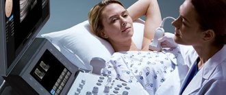
How is a tomography examination performed?
There is no need to prepare for scanning; it is enough to remove metal elements from the body or clothing, as they darken the area being studied and weaken the effect of X-rays. At the invitation of the medical staff, the patient goes into the CT room, lies down on the retractable tomograph table and tries not to move throughout the entire procedure. At this time, the ring part of the device rotates around the chest area, taking multiple images, which are transmitted to the expert's computer in the next room. Some scans are done while inhaling, so you need to hold your breath on command. When the scan is completed, the radiologist has not only static images, but also a three-dimensional model of the organ on which to prepare a conclusion.
Lobar pneumonia in children: special signs
With this form of pneumonia in children, the entire lobe of the lung is affected. Croupous inflammation practically does not occur in infants; it is most often diagnosed in children aged 2 to 5 years, while the more typical form of this disease is observed at an older age from 5 to 15 years. For some pathogens, such as pneumococcus, symptoms appear on the first day after infection.
The disease in children differs from that in adults:
- there is no “rusty” sputum;
- in most cases, not the entire lobe of the organ is affected, but one or more segments;
- When listening in the acute phase, wheezing is detected in only 15% of children, and in all of them during the period of resolution.
Differentiation of pneumonia from other lung pathologies
An important stage in making a diagnosis remains to distinguish it from other lung diseases, coronavirus. So, with bronchitis there will be no darkening in the image, but instead an increase in the pulmonary pattern.
Strengthening the pulmonary pattern
In the presence of a foreign body, a darkening with clear edges is observed, localized in the lower lobe of the lung. It is difficult to confuse it with inflammation of a typical nature.
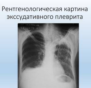
With pleurisy, the picture shows an accumulation of exudate in the affected area. Pleurisy acts as a complication of untreated pneumonia.
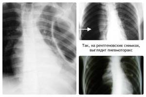
In pneumothorax, a characteristic level of fluid is present. The image shows a clearing; the pulmonary pattern is not visible.
An X-ray is the main examination, and if necessary, a computed tomography scan is prescribed.
Age-related features of pneumonia in children
There are several forms of the disease:
Intrauterine transplacental
The source is the mother, infection occurs in the womb. The infection reaches the fetus through the hematogenous route, that is, through the blood. The presence of a focus of infection in the maternal body is characteristic. Often this type of disease can be accompanied by sepsis. Infants in the first hours of birth, in the presence of intrauterine pneumonia, may experience the following symptoms:
- respiratory failure;
- dyspnea;
- blue discoloration of the skin;
- wet rales.
An x-ray can immediately after birth confirm the presence of lesions.
Intrauterine amniotic
The source of infection is the mother; the pathogens enter the fetus's body along with infected amniotic fluid, which the baby swallows. One of the signs of the presence of a lesion is the diagnosis of polyhydramnios in a woman.
Intrapartum
The source is the mother's body. Infection occurs during the movement of the fetus through the birth canal. The route of transmission of infection is contact - contamination with pathogens contained in the amniotic fluid (amniotic fluid) and mucous secretions. Signs do not appear immediately, but after 2-3 days after birth. The clinical picture is similar to intrauterine infection.
The initial stage is characterized by:
- respiratory failure;
- toxicosis;
- weakness, lethargy;
- the baby refuses the breast;
- foam is released from the oral cavity.
The following signs are common:
- diarrhea;
- otitis;
- conjunctivitis
Postnatal pneumonia (domestic)
The source of infection is the people around the baby at home. Infection occurs after discharge from the hospital. The route of transmission of the infectious agent occurs through the air - aerogenic method. Most often, the disease is associated with the presence of pneumococcal pathogens and massive contamination of the upper respiratory tract.
The disease begins acutely and the following symptoms are observed:
- cough;
- heat;
- sputum discharge;
- breathing may be weak or harsh;
- fine bubbling rales are heard.
Postnatal nosocomial (in-hospital)
The source of infection is medical workers and equipment. Infection occurs after birth. There are several ways of infection - bronchogenic (the disease originates in the bronchi), contact, aerogenic (rare infection through the air). More often they begin in the first week of life with general symptoms of toxicosis.
It is characterized by signs of acute toxicosis and a change in the acid-base balance towards increasing acidity. More often, the symptoms have blurred signs and can have a rapid course with a fatal outcome. Infants on mechanical ventilation (artificial pulmonary ventilation) may develop early pneumonia - in the first 5 days, and late pneumonia - in the next 5 days.
What is better to do in case of pneumonia: CT scan or x-ray?
Only a doctor can prescribe a CT scan or x-ray of the lungs for pneumonia after studying the symptoms, laboratory tests, and the patient’s individual clinical picture. The presence of fluid or pus in the alveoli, as well as fibrosis, is visualized on both x-ray and CT scans. However, traditional X-rays for stage I-II pneumonia may not be enough, while on CT it is more clearly visible as “ground glass”. For SARS and coronavirus, it is recommended to do a CT scan of the lungs.
Symptoms of inflammation in children over two years of age
More often, pneumonia develops in the presence of a cold. And a sharp jump in temperature during the improvement stage should worry parents. During the same period, intoxication symptoms and cough may increase.
The child may experience prolonged, up to a week, lethargy and sleep disturbances. Shortness of breath appears, breathing becomes more frequent. Possible pale skin. A high temperature that lasts longer than 4 days and is not brought down by traditional means can also signal the presence of an inflammatory process in the lungs.
Methods for diagnosing the disease in children
The primary method for identifying the disease is an initial appointment, which includes a survey, listening with a stethoscope, measuring body temperature, examination and palpation (palpation). If certain symptoms are present, the patient is sent to confirm the diagnosis - an x-ray examination. The image will indicate the exact location of the affected source of infection. This information is especially important for repeated cases of pneumonia.
To identify the type of pathogen, a laboratory test of mucous discharge from the nose and sputum is prescribed. For these purposes, the following methods are used:
- immunoenzyme;
- immunofluorescent;
- DNA polymerase.
A general blood test shows the number of leukocytes, acceleration of ESR and toxic granularity of neutrophils.
Causes of pneumonia
Infection occurs when a pathogenic agent enters the lungs.
In 40% of cases the disease is caused by pneumococci. Other bacterial pathogens include streptococci, staphylococci and Haemophilus influenzae, gram-negative microorganisms, and opportunistic microflora. Viral pneumonia develops when affected by influenza, parainfluenza, cytomegalovirus, adenoviruses, and coronaviruses. Atypical pathogens include mycoplasma, chlamydia, legionella, and fungi. Risk factors are considered:
- reduced immunity;
- cancer, including undergoing chemotherapy;
- autoimmune diseases;
- chronic lung diseases;
- diseases of the digestive tract;
- diseases of the heart and blood vessels;
- unhealthy lifestyle (smoking, alcohol abuse);
- hypothermia;
- infancy or old age;
- postoperative period.
There are four main mechanisms that cause pneumonia:
- Inhalation of the contents of the oropharynx and digestive tract, since these secretions contain a high concentration of various pathogenic agents.
- Inhalation of air in which the pathogen is present (through contact with a sick person, as well as rodents and birds).
- Entry of the pathogen into the blood from foci of infection located outside the lungs (for example, from the gastrointestinal tract).
- Infection from other internal organs, as well as from the outside with a penetrating wound to the chest.