Despite its relative simplicity and accessibility, the method is very informative, so it is by default included in the standard gynecological examination, being an important part of the screening examination of women for the purpose of early detection of background and precancerous diseases.
It is a safe and painless procedure that can be performed on a regular basis. Biological material is collected by a gynecologist using special spatulas and brushes (in the presence of visibly altered areas, selectively), then the resulting material is transferred to glass slides, the smears are dried and labeled. Smears prepared in this way are sent to a clinical diagnostic laboratory, where microscopy is carried out by a laboratory assistant.
Indications for the study
- screening during gynecological examination - it is recommended to conduct a cytological examination annually for all women over 25 years of age, as well as for those who are sexually active under the age of 25 years;
- dynamic observation of patients whose smears had previously revealed pathological changes, as well as patients with a history of human papillomavirus infection;
- monitoring the effectiveness of treatment.
The material is not collected during menstrual bleeding; the optimal time is no earlier than the 5th day of the menstrual cycle and no later than 5 days before the start of menstruation.
Preparing for scraping
Endocervical curettage is the final stage of diagnosis. The main goal of this stage is the morphological verification of the putative clinical diagnosis. The procedure is performed if there are indications, which are determined based on the results of a cytological smear and colposcopic examination. The patient undergoes simple and extended colposcopy. The detection of abnormal colposcopic signs serves as an indication not only for curettage of the cervical canal, but also for a targeted biopsy of the cervix.
Using ultrasound scanning of the pelvic organs, diseases of the uterus and appendages are excluded. In case of combined pathology of the endometrium and endocervix, hysteroscopy with RDV is prescribed. The patient is also recommended to undergo cervicoscopy, which allows visualization of the canal along its entire length. General clinical examination includes general blood and urine tests, blood tests for HIV, hepatitis B and C, RW. The gynecologist prescribes an examination for urogenital infections (smear microscopy, flora culture), diagnosis of STIs using PCR, cytological examination of smears (PAP test), testing for HPV infection using qualitative and quantitative methods.
Before the procedure of curettage of the cervical canal, it is necessary to shave the area of the external genitalia. For 2 days before the procedure, sexual intercourse, the use of vaginal tampons, spermicides, and douching should be avoided. Curettage of the cervical canal is carried out in the first phase of the menstrual cycle. Preparation also includes emptying your bladder immediately before the procedure. If the pain threshold is reduced, it is recommended to take an anesthetic drug 30-40 minutes before the procedure.
Research results
The characteristics of the cellular material of the smears largely depend on the age of the patient and the phase of the menstrual cycle in which the smears were made. Normally, smears from the vaginal part of the cervix contain squamous epithelial cells, and smears from the cervical canal contain columnar epithelial cells. Cytological examination helps to identify signs characteristic of acute and chronic inflammatory processes, infectious lesions, as well as diagnose precancerous changes and even malignant neoplasms in the early stages.
Unsatisfactory results of a cytological examination are a reason for colposcopy and biopsy of the cervix (if precancerous changes are detected, cancer is suspected), as well as for further bacteriological studies, PCR diagnostics (if signs of infection are detected). Based on the data obtained, as well as the results of a gynecological examination and other research methods, the gynecologist makes a final diagnosis and develops a treatment plan.
The main value of the method is that it allows you to identify precancerous changes and prevent the development of cervical cancer, identify cancer at early, preclinical stages and begin timely, gentle treatment, which will avoid disability and premature death.
Liquid cytology of the cervix and cervical canal
Cytological examination of scrapings
from the cervical canal and from the surface of the cervix has long been included in mandatory screening recommended annually for women who are sexually active. This need arose as a result of the increased risk of developing cervical cancer in fairly young women. Worldwide, cervical cancer is the third most common cancer in women, second only to breast and colon cancer. It should be noted that recently cancer has become “younger”, that is, cases of cervical cancer pathology have begun to be observed more often in women of the reproductive period, while previously women during menopause were at risk. This trend is largely explained by an established predisposing factor for the development of cervical cancer, namely the presence of the human papilloma virus. This virus is transmitted sexually and can go undetected for a long time, since it does not cause anything other than changes in the mucous membranes of the cervix and vagina. A doctor can evaluate such changes during an examination, but even in this case, the conclusion is subjective - the doctor suspects the presence of certain changes, but they can only be talked about for sure by assessing the material obtained from the cervix. It is this type of study that has become part of the standard for cervical cancer screening.
Previous cytological examination
ecto- and endocervix material was carried out by applying it to a glass slide immediately after collection. Currently, it is possible to collect material in a special environment, which promotes better preservation of cells and cleanses them of mucus and impurities. This method is called liquid cytology because the cells are in liquid. In laboratory conditions, a sample with material is centrifuged and the cells form a certain layer, by assessing which a cytologist can give a more accurate description of the cytogram, reducing the possibility of errors due to impurities in the smear.
This method makes it possible to ensure high quality of biomaterial collection and its long-term preservation without changing the morphological properties of cells, excellent quality of cytological preparations and the ability to perform additional studies (HPV test, immunocytochemical studies) without resorting to repeated smear taking. In this way, it is possible to minimize possible technical errors during the study - a cytobrush is placed in the liquid, on which all the resulting cells are stored. This allows laboratory diagnostic doctors to most accurately evaluate the obtained material. The result of the study is not descriptive in nature, but according to the generally accepted Bethesda Terminology System (TBS). This smear scoring system is used throughout the world and is used to make clinical diagnoses.
It should be noted that the drug is stained using the Papanicolaou method, therefore this analysis is also called a liquid Pap test. The smear contains cells of the exo- and endocervix, with special attention paid to whether cells from the transformation zone are present in the material. This zone is a transition zone between two types of epithelium; most of all changes begin precisely from the transformation zone.
The adequacy of the material is also assessed, that is, how correctly the doctor performed the sampling. For example, with heavy mucous discharge, if the cervix was not prepared for collection, the material may contain an insufficient number of cells for evaluation, and the result may be unreliable. To avoid this, in a separate column the laboratory doctor notes how high-quality the material was received.
Statistics
The incidence rate of cervical cancer fell by more than 50% from the mid-1970s to the mid-2000s, in part due to an increase in screening tests to detect cervical changes before they become cancerous. The decline in incidence rates among young women may be due to the use of the HPV vaccine. CC is most often diagnosed between the ages of 35 and 44 years. The average age of diagnosis is 50 years. About 20% of cancer cases are diagnosed after age 65. Typically, these cases occur in people who did not undergo regular screening for CC before age 65. The 5-year survival rate measures what percentage of people live at least 5 years after their cancer is diagnosed. The 5-year survival rate for all people with cervical cancer is 66%.
After scraping
Within 1 hour after the manipulation, the patient is under medical supervision; if her condition is satisfactory, the woman is allowed to go home. Nagging pain and scanty discharge from the genital tract after curettage of the cervical canal may bother you for several days. During this period, sexual intercourse and thermal procedures (visiting baths, saunas) should be excluded. The question of the need to use antibacterial drugs is decided on an individual basis. After 10-15 days, a visit to the treating gynecologist is necessary to discuss the results of the histological report and determine further treatment tactics.
Complications after curettage of the cervical canal occur in extremely rare cases. In the long term, there is a possibility of developing cervical endometriosis, cicatricial canal stenosis, and isthmic-cervical insufficiency.
Risk factors
- Infection caused by the human papillomavirus (HPV).
The most important risk factor for cervical cancer is HPV infection. HPV is common. Most people become infected with HPV when they become sexually active, and most people clear the virus without problems. There are more than 100 different types of HPV. Not all of them are related to cancer. The types or strains of HPV that are most often associated with CC are HPV16 and HPV18. Having sex at an earlier age or having multiple sexual partners increases your risk of contracting high-risk HPV.
- Weakened immune system.
People with weakened immune systems have a higher risk of developing cervical cancer. A weakened immune system can be caused by immune suppression from corticosteroid drugs, organ transplantation, treatment for other types of cancer, or the human immunodeficiency virus (HIV), which is the virus that causes acquired immunodeficiency syndrome (AIDS). When a person is infected with HIV, their immune system is unable to fight early stage cancer.
- Herpes.
Women with genital herpes have a higher risk of developing CC.
- Smoking.
Women who smoke are approximately twice as likely to develop CC as women who do not smoke.
- Age.
People under 20 rarely get cervical cancer, but the risk increases in their 30s. Women older than this age group remain at risk and need regular screenings, which include Pap smears and/or HPV.
- Oral contraceptives.
Some studies suggest that oral contraceptives, which are birth control pills, may be associated with an increased risk of CC.
About abnormal cervical cells that can cause cancer
CC begins when healthy cells on the surface of the cervix are changed or infected by the human papillomavirus (HPV) and grow out of control, forming a mass called a tumor. Long-term HPV infection of the cervix can lead to cancer, which will further lead to the oncological process. The tumor can be malignant or benign. At first, the changes in the cell are abnormal, noncancerous, and are sometimes called “atypical cells.” Scientists believe that some of these abnormal changes are the first step in a series of slow changes that can lead to cancer. Some abnormal cells disappear without treatment, but others can become cancerous. This precancerous phase is called cervical dysplasia, which is abnormal cell growth. Sometimes dysplasia tissue needs to be removed to prevent cancer from developing. Often, dysplasia tissue can be removed or destroyed without harming healthy tissue, but in some cases, a hysterectomy—removal of the uterus and cervix—is required to prevent cervical cancer.
Indications for the procedure
According to the recommendations of gynecologists, a Pap test should be performed on women after three years of sexual activity and then annually for preventive purposes. If the test shows a negative result 3 times in a row, it can be performed once every 2-3 years. There are so-called risk groups of women prone to developing cancer of the reproductive organs.
These include women:
- using hormonal contraception methods;
- diagnosed with infertility;
- diagnosed with genital herpes;
- those suffering from obesity, metabolic disorders;
- in preparation for gynecological surgery;
- smokers
Sometimes this examination is prescribed unscheduled. For example, it is advisable to conduct a Pap test for a woman planning a pregnancy and a pregnant woman before registering with a antenatal clinic. Gynecologists recommend taking a smear from the cervical canal before installing an intrauterine device.
How is cervical oncocytology done?
The resulting smears are studied in the laboratory. For Pap staining, the evaluation is performed by staff using a microscopic examination. If there are a lot of cells and they are layered on top of each other, it is not always possible to draw correct conclusions, so laboratory doctors recommend repeating the test.
Liquid oncocytology is more informative, since biological samples are more “clean” (there is no accumulation of cells, blood elements, etc.). They are prepared and processed by a special computerized system. If necessary, biological material can be subjected to additional diagnostic methods, for example, immunohistochemical analysis. Therefore, it is liquid oncocytology that is considered the standard in the diagnosis of cervical diseases, in particular cancer.
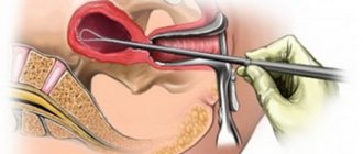
Methodology
Curettage of the cervical canal is carried out on an outpatient basis under a local paracervical block. The procedure is carried out in compliance with all the rules of asepsis and antisepsis without prior dilatation of the cervix and probing of the uterine cavity. After inserting double-leaf speculum into the vagina, the cervix is disinfected with an alcohol iodine solution. To straighten the uterine canal, the anterior lip of the cervix is grabbed with a single-tooth holder and slightly lowered to the entrance to the vagina. Curettage of the cervical canal is carried out with a small sharp curette No. 2. The instrument is inserted only to the internal pharynx. When the curette penetrates the uterine cavity, endometrial cells enter the scraping of the endocervix, which reduces the diagnostic value of the method.
The pointed surface of the curette is pressed tightly against the wall and with movements from top to bottom in the direction from the internal pharynx to the external pharynx, the canal is scraped along the entire circumference without removing the instrument from it. After scraping all the walls, the material, consisting of blood, mucus, cellular and tissue detritus, is placed in a container with formaldehyde and sent for histological examination. A smear is applied to a glass slide using a curette for cytological examination. The instruments are removed and the cervix is re-treated with an antiseptic. The duration of the cervical canal curettage procedure is 3-5 minutes.