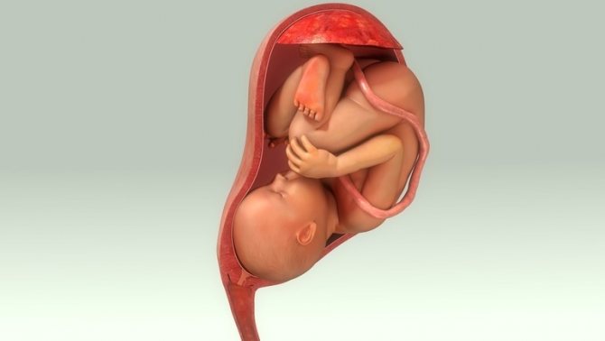
The third trimester is the home stretch. The baby is growing by leaps and bounds. He actively moves, frowns and smiles, and practices sucking and breathing movements. Soon he will be ready to be born.
In the third trimester of pregnancy, both a routine ultrasound and a planned third screening are performed. The best time to conduct a screening ultrasound is 30-32 weeks. It is at this time that the readings will be as informative as possible. However, closer to childbirth, an ultrasound examination may also be required to assess the position of the baby, its development, and the condition of the placenta.
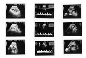
What to look for and when to do the third ultrasound during pregnancy
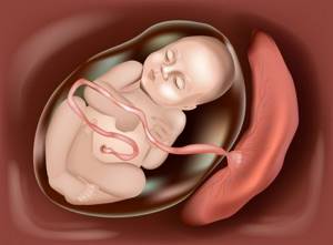
During pregnancy ultrasound in the 3rd trimester, the attention of specialists is focused on the placenta. They evaluate the condition of the child's seat for its position, thickness and maturity. These indicators are of a very high degree of importance for a number of reasons, the main one of which is the function of the placenta in feeding the child and saturating his body with oxygen.
In addition, at the third screening, the diagnostician must:
- carry out fetometry - determination of the main physiological parameters of the fetus, including the height and weight of the child;
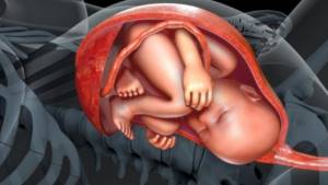
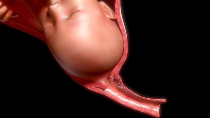
- assess how the baby is positioned in the uterus in order to develop tactics before childbirth (naturally or via cesarean section);
- examine amniotic fluid, breathing rate and motor activity of the baby;
- determine the date of birth of the child;
- examine the female organs to exclude the manifestation of dangerous disorders in the form of neoplasms or infectious processes.
Thus, the main purpose of a screening study in the 3rd trimester is to finally establish and record the baby’s health status, with which he will approach his birth.

It is during this period that symptoms of some pathologies may be detected later. From the point of view of the accuracy of expert diagnostics, the week in which it is carried out is of great importance. Therefore, gynecologists take a responsible approach to the question of when to do the third planned ultrasound.
Our advantages
- A large number of clinics. Alfa Health Center clinics are located in many large cities of Russia. In each of them we offer modern diagnostic methods and affordable prices.
- Taking care of clients. Our staff does everything to make patients feel comfortable. Reception is by appointment only. When you arrive at our clinic at the appointed time, you will not have to wait in line. Survey results are available within 15 minutes.
- High-quality equipment for precise results. The General Electric LOGIQ S8 ultrasound machine allows you to obtain accurate results and identify possible pathologies even in the early stages.
At what time is the third ultrasound performed?

The Russian Ministry of Health has established standards that determine at what stage of pregnancy the third ultrasound is performed. Often sent for screening from the 32nd week of pregnancy to the 34th. But in some cases, it is possible to prescribe an earlier date - from 28 weeks or from 30 weeks. This happens in situations where developmental abnormalities were previously identified, treatment was prescribed, and the doctor wants to make sure that it produced results.
It is difficult to say when it is better to do an ultrasound in the 3rd trimester of pregnancy. The duration depends on the doctor’s decision.
Key indicators and standards

We list the main indicators and norms of ultrasound in the 3rd trimester of pregnancy, which will be included in the screening protocol:
- Fetometry criteria.
These include: bipariental and fronto-occipital dimensions; circumference of the head, chest and abdomen; length of bones (hips, legs, shoulders and forearm); weight and height of the baby. The normal weight at the time of examination is 1700-2150 grams, the normal height is 42-45 cm. Based on bone length, the doctor can diagnose many genetic diseases, including Down syndrome. - Number of fetuses (singleton pregnancy, twins, triplets).
- Development of internal organs.
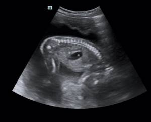
Their compliance with the gestational age is determined. Particular attention is paid to the fetal lungs to assess the possibility of spontaneous breathing after birth). - Longitudinal section of the spine.
Using this element of the study, the diagnostician determines whether there are disorders in the development of the musculoskeletal system. - Thickness, presentation and maturity of the placental sac.
- Motor activity and heart rate.
In the last weeks of pregnancy, there is a decrease in motor activity, given that the free space in the uterus is becoming less and less. This moment can cause great anxiety for the mother. During an ultrasound, the doctor will reliably tell you whether there is cause for alarm. He will evaluate the frequency of his movements and listen to his heartbeat. - Condition of the umbilical cord.
During the examination, the specialist evaluates not only the structure of the blood vessels that provide nutrition to the child, but also their location. In the later stages, doctors need to know whether there is an entanglement in the umbilical cord. If there is repeated entanglement, a caesarean section may be prescribed. - Condition of the cervix.

What is studied at 30 – 34 weeks?
At this stage, the fetus is already almost formed, the time of delivery is steadily approaching, and therefore the doctor will pay attention during the third planned ultrasound examination to the following important criteria:
- location of the fetus (or fetuses) in the uterus;
- placement of the placenta, its thickness, density, presence of abnormal changes, as well as its degree of maturity;
- umbilical cord condition;
- baby size;
- parameters of internal organs;
- volume of amniotic fluid, its purity;
- the degree of tension of the uterus (tone), the smoothness and length of its cervix, the opening of the pharynx.
At the conclusion of the ultrasound, the doctor indicates all the studied parameters and confirms their correspondence to the gestational age. The doctor is obliged to exclude and provide for all potential complications of the last weeks of gestation and the birth process itself.
Interpretation of ultrasound 3 trimesters
Polyhydramnios
Amniotic fluid plays an important role, throughout pregnancy, protecting the baby from adverse external influences and allowing him to move freely in the intrauterine space.
The state of his health directly depends on how much amniotic fluid is present around the baby. An ultrasound specialist, using good modern equipment, will be able to determine the quantity and quality of amniotic fluid.
It is a bad sign if there is too much amniotic fluid. In this case, the doctor diagnoses polyhydramnios. A woman will definitely have to undergo serious drug treatment, otherwise complications are possible, including hypoxia and infection of the fetus, gestosis, premature rupture of water and placental abruption, and weakness of labor.
Low water
For oligohydramnios, urgent treatment in a hospital setting is also indicated. A reduced amount of amniotic fluid in the 3rd trimester is fraught with serious consequences. It can lead to defects in the intrauterine development of the child and provoke the manifestation of anomalies: curvature of the spine and bone tissue, clubfoot. In later stages, oligohydramnios becomes a consequence of complications in the placenta.
Cervical length
When deciphering the length of the cervix, the doctor will take into account that it should be at least 3-4 centimeters. If it is shorter, then this can lead to isthmic-cervical insufficiency, when the cervix, under the pressure of an enlarged fetus, simply cannot hold it any longer. It is easy to guess what consequences this may cause. For ICI, two treatment options are possible: surgical (up to 27 weeks) or the application of a pessary - a silicone or plastic device that artificially supports the uterus.
Placenta
This temporary organ, which appears in the womb of a woman only during the gestational period, goes through different stages during its existence - from maturation to aging closer to the birth process. An ultrasound diagnostic doctor looks at the condition of the placenta in the third trimester in order to exclude the possibility of fetal hypoxia and identify signs of pathological phenomena.
The normal thickness at 32 weeks should be within the normal range - 25-42 mm, and maturity should be of the second degree. If the third degree is recorded, this is a sign of premature aging of the child’s place.
Fetal heart rate
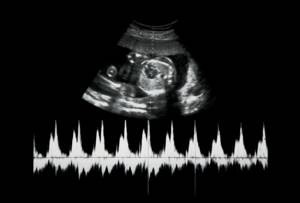
On an ultrasound of the 3rd trimester, the baby’s heart rate is 120-160 beats per minute. By comparing the indicators obtained during screening with the heart rate norm table, the specialist must determine whether there are any deviations. If they are found, the doctor makes a diagnosis - tachycardia (rapid heartbeat) or bradycardia (slow heartbeat). Such diseases can be caused by various reasons: disturbances in the internal blood flow of the uterus and placenta, inflammation, high intracranial pressure in the fetus, heart defects, etc.
Respiratory system
Already in the womb, the baby begins to train its lungs by making breathing movements. Ultrasound can record their frequency and assess the functional state of the fetus. During breathing movements, the baby's lungs do not expand, but such exercises help blood flow to the heart and also promote the exchange of amniotic fluid. During the examination, the diagnostician examines the degree of maturation of the organs of the respiratory system and interprets the ultrasound and gives a conclusion about their development.
First degree violations

If the expectant mother was diagnosed with a first-degree disorder in custody, this means that the doctor found some abnormalities in the development of the fetus, but they are minor. In this case, the gynecologist prescribes treatment aimed at blocking the development of the disease and preventing the occurrence of complications. In addition, the woman will be sent for re-screening to determine the effectiveness of the treatment.
Second degree violations
The interpretation of the ultrasound indicates second-degree violations if the pregnant woman has pathologies in the movement of blood through the vessels of the placenta. The mother's body cannot provide the child with the necessary nutrition, which is why he may be delayed in development or have birth defects. In this case, the woman is not sent to the hospital, but she will need medical care at the clinic.
Third degree violations

Third degree disorders are characterized by serious abnormalities in the blood flow of the placenta. In this case, there is a disruption of the processes that ensure the viability of the baby. As a rule, in such cases, fetal death is likely due to the occurrence of severe malformations. In some situations, it is possible to maintain the pregnancy, so the woman is urgently hospitalized.
Types of ultrasound
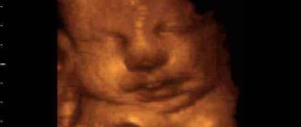
3D
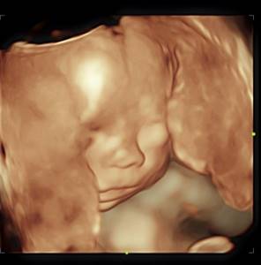
Three-dimensional ultrasound during late pregnancy makes it possible to see the smallest details in the development of the child’s body parts and organs. A high-quality three-dimensional image allows not only to carry out an accurate diagnosis, but also to take photographs and videos of the baby’s intrauterine life.
Using 3D ultrasound, it is possible to detect abnormalities that are sometimes not visible during regular screening. The study is paid.
4D
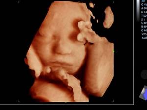
4D ultrasound, which includes four dimensions (width, height, depth and time), allows you to record your baby's movements in real time. The diagnostician has the opportunity to record the clip on disk and provide the recording to parents. Of course, this service, like the screening itself, is provided for a fee.
Most likely, at previous screenings, mom and dad were already told whether they were having a boy or a girl. At the third study, they will definitely confirm the sex of the child.
Additional ultrasounds
According to indications, the doctor may prescribe additional ultrasounds in the 3rd trimester of pregnancy:
- Ultrasound amnioscopy – assessment of the state of amniotic fluid. This procedure provides information about the amount of amniotic fluid in the uterus. Based on the results of the study, a diagnosis is made - oligohydramnios or polyhydramnios.
- Doppler testing is a method that allows you to calculate the speed of blood flow through the vessels of the umbilical cord and the child itself, as well as the resistance of the blood vessels. Using Doppler, you can judge the functioning of the placenta.
If irregularities have been identified, the gynecologist may prescribe a re-examination to rule out medical error.
Ultrasound in the ninth month of pregnancy
In the ninth month, birth can occur at any time. An ultrasound at this stage is necessary in order to determine possible contraindications for natural childbirth. On the part of the fetus, contraindications may be as follows:
- acute hypoxia;
- transverse position;
- breech presentation with a large fetal weight or multiple pregnancy;
- foot, frontal and facial presentation.
An ultrasound examination will also help diagnose some maternal emergencies that require a cesarean section (placental abruption, uterine scar rupture, and others).
How to prepare for your third ultrasound
In order for the third ultrasound during pregnancy to go smoothly and without problems, you need to prepare for it in advance.
To prepare the body, experts recommend not eating foods that cause increased gas formation a day or two before the procedure.
It is also necessary to wash the genitals.

It is better to come to the clinic in advance so that the body is not overstimulated. If an ultrasound is performed on a paid basis, then, as a rule, you do not need to bring anything with you. If in a clinic, you may need a diaper, towel and shoe covers.

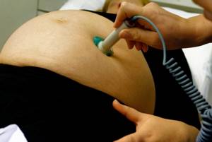
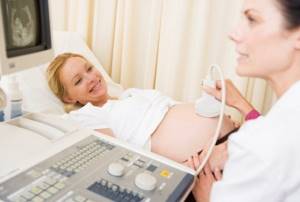
At the last scheduled ultrasound, the examination is carried out through the abdominal probe. A woman needs to take a comfortable position on the couch and listen to the recommendations of the diagnostician.
How is the procedure performed?
At all stages of pregnancy, the ultrasound procedure is approximately the same. In the doctor’s office, the woman, depending on the research method, frees her stomach from clothes or undresses to the waist, then lies down on the couch. With the abdominal method, a conductive, harmless gel is applied to the skin. After this, the specialist applies the ultrasound machine sensor to the patient’s abdomen and moves it along the anterior wall of the peritoneum. During a transvaginal examination, a condom is placed over the probe and then inserted into the vagina. The procedure does not cause discomfort to either the expectant mother or the fetus. The description is drawn up according to the protocol. The data is assessed in relation to ultrasound standards by week. To sign up for an ultrasound scan at Alpha Health Center, contact the specialists of our call center. We will select a time convenient for you, tell you about preparation for the procedure and answer all questions about it. Also in our Center you can get a consultation with a gynecologist based on the results of an ultrasound and other necessary examinations.