The twentieth week ends the fifth month and is the midpoint of pregnancy. At this stage of development, the child’s body is already formed so much that after 3-4 weeks, in emergency cases, it will be able to survive outside the mother’s body. But normally he will remain under his protection for another 4 months.
At the twentieth week, a woman may experience unpleasant symptoms caused by an enlarging uterus. In general, this time is favorable for her. It can be devoted to shopping and personal development.
Interesting Facts
| Options | Indications |
| Time from conception | 18 weeks |
| Period by month | 20 weeks |
| What month | 5 |
| Dimensions and weight of the fetus | 250 mm, 290 g |
| Uterus dimensions | At or above the navel |
| Pregnant weight | The increase over the last week is no more than 400-500 g |
Your baby is the size of
small coconut
250mm Size
290 g Weight
Congratulations! You are exactly halfway there. At week 20, a woman is usually full of strength and joyful, including because she can feel the baby’s movements in the womb. This is the first interaction between mother and child. At this time, you can find out his gender, start choosing a name and planning a dowry for the birth.
Recommendations
At the 20th week of pregnancy, all previous recommendations remain valid. You already know that you need to monitor the quality of your diet and follow a regimen: eat in small portions, do not overuse foods that cause allergies (citrus fruits, etc.). Surely you walk a lot, and maybe you have already started attending special classes for pregnant women.
If you experience dizziness at 19–20 weeks of pregnancy, try not to fast. Feel like you need to sit down or lie down? Don't be shy and do it! Move carefully, without sharp turns or climbs, try not to stay in the same position for a long time.
If your legs swell during the 19th – 20th week of pregnancy, keep them elevated more often. Make a habit of doing a massage with cream in the evenings. Before that, you can pamper your feet with a cool bath with your favorite essential oil.
Feelings of the expectant mother
By the end of week 20, your weight should normally increase by 3-5 kg. The belly becomes more and more rounded and rises higher. The fundus of the uterus is now at the level of the navel. Every week its size will grow by 1 cm. Due to the growth of the uterus and the pressure it puts on the internal organs, a woman may experience shortness of breath and heartburn.
Abundant mucous vaginal discharge at 20 weeks of pregnancy should not bother you. This is normal, this is how the body protects the fetus from infections. An alarming symptom is bloody or cheesy discharge with an unpleasant odor. The first indicate the threat of interruption, the second indicate the appearance of candidiasis. In both cases, urgent treatment is necessary.
Belly seems too big
It makes no sense to give norms for abdominal volumes here, because each woman has her own initial volumes, but there are certain conventional guidelines: if at 20 weeks of pregnancy the abdominal volume was 70-75 cm, then at 30 weeks – 82-87 cm and 95-100 cm by the 40th week. An increased abdominal size may indicate:
- multiple pregnancy (twins or triplets),
- polyhydramnios (its cause may be infection, endocrine diseases and Rhesus conflict),
- large fetus (the most common reason here is endocrine diseases of the mother and the high growth of its parents)
- fetal development abnormalities,
- chorionepithelioma, or hydatidiform mole (this is a malignant neoplasm from the cells of the chorion, that is, the embryo),
- breech presentation of the fetus (when the pelvic end of the baby is below his head). In this case, it is important to timely and competently decide the question of how best to give birth - by yourself or by caesarean section.
How the fetus develops
He has a lot to do now:
- it floats and pushes off the walls of the uterus;
- hiccups;
- flinches if mom is worried;
- thumb sucking;
- swallows amniotic fluid.
The latter contributes to the development of the digestive system. Already now, meconium is forming in the intestines - original feces, which will pass in the first day of the baby’s life.
By the 20th week of pregnancy, the fetus weighs an average of 290 grams, its body length is 16-17 cm from crown to tailbone and 25 cm from head to toe. The active development of the nervous system continues, the senses are improved, and taste buds develop.
The girls have a formed uterus, fallopian tubes and ovaries, in which 7 million eggs have already been formed. In the future, there will be 3.5 times less of them, this is a reserve for the rest of life. In boys, the external genitalia grow, the testicles are still in the abdomen and will descend into the scrotum in the 3rd trimester of pregnancy.
Preparation for the procedure
An ultrasound scan for the twentieth week of the pregnancy cycle will require a little preparation from the woman:
- Before the examination, you should definitely carry out hygiene procedures. It is better to wear clothes for an ultrasound in such a cut that it is easy to expose the stomach.
- Since the uterus has already enlarged due to its growth, there is no need to drink water before the procedure. It will be possible to distinguish the child’s organs on the screen.
- The day before diagnosis, you should exclude: citrus fruits, seafood, carbonated drinks. To reduce the amount of gas in the intestines, you can drink activated charcoal two to three hours before your visit to the doctor.
For examination by a diagnostician, the expectant mother should bring:
- pregnant woman's card;
- all previous tests;
- medical insurance;
- passport.
Screening can be carried out both in a private clinic and in a district clinic. If the expectant mother prefers to go to the clinic, it is worth taking a diaper with her, which will be needed for the ultrasound.
Tests and ultrasound
The 20th obstetric week of pregnancy is the last possible for 2 screenings. Go through it if you haven't already. The study will show the possible risks of developing genetic pathologies and structural anomalies of the fetus. The study includes a biochemical blood test and ultrasound.
What will the ultrasound show?
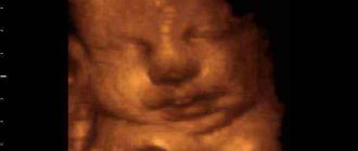
Using ultrasound, the doctor will determine the correspondence of the size of the fetus to the gestational age, assess the amount of amniotic fluid and the condition of the placenta. He will study the structure of internal organs, with special attention paid to the heart and circulatory system.
During an ultrasound at 20 weeks of pregnancy, you will find out the gender of the unborn baby. The risk of error is now minimal, because the fetal genitals are formed. The exception is when the child is “hiding” and there is no way to examine him in detail. In this case, 3D research can help.
If a specialist tells you that the fetus is breech or transverse, do not rush to worry. The most favorable position for childbirth is the head position, which the baby can take even at 38 weeks of pregnancy.
Accuracy of the study
An error during an ultrasound examination occurs in 5% of 100 cases.
There are several reasons why the study is wrong in its data:
- outdated equipment;
- unqualified doctor;
- short gestational age;
- features of maternal anatomy.
Diagnoses that can be mistaken by ultrasound:
- frozen pregnancy;
- ectopic pregnancy;
- false absence of pregnancy;
- genetic diseases of the fetus;
- gender of the child.
Possible complications
The twentieth week of pregnancy is a relatively calm time for the mother’s health. But even at this stage, pathologies can develop that significantly complicate the course of gestation.
Isthmic-cervical insufficiency
This diagnosis is made if, according to ultrasound data at 16-24 weeks of pregnancy, the length of the cervix is less than 25 mm. This condition is dangerous because it causes miscarriage or premature birth. Occurs as a result of surgical trauma or congenital genetic diseases. For a short cervix, hormonal therapy, wearing a pessary, and suturing are effective.
Placenta previa
Normally, the placenta is usually located along the posterior wall of the uterus or in the fundus, sometimes on the anterior wall. Here she receives better blood supply and is protected from accidental injuries. Previa is said to occur if the placenta is located in the lower parts of the uterus and partially or completely covers the area of the internal os. There is also low placentation, when the placenta does not cover the pharynx, but is located lower than it should be normally.
This complication occurs in only 1% of cases and usually develops against the background of endometrial inflammation after surgery, multiple difficult births, or uterine fibroids.
Late toxicosis, or gestosis
Occurs after 20 weeks of pregnancy. Its symptoms are swelling, increased blood pressure and the presence of protein in the urine. The condition is considered dangerous and requires hospital treatment. To prevent its development, do not miss scheduled appointments with the gynecologist, take a urine test on time, and monitor your blood pressure yourself.
Main stages
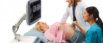
In the second trimester of pregnancy, an ultrasound examination is usually performed using the abdominal method; the procedure consists of several stages:
- The doctor applies a special gel to the woman’s stomach and runs a special ultra-wave sensor along the outside of the uterus.
- After this, an image of the fetus is displayed on the screen.
- The built-in program measures the child’s indicators.
- The doctor collects this data and makes an analysis based on it.
- After the procedure, the doctor gives the mother a protocol with a transcript of the study results.
If a woman has excess weight concentrated in the abdominal area, doctors prefer to use a vaginal probe to get a full view of the inside of the uterus.
Step-by-step ultrasound transvaginal method:
- A condom is placed on the intravaginal sensor. After this, the sensor is lubricated with gel.
- The doctor inserts a probe into the woman's vagina, examining the reproductive organs and the development of the fetus in the womb.
- The data is displayed on the monitor screen. The ultrasound doctor begins to decipher the obtained parameters and, upon completion of the analysis, announces them to the expectant mother.
Do's and Don'ts
In addition to the well-known and understandable prohibitions on alcohol and nicotine during pregnancy, we also do not recommend:
- consume more than 300 mg of caffeine per day, that is, about 2 cups, since large doses of caffeine harm the development of the fetus;
- engage in horse riding, tennis, diving, football and other traumatic sports;
- eat thermally unprocessed fish and seafood, eggs and meat, soft blue cheeses, drink unpasteurized milk, as these products sometimes cause parasitic diseases and intestinal infections.
But contrary to stereotypes, during pregnancy you can eat chocolate and citrus fruits, dye your hair, and, in the absence of contraindications, fly on an airplane and have sex.
Factors influencing examination results
Deviations from accepted ultrasound standards are most often observed in women, the so-called risk group. The main factors influencing the study indicators are the discrepancy between the parents in terms of the Rh blood factor (the mother has a negative indicator), the age of the pregnant woman 35+, the woman’s antisocial behavior and lifestyle (alcohol, drugs), and the patient’s hormone dependence.
The list continues with exacerbation of chronic renal pathologies, parasitic infestations, in vitro fertilization, and the presence of several embryos. Based on the results of the second screening, the doctor predicts the further course of the perinatal period and possible complications during delivery. Regular monitoring allows you to prevent complications in a timely manner, and during a normal pregnancy, provide psychological comfort to the expectant mother.
Checklist for 20 weeks of pregnancy
- Update your wardrobe: your belly quickly increases in size in the second trimester.
- Continue to lead an active lifestyle: this will help you cope more easily with all the hardships of pregnancy and prepare for childbirth.
- If colostrum appears, you should stock up on absorbent breast wipes.
You can undergo all types of ultrasound during pregnancy, including 3D or with Doppler, at the Women's Medical Center. We have the most modern equipment with highly sensitive sensors. Our specialists have many years of practical experience, which means that the likelihood of errors during the study is excluded.
Where to do an ultrasound in the second trimester of pregnancy in St. Petersburg
Ultrasound in the second trimester is an important and necessary procedure to monitor the normal course of pregnancy. It needs to be done on a good machine, otherwise the examination will be useless. This can be done at the specialized gynecological Center Diana in St. Petersburg.
We purchased the latest 3D, 4D ultrasound machine WITH DOPPLER FROM SAMSUNG MEDISON. The cost of examination of pregnant women is from 1300 rubles. A disc with the recording is given as a gift.
If you find an error, please select a piece of text and press Ctrl+Enter