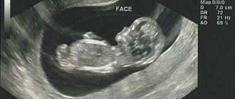
According to the decree of the Ministry of Health, at 11-13 weeks of pregnancy, a woman should undergo the first scheduled ultrasound. During this period, various abnormalities in the development of the fetus, some congenital defects, can be detected.
Many women do not understand why an ultrasound examination is prescribed, so they skip the diagnosis. But it is the first ultrasound that may be decisive in deciding whether to continue the pregnancy.
Purposes of ultrasound
Ultrasound examination (ultrasound) is recognized as the safest and highest quality method of monitoring the course of pregnancy throughout the world. Gynecologists strongly recommend that you undergo it on time in order to track problems with pregnancy and fetal pathologies in a timely manner.
The need for an ultrasound scan in the early stages is due to the following indications:
- assessment of the condition of the uterine walls, ovaries and cervix;
- establishment of intrauterine and ectopic pregnancy;
- determining the number of fertilized eggs (determination of multiple pregnancy);
- age over 35 years;
- monitoring the growth process , size and structure of the fertilized egg or embryo;
- fetal viability assessment;
- identification of tumors of the internal genital organs that can affect pregnancy.
You must undergo an ultrasound if:
- there is a presence of drug or alcohol addiction ;
- there is a threat of spontaneous miscarriage ;
- during previous pregnancies, genetic abnormalities or anomalies in the development of the fetus ;
- suffered infectious diseases in early pregnancy;
- there is a suspicion of a diagnosis of a regressing or frozen pregnancy (the fetus has stopped developing).
Ultrasound is mandatory in the early stages of women who are forced to take medications prohibited during pregnancy due to health conditions;
Analysis for fetal chromosomal pathology
During pregnancy, a woman undergoes many studies that allow her to monitor the process of fetal development, promptly detect pathology and prevent its development. The most important stage of the examination is an analysis of fetal chromosomal pathology (karyotype). This is especially true for pregnant women with hereditary diseases and a high risk of premature birth. Such patients should be consulted by a geneticist: a qualified specialist is able to determine the likelihood of the child developing congenital anomalies or genetic diseases. However, all pregnant women should undergo routine examination - genetic tests, the so-called NIPTs (non-invasive prenatal tests).
Parameters studied in the fetus
Ultrasound examines individual organs and parameters of the fetus:
- belly size;
- size of the heart and outgoing vessels;
- the bones of the thigh , forearm, lower leg, shoulder, and long tubular bones are measured;
- coccygeal-parietal size and length of the fetus;
- head (circumference, length from occiput to forehead, biparietal diameter).
The doctor finds out where the stomach and heart are located. Determines a number of brain structures that should be present at a given stage of pregnancy.
How are the risks of chromosomal abnormalities calculated?
Each screening is calculated according to the formula included in the methodology: for example, the “Priska” formula differs from the “Astraia” formula. In the case of the “Priska” analysis, an indicator of 1: 1000 is considered the threshold; Anything higher is high risk, anything lower is low risk. Both methods give the likelihood of the presence of chromosomal pathology; in fact, the presence of an increased probability speaks only of probability and requires clarification. Getting an accurate answer is the goal of invasive prenatal diagnosis. In cases of increased risk of chromosomal pathology, a consultation with a geneticist is prescribed.
The tasks of a genetic specialist are:
- collecting a detailed history of the pregnant woman’s life;
- studying the medical history of the pregnant woman and all close relatives in order to identify hereditary diseases (diabetes mellitus, heart disease, etc.);
- determining the likelihood of whether the lifestyle of the expectant mother can affect the intrauterine and subsequent development of the child;
- deciphering the results of prenatal tests.
Diagnosable disorders
Thanks to this examination, it became possible to identify groups of pregnant women who have an increased likelihood of having children with various chromosomal abnormalities.
An ultrasound can detect the following disorders and malformations:
- Down, Patau, Edwards, Smith-Lemli-Opitz, Corneli de Lange, Shereshevsky-Turner syndromes
- cystic hygroma (tumor formation in the neck);
- triploidy (cells have three chromosomes instead of two);
- defectiveness of the anterior abdominal wall , prolapse of internal organs and intestinal loops;
- asymmetry of the cerebral hemispheres;
- irregular shape of the vascular plexus in the fetus;
- violation of the integrity of the skull bones;
- diaphragmatic hernia of the stomach.
Preparation for ultrasound diagnostics
The first pregnancy examination can be carried out using transabdominal (TAUS) and transvaginal (TVUS) methods.
Preparation for TAUS includes a number of rules:
- Three days before the ultrasound, it is recommended to start following a diet that involves avoiding heavy foods, such as: fatty meat and fish, milk and milk-based products (except yogurt and hard cheese), black bread, cabbage, peas, prunes, pears, apples, beans, baked goods, carbonated and alcoholic drinks, coffee. This diet reduces gas formation in the intestines, making the examination more informative;
- You can eat yoghurts , which are rich in bifidobacteria and enzymes, mint, and cinnamon. These products reduce gas formation;
- An hour and a half before the procedure, you need to fill the bladder with water or other non-carbonated liquid. The lower the gestational age, the more the bladder should be filled, raising the uterus, which is located deep in the pelvis;
- You should bring a towel with you to the examination
TVUS requires only hygienic preparation and an empty bladder.
TAUZI:
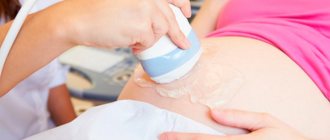
- The woman lies with her back on the couch and frees her entire stomach from outer clothing;
- The sonologist applies an ultrasound gel to the abdominal wall, which increases the mobility of the ultrasound sensor and improves the passage of waves through body tissue;
- An ultrasound sensor is applied to the body, after which the doctor begins to smoothly move it over the surface of the body, taking readings and logging them;
- During the examination, the patient may be asked to lie on her side, place her fists under her lumbar spine, or bend her knees;
- Upon completion of the procedure, the gel is removed from the abdomen with a towel or dry cloth.
Why is prenatal diagnosis needed?
The main task that the prenatal diagnostic test solves is to determine the presence of genetic disorders of the fetus before its birth.
There are non-invasive (i.e. without taking fetal tissue, by blood/ultrasound) prenatal diagnosis and invasive: taking umbilical cord blood, a piece of chorion/placenta and/or amniotic fluid.
Non-invasive diagnostics can be performed on any pregnant woman: from minimal (mandatory in the Russian Federation) to extended, carried out at the request of the spouses and providing information on a larger number of chromosomal disorders.
TVUSI:
- The woman removes clothing from the lower part of her body, sits on the couch and assumes the position for a standard gynecological examination;
- A condom will be placed on a special ultrasound sensor, then it will be lubricated with sound-conducting gel;
- The doctor inserts an ultrasound probe into the vagina and begins scanning, at the same time taking readings from the screen and recording it in the study protocol.
Using TVUS, you can determine the presence of pregnancy even with a five-day delay in menstruation (if the pregnancy lasts more than 2 weeks).
This examination method is preferable in the first trimester.
IS IT POSSIBLE TO PREDICT THE DEVELOPMENT OF PRE-ECLAMPSIA?
Yes, I have. When conducting combined screening in the 1st trimester, a pregnant woman can undergo additional research to calculate the risks of developing preeclampsia.
Based on medical history, blood pressure measurements, and data obtained during ultrasound prenatal screening, the risk of developing preeclampsia in each specific pregnant woman is calculated. This predicts about 80% of cases of early preeclampsia (<34 weeks), 65% of cases of premature PE (<37 weeks) and 45% of cases of full-term PE (≥37 weeks).
How to decipher an ultrasound result
Assessment of fetal vital activity includes only two main indicators: the ability to move and heartbeat. Ultrasound allows the specialist to observe the fetal heartbeat from the 6th week of pregnancy. The presence of contractions with good rhythm is considered the norm.
For each stage of pregnancy, the following frequencies of contractions per minute are considered normal:
- 6-8 weeks – 130-140;
- 9-10 weeks – 190;
- remaining time until birth – 140-160.
Heart rate measurement is mandatory. Based on this indicator, the doctor can determine the presence of problems in bearing a child. If it is underestimated or exceeded, the woman will be included in the risk group.
At 7-9 weeks, active mobility of the embryo should be noticeable. At first, the fetus moves its whole body chaotically; over time, flexion and extension movements can be seen (due to the fact that the fetus is often in a state of rest, the indicator of motor activity does not have much weight in assessing its viability).
The parameters of the embryo and ovum, as well as the progression of their growth, are determined by the following indicators:
- The diameter of the fetal egg (at the moment when the diameter is 14 mm, and the doctor is not able to detect an embryo on an ultrasound, it is considered that this pregnancy has stopped developing);
- Coccygeal-parietal size (length of the fetal body from the coccyx to the crown). This indicator is measured first, and thanks to it you can find out the age of the fetus with an error of up to 3 days.
Happy stories
Polina
Sep 30 2021
Many thanks to your clinic and especially to Dyana Omarovna!! IVF I got pregnant on the first try, my son is already 5 years old!!! I'll come again!!!
Read the story
Svetlana
11 Sep. 2021
We would like to express our gratitude to the Fertimed clinic and in particular to the doctor from God Efimova Maria Sergeevna, she is our guardian angel thanks to her professionalism
Read the story
Sadovskaya-Shmeleva Tatyana Viktorovna
15 Jul. 2021
On May 4, 2021, a baby weighing 3150 grams and height 50 cm was born. This happiness cannot be expressed in words. Especially when the path to it is very long and difficult.
Read the story
Dzagoev family
May 03, 2021
Now I want to write my own story of happy motherhood. I have 9 years of infertility behind me, 4 IVF attempts, 3 of them were unsuccessful! 1 attempt was
Read the story
Svetlana
23 Feb 2021
I would like to express my deep gratitude to Tatyana Evgenievna Samoilova! Thanks to her, I am a happy mother! Our acquaintance began back in 2010, when
Read the story
Grineva Gulnaz
19 Feb 2021
I would like to express my deep gratitude to the doctor from God, Anna Anatolyevna Smirnova! It is thanks to her that we have been happy parents for five years now.
Read the story
Olga
23 Jan 2021
Our boy’s life began in Fertimed. Exactly 5 years ago they gave us his first photo, in it he was 8 weeks and 1 day old. What about us, future parents?
Read the story
Oksana
23 Dec 2020
I would like to express my gratitude to the MOON AND BACK to Diana Omarovna, Margarita Beniaminovna, Mikhail Yuryevich for our son, whose birth we are almost
Read the story
Natalia
29 Nov. 2020
I am 40, my husband is 49. We have a male factor, which makes it very difficult to get pregnant: most embryos stop on the 5th day of development. Except
Read the story
Elena
13 Nov. 2020
Our Moscow holidays were spent at the Fertimed clinic to fulfill our deepest desires. After endless attempts to get pregnant with...
Read the story
Marina
Oct 11 2020
It is impossible to express in words all the gratitude to our wizard - Torchinov Aslanbek Ruslanovich!!! Thanks to him, our baby was born on March 29, 2020.
Read the story
Irina
12 2020
I would like to express my deep gratitude to all the employees of this center for their professionalism. I would like to say a special HUGE THANK YOU to our doctor
Read the story
Orlova Olga Aleksandrovna and Chemodurov Dmitry Leonidovich
24 Jul. 2020
We would like to express our deep gratitude to all the doctors at this clinic! Thanks to you, we have two wonderful children growing up in our family - Zakhar (24.0
Read the story
Oksana
29 Jun 2020
Hello! We have an unusual story. My husband and I had our first children born in EB. Because We always wanted a very large family, so we decided not to stop..
Read the story
Evgeniya
May 16, 2020
My children are already 11 and 8 years old, but I periodically remember you with gratitude, dear Fertimed. Especially Anna Anatolyevna, Diana Omarovna, Margar
Read the story
Irina
17 Apr 2020
We owe our happy story to Anna Anatolyevna Smirnova! Thanks to her professionalism, our son was born. Anna Anatolyevna - they will notice
Read the story
Nikolai and Ekaterina
19 Feb 2020
Dear FertiMed, my wife and I want to sincerely thank the wonderful doctor, smart woman and great professional - Anna Anatolyevna Smirnova and sk
Read the story
Maria
10 Dec. 2019
Two years of unsuccessfully visiting doctors with my husband. We were advised to contact Zhordanidze Diana Omarovna in Fertimed. Wonderful doctor! Rezul
Read the story
Alipat
Oct 21 2019
Thanks to you, your clinic and Anna Anatolyevna Smirnova, I came to my cherished dream after 7 years of wandering from one doctor to another. Thankfully, I found out about
Read the story
Natalia
09 Oct 2019
I'll write a story too. 7 years of infertility, 3 IVF attempts. Out of desperation, I go and ask for a referral for laparoscopy. And in the intensive care unit I got into a conversation with a woman. TO
Read the story
Natalia
20 Feb 2019
Hello. I want to express my deep gratitude to your center. I got the impression of a close-knit team focused on results. Were treated by
Read the story
Andrey and Alexandra
14 Feb 2019
Many thanks from our entire large family to all the employees of the center for your work, for the smile on your face and faith in a positive result! Mihai
Read the story
Valentina
15 Jan 2019
My husband and I’s path to the happiness of being parents began in 2012 after the sounds of Mendelssohn’s wedding march died down. Like everyone else, we thought
Read the story
Catherine
May 16, 2019
After marriage, I couldn’t get pregnant at the age of 22; it turned out to be problems with the fallopian tubes. When I found out that I could never have anything on my own
Read the story
Olga
02 Mar 2019
I would like to express my deep gratitude to the FertiMed clinic, and especially to doctor Aslanbek Ruslanovich, for his good, caring attitude, and also for his
Read the story
Irina
23 Jun 2018
I would like to express my deep gratitude to the wonderful doctor of the FertiMed clinic, Aslanbek Ruslanovich Torchinov, for our long-awaited son. On hold
Read the story
Yuri
May 15, 2018
It will probably be an unusual review, since this is a review from a new father, and in general, I don’t really understand how I can express ours to you,
Read the story
Catherine
15 Dec. 2017
We first turned to FertiMed back in 2010, after 3 years of visiting doctors and undergoing all sorts of exotic tests. Unfortunately, the first
Read the story
Catherine
25 Sep. 2017
I got married at 22, my husband was 23, I never thought that there might be problems with conception, it turned out after I had appendicitis in childhood.
Read the story
Elena
May 23, 2017
My husband and I dreamed of having a child for 6 years. I have an adult daughter (20 years old) from my first marriage; my husband had no children. Passed several attempts at Eco in p.
Read the story
Elena
21 Feb 2017
My husband and I wanted more than anything to become parents, but time passed, wandering around to different doctors led to nothing, constant tests and treatment
Read the story
Nina and Peter
27 Sep. 2017
My husband looked at his spermogram as if it were a death sentence - not a single sperm count. And the reason is unclear. There was only one hope - for TEZU. It was explained to us that
Read the story
Faith
18 Jul 2016
I was ready to do anything to have a baby. By the age of 32, I had already undergone five operations on my ovaries and only one tiny piece remained from them. Nasty
Read the story
Maria
27 Jun 2016
I had two ectopics and no chance of having my baby. The irony was that my closest
Read the story
Valentina and Mikhail
Oct 14 2016
My husband and I flew from Kamchatka. They really wanted a second child. FertiMed was recommended by friends. They say that small and thin women like me have
Read the story
Sophia
27 Jul 2016
At FertiMed, at first they didn’t believe that I had 43 IVF attempts. There was clearly distrust and sympathy in the eyes of the staff - they say the woman went crazy.
Read the story
Alya
30 Sep. 2016
When I left my husband, with whom we had three children, no one, of course, could understand this, and no one wanted to. And even more no one could understand what m
Read the story
Valentina and Victor
01 Nov 2016
Our Dasha is growing up, at 1 year and 9 months she is a very smart girl, learning the alphabet on her own tablet (a real one, not a toy one)
Read the story
Christina
05 Apr 2016
I was 45 when I was ready for IVF. At the first meeting, the doctor at FertiMed said: there is no chance of having a child with my own eggs. At first there were sh
Read the story
Yana
13 2016
I am 29 years younger than my husband. He has three adult children, but I really wanted my child and ours together. Over the past few years, my husband has been very
Read the story
Anna
01 Mar 2016
I'm a happy mom! 5 years of standing with a birch tree, measuring basal temperature, tracking ovulation, hysteroscopy, laparoscopy (for my husband too), clostil
Read the story
Normal embryo size
| Fetal age in weeks and days | Normal size, mm |
| 7 weeks | 8±3 |
| 7 weeks 3 days | 11±3 |
| 8 weeks | 14±4 |
| 8 weeks 3 days | 17±5 |
| 9 weeks | 22±5 |
| 9 weeks 3 days | 25±6 |
| 10 weeks | 31±7 |
| 10 weeks 3 days | 35±8 |
| 11 weeks | 42±8 |
| 11 weeks 3 days | 45±8 |
| 12 weeks | 51±8 |
| 12 weeks 3 days | 57±10 |
| 13 weeks | 63±12 |
| 13 weeks 3 days | 68±12 |
| 14 weeks | 76±13 |
When performing an ultrasound, the doctor must pay special attention to the anatomy of the embryo. At a period of more than 12 weeks, it is possible to determine the presence of pathologies incompatible with life, for example, a hernia or absence of the brain, and it is already possible to assess the correctness of the structure of the skeleton.
The specialist must evaluate the collar space and determine its thickness (based on these indicators, the presence of chromosomal diseases of the fetus can be detected). The maximum permissible expansion of the collar space is 3 mm. Exceeding the boundaries of this indicator in 80% of cases indicates the presence of chromosome pathology.
| Fetal age in weeks and days | Thickness of collar space, mm | ||
| 5th percentile | 50th percentile | 95th percentile | |
| 10 weeks 0 days – 10 weeks 6 days | 0,8 | 1,5 | 2,2 |
| 11 weeks 0 days – 11 weeks 6 days | 0,8 | 1,5 | 2,4 |
| 12 weeks 0 days – 12 weeks 6 days | 0,7 | 1,5 | 2,5 |
| 13 weeks 0 days – 13 weeks 6 days | 0,7 | 1,7 | 2,7 |
During an ultrasound, the chorion, amnion and yolk sac are necessarily examined.
- The chorion is the outer fleecy membrane of the fertilized egg. On ultrasound it looks like a white ring with wave-shaped contours. The thickness depends on the stage of pregnancy. Developmental disorders often lead to miscarriage.
- Amnion is a watery membrane that is a bubble. It contains an embryo surrounded by amniotic fluid. Developmental disorders lead to pregnancy termination. An increased size of the amnion indicates the presence of an internal infection.
- The yolk (gestational) sac is an organ that significantly helps the development of the embryo in the early stages. Responsible for respiratory and nutritional functions until the organs necessary for these actions are developed. On ultrasound it is noticeable from the 6th week of pregnancy, after the 11th week it ceases to function. Lack of visualization during this period of time may indicate a possible miscarriage.
An early ultrasound examination by a qualified specialist can help identify:
- Bubble drift . A rare complication caused by disruption of the chorion. When it occurs, it often leads to the death of the embryo;
- Corpus luteum cyst . Common pathology. The structure is heterogeneous. Has a tendency to self-resorption.
Double and triple tests
Most pregnant women in the Russian Federation, according to standards, take the first genetic test (the so-called first screening) to exclude serious chromosomal pathology of the fetus. Diagnosed diseases include Down, Edwards and Patau syndromes; The test is carried out at 10-12 weeks of pregnancy to enable termination of the pregnancy. This is a so-called double test - the patient takes a biochemical blood test and undergoes an ultrasound examination (the so-called “collar zone” is measured). Based on the results obtained, the probability of having a child with a chromosomal abnormality is calculated.
The calculation can be carried out taking into account several (three or even four) indicators, in 16-18 weeks. Mandatory passing of such tests has been canceled due to their low diagnostic value, however, we conduct ultrasound screening at this time in our clinic. Special mention should be made of advanced tests that can be performed as early as 9 weeks of pregnancy. Such tests can provide information on most fetal chromosomes, comparable in value and diagnostic accuracy to invasive tests. There are tests that can work for surrogacy, donor programs, and even for twins (standard screenings do not provide such capabilities).
