Interesting Facts
| Options | Indications |
| Time from conception | 33 weeks |
| Period by month | 35 weeks |
| What month | 8 |
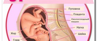
| Dimensions and weight of the fetus | 460 mm, 2400 g |
| Uterus dimensions | VDM - 33-35 cm |
| Pregnant weight | Gain from the beginning of pregnancy is 10-14 kg; over the last week 400-500 g |
Your baby is the size of
Melon Torpedo
460mm Size
2400 g Weight
The closer the date of birth, the more often a pregnant woman thinks about when she can finally return to her usual lifestyle. This is not surprising and quite understandable. A large belly makes it difficult to move, you get tired faster, and sleep sometimes does not bring long-awaited restoration of strength. But don't be discouraged! Very soon you will become a mother!
We will find out how pregnancy proceeds at 35 weeks, how the fetus develops and how a woman’s feelings change during this period. But first, a few words about calculating gestational ages.
What is the placenta?
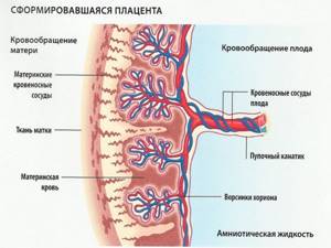
This is the only organ that is formed only during pregnancy and is removed from the mother’s body immediately after the birth of the child. The functions of the placenta include providing the fetus with oxygen and nutrients, removing carbon dioxide and metabolic products, protecting against infections, as well as releasing hormones necessary for the normal course of pregnancy and subsequent childbirth. The development of the embryo depends on the functionality of the child's place, so even minor disruptions in its functioning can lead to serious perinatal complications.
Feelings of the expectant mother
At 35 weeks of pregnancy, the stomach may already drop, so breathing will become easier, shortness of breath and heartburn will disappear. But the urge to urinate will become more frequent: the uterus will put more pressure on the bladder.
The production of colostrum is activated in the mammary glands. If you might not have noticed it before, now you may notice stains on your laundry, especially after sleep. This is fine. Use special bra pads.
35th week of pregnancy: lower back pain or abdominal pain
These sensations are associated with a growing belly and significant stress on the back. The baby's head is now pressing directly on the pelvic bones. In addition, he is quite cramped in the womb, and sharp jolts can be very painful. Wearing a bandage will help cope with discomfort. If the pain is unbearable and is of a regular nature, be sure to inform your gynecologist about this: perhaps we are talking about increased tone or the onset of labor.
How you feel
Most likely, during the 35 weeks of pregnancy you have already become accustomed to the daily presence of a variety of unpleasant sensations in your life - in the spine, lower back, legs. Pain appears due to the enormous load on the body and a shift in the center of gravity. Don't forget to wear a bandage, avoid standing for long periods of time, and periodically unload your spine in a lying or reclining position. Regular warm-up (every 15 - 20 minutes), in which it is advisable to include circular movements of the pelvis: they not only reduce, but also prevent pain in the hips and sacrum, will also help to cope with exacerbations of pain.
Another trouble at 34–35 weeks of pregnancy is possible pain in the wrists and fingers, which occurs in those who sit at the computer for a long time. This is the so-called tunnel syndrome. If you feel heaviness in your hands, numbness and pain in your hands, tingling in your fingers, periodically do relaxation exercises and rub your hands until you feel warm.
You may have a headache from time to time. Cool compresses and rest in a dimly lit room with an open window will help. In addition to general discomfort, nausea sometimes appears at the 35th week of pregnancy. Due to the fact that almost the entire abdominal cavity is occupied by the uterus, digestive function is impaired. Try not to overload your stomach: eat small portions, but often. This way you will avoid overeating and reduce unpleasant symptoms.
At the 34th - 35th week of pregnancy, in the supine position, compression syndrome of the inferior vena cava may occur, when the woman feels dizzy and short of air. In this condition, the fetal heart rate drops sharply, and this is very dangerous. During rest, a semi-sitting position is acceptable, in which the back deviates horizontally by 45 - 30 degrees. It’s better to sleep on the left side or in an intermediate position, with a couple of pillows under your back. The most correct position for blood circulation in the uterus is lying on your side with a pillow between your legs. For convenience, you can place a flat bag under your stomach.
At 35 weeks of pregnancy, you may notice that the nature of your discharge has changed. They are still light milky, but mucus impurities appear in the creamy mass. These are nothing more than particles of a plug that “clogs” the cervix. Shortly before birth, the cervical canal opens slightly and the mucus plug gradually separates. In some women, it comes out not in parts, but entirely - in this case, you will see a clot of mucus streaked with blood. If this happens, you need to be prepared for the fact that labor may begin at any moment, and collect packages for the maternity hospital in advance.
What happens to the fetus
At week 35, the baby’s body acquires the cute roundness and folds characteristic of newborns due to the growth of adipose tissue. The average weight of a child is 2.4 kg, height - 46 cm. He perfectly distinguishes sounds and reacts to light, stroking the belly and other external stimuli.
The baby sleeps most of the day, but within 12 hours you should normally count about 10 movements. Sometimes, after swallowing too much amniotic fluid, he hiccups and you feel your stomach flutter. This is absolutely normal, there is no need to be alarmed. You can help your child by changing your body position or breathing deeply.
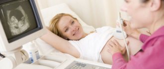
Ultrasound technique of this period
Ultrasound examination at this stage is carried out transabdominally. The woman lies down on the couch, the doctor applies a special gel to her stomach and conducts an examination using a sensor. Sometimes it may be necessary to perform the examination in the lateral position.
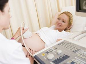
There is no need to prepare for such a procedure. It is advisable that the child behave calmly during the study, because its activity may interfere with accurate measurements.
Tests and ultrasound
In the third trimester, each of your visits to the gynecologist will be accompanied by a study of the fetal heartbeat - cardiotocography. By assessing the number of beats per minute at rest and during movements, the gynecologist can judge the child’s condition, promptly identify complications and give a referral to the hospital.
An ultrasound examination is performed at week 35 if it was previously detected or the doctor suspects:
- umbilical cord entanglement;
- disturbances in placental blood flow or premature aging of the placenta;
- insufficient or excessive volume of amniotic fluid.
Conclusion Ultrasound largely determines the tactics of childbirth. For example, in difficult cases of entanglement, a caesarean section is prescribed.
Medical observation
In some cases, at 35 weeks of pregnancy, the gynecologist may prescribe an ultrasound. The study will allow you to determine the weight and position of the child, as well as identify possible complications, for example, anomalies of the placenta, oligohydramnios or polyhydramnios, entanglement of the fetus with the umbilical cord, etc. The information obtained will help the specialist develop tactics for future childbirth.
This week the doctor will definitely measure your blood pressure and determine your weight and size. In addition, as usual, you will need to do a general urine and blood test.
Other signs of impending labor include:
Weight loss
You may suddenly feel like you've lost some weight. This is due to the removal of excess fluid from the body, swelling is reduced. Changes in the nature of bowel movements are also associated with this. You may need to go to the toilet more often, the consistency of your stool becomes thinner, and many women complain of diarrhea.
Prolapse of the uterine fundus
The baby presses its head against the lower part of the uterus and pulls it down to the entrance to the pelvis. He himself occupies the most convenient position for birth. As a result of these preparations, you will feel how it has become easier to breathe and heartburn has disappeared.
Note that even the most experienced gynecologist will not be able to tell you how soon labor will begin after the appearance of these symptoms.
Child
Active “polishing” of all the baby’s organs continues. The little nails have now grown to the edge of the pads and it is likely that the baby will be born with a long “manicure”. Subcutaneous fat continues to be actively deposited, especially in the peri-shoulder area, and the tiny shoulders become plumper.

What am I doing there
I listen to what is happening outside, hiccup and blink
Celebrity moms who aren't shy about wearing bikinis during pregnancy
Checklist for 35 weeks of pregnancy
- Even if you do not feel the former lightness in your body, do not forget about physical activity. It will help keep your body in shape. A simple walk in the fresh air promotes blood circulation, improves sleep and overall well-being.
- Make a list of things you will need in the maternity hospital. Attend an open day: such events are regularly held in maternity hospitals. You can clarify all the questions of your stay: how the rooms are equipped, is it possible for a partner to be present, who can visit you and when, what can and cannot be taken with you.
- Pay special attention to foods high in iron in your diet. In recent weeks, the baby has been stocking up on this microelement. Its sufficient consumption will prevent the development of anemia in the first year of life.
The thirty-fifth week of pregnancy is the time when you can still have time to attend courses for expectant mothers at the Women's Medical Center. Prepare for childbirth correctly with our specialists - experienced gynecologists and pediatricians.
Good to know
Childbirth with a scar on the uterus
What awaits a premature baby immediately after birth in the hospital?
How to choose a nursing bra
Is a pregnant woman beautiful?
What worries a woman during pregnancy?
What are the methods of preparing for childbirth?
All texts for pages about mother and baby were kindly provided by RAMA Publishing - these are chapters from the book by Svetlana Klaas “Your Favorite Little Man from Conception to Birth”, reviewer Irina Nikolaevna Kononova, Candidate of Medical Sciences, Associate Professor of the Department of Obstetrics and Gynecology of the Ural State Medical Academy (Ekaterinburg).
Diagnostic screening ultrasound during pregnancy: 1, 2 and 3 screenings
Ultrasound during pregnancy is done with one main goal - accurate diagnosis of pathologies in the fetus, diagnosis of pathologies in the mother. Screening ultrasounds are performed precisely at certain times, this is due to the peculiarities of the development of the fetus and placenta and the ability to obtain the most accurate data.
If necessary, additional ultrasounds are prescribed, the method is completely safe, ultrasounds can be done as often as necessary. In some cases, ultrasound during pregnancy is prescribed quite often - this allows you to control the situation and make the right decision for the obstetrician about further management of the pregnancy.
Reproductive health
For couples who are planning a child, we have developed a diagnostic program for a quick health check. Based on the results, either a more in-depth diagnosis will be prescribed or recommendations will be given. The program allows you to quickly and inexpensively find out your reproductive health status.
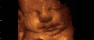
Today you can do a 3D ultrasound during pregnancy; the technique allows you to get better visualization and see your unborn child in more detail.
First screening ultrasound at 11-13 weeks of pregnancy
Screening ultrasound at 12-13 weeks is already capable of identifying the main part of genetic and chromosomal pathologies. In addition, the doctor looks at the development of the fetus as a whole, and the features of the development of the placenta. What is determined and what data is included in the protocol of the first screening ultrasound during pregnancy:
- CTE (length from the coccyx to the crown) at 11-13 weeks in the range of 43-65 mm. An increase in size may indicate a large fetus or a diagnostic error, while a decrease in size may be due to anatomical features, developmental delays, infectious diseases suffered by the mother, or genetic pathologies.
- BDP (biparietal size) - the distance from temple to temple. Normally, this value is 17-24 mm. However, minor changes are allowed if they are increased proportionally. High values of BPR with normal others often occur with hydrocephalus or brain hernias, low values with slowed brain growth. This indicator is not absolute when decoding the protocol, but the BPR values are taken into account.
- TVP (nuchal translucency thickness) is one of the most important ultrasound indicators for pregnant women at 11-13 weeks. A value of 1.6-1.7 mm is considered normal. If an ultrasound reveals a TVP of more than 3 mm, then this most often indicates severe chromosomal disorders - Down syndrome, Edwards syndrome, etc. But ultrasound alone does not make a diagnosis and additional examinations are carried out.
- The size of the nose bones. The length of the nasal bone should be 2-4.2 mm. Too low a value may indicate pathology or individual characteristics of fetal development.
Additionally, the presence and condition of the vessels in the umbilical cord are taken into account, and an analysis is done for markers of chromosomal abnormalities. Here you can read more about the first trimester of pregnancy.
What is examined during the first ultrasound at 4-6 weeks:
Usually the first diagnostic ultrasound is preceded by a study that confirms the fact of pregnancy.
- the size of the chorion, amnion and yolk sac. If the chorion (this is the future placenta) is located extremely low, there is a risk of miscarriage, and subsequently the risk of placenta previa; the volume of amnion with amniotic fluid is about 50-100 ml; Normally, the yolk sac should already be absent, but if the ultrasound report says that it is visualized, there is a risk of pregnancy pathologies.
- examine the cervix, its length should be 35-40 mm; if the cervix is shortened, there is a risk of miscarriage, and subsequently premature birth;
- Heart rate. The fetal heart beats 140-160 times per minute, with deviations of no more than 40 beats in both directions possible.
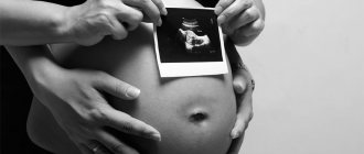
You can do a 3D ultrasound and you will be left with a beautiful image - the first photographs of your baby. The results of screening ultrasound must be interpreted by doctors, since all data must be taken into account in totality, and a deviation in any one value does not speak in favor of pathology.
Second screening ultrasound during pregnancy
The second screening ultrasound is performed at 19-21 weeks of pregnancy. All organs and tissues are already formed, pathologies and developmental anomalies are clearly visible. The main task of this ultrasound is to identify malformations and, if they are detected, to refer the woman to a geneticist for consultation. A geneticist can suggest premature birth, but only after cordocentesis - a chromosomal analysis of umbilical cord blood.
What is examined during the second ultrasound during pregnancy
First of all, the doctor evaluates the correct formation of body parts, internal organs and the brain. The ultrasound report must contain the following data:
- Biparietal head size at 20 weeks 39-56 mm;
- Fronto-occipital size at 20 weeks 53-75 mm;
- Femur length at 20 weeks 23-38 mm;
- The diameter of the chest at 20 weeks is 24-36 mm;
- The length of the humerus at 20 weeks is 24-36 mm;
- Breast circumference at 20 weeks - 154-186 mm;
- Abdominal circumference at 20 weeks - 124-164 mm;
- The size of the cerebellum at 20 weeks is 18-22 mm;
- Amniotic fluid index at 20 weeks - 86-230 mm;
You need to understand that the sizes greatly depend on the timing of pregnancy and the individual characteristics of the fetus. But, if the sizes are significantly smaller than normal and are not proportional to each other, this often indicates a delay in fetal development and requires clarification of the reasons and their subsequent elimination. The ultrasound protocol should be deciphered by an obstetrician, taking into account the data in conjunction with the medical history and individual characteristics of the pregnant woman. Here you can read more about the second trimester of pregnancy.
It is recommended not to neglect screening ultrasounds during pregnancy, especially if the first ultrasound revealed any risks or the pregnant woman has a complicated medical history. This will allow timely treatment to begin, and if fetal pathologies that are incompatible with life are detected, there will be time for detailed diagnosis and making an informed decision.
Modern equipment provides detailed visualization, the risk of errors is almost completely eliminated, so if you have had an ultrasound and doubt the reliability of the data, we recommend that you do the ultrasound again. And, it is always recommended to repeat the ultrasound after 5-7 days, if there are doubts about the accuracy, deviations from the norm have been identified. For better visualization, you can do a 3D ultrasound.
Third screening ultrasound during pregnancy
The third screening ultrasound during pregnancy is performed at 30-34 weeks. Its task is to assess the degree of maturation of all organs, to identify abnormalities that are not visible at 20 weeks - anomalies in the development of bones and internal organs. If necessary, a 3D reconstruction of the bones of the face and skeleton is performed. This allows you to adjust pregnancy management tactics, since some children need surgical care immediately after birth. The condition of the placenta, amniotic fluid and other data are also assessed, and in some cases the doctor may decide on early delivery. Here you can read more about the third trimester of pregnancy.
What is examined during the third ultrasound during pregnancy
- The degree of maturity of the placenta is first or second, if premature maturation of the placenta is diagnosed - that is, there is a high risk of hypoxia and developmental delays;
- Cervix - the os of the cervix must be closed, if it is open, then medical attention is needed - a special ring is applied to avoid early labor;
- The amniotic fluid index is 81-278 mm, lower values indicate oligohydramnios - the reason may be previous infections or the presence of chronic foci of infection, moderate oligohydramnios is not dangerous for the fetus, with significant oligohydramnios there is a risk of fusion of the fetal toes and developmental anomalies - in these In cases, ultrasound is repeated every 5-7 days and earlier fertilization is often recommended.
- BDP (biparietal size) - 85-89 mm;
- Head circumference - 309-323 mm;
- Fronto-occipital size - 102-107 mm;
- Abdominal girth - 266-285 mm;
- Shoulder length - 55-59 mm;
- Thigh length - 62-66 mm.
In addition, the condition of the umbilical cord and the vessels in it is assessed, the presence of hypoxia is determined and, if it is detected, its degree is determined.
If the fetal weight is significantly higher than normal, it is recommended to perform another ultrasound immediately before birth to decide on the possibility of vaginal delivery. If a low placement of the placenta or its presentation is detected, then operative delivery is almost always recommended. If premature aging of the placenta has been detected, the doctor may decide on early delivery.
These examinations can be carried out at the medical center named after. Speransky. We have installed high-quality equipment that allows us to perform both conventional ultrasound and 3D visualization. There is a license to conduct pregnancy, all narrow specialists work and there is a laboratory diagnostics department. At the same time, prices for ultrasound of pregnant women are 10-15% lower than the city average. If you are planning to do a 3D ultrasound, you need to keep in mind that the procedure will last more than an hour. In addition to 3D ultrasound, we make a video recording for future parents; a spouse can be present during the procedure.
You can sign up for an examination by phone or through the online registration.
Make an appointment by phone
Nutrition
At the 35th week, you should monitor your weight more carefully - gaining excess weight now is not at all beneficial. But you shouldn’t go on a diet either, just balance your diet - more fruits, vegetables, cereals, less fatty foods, baked goods, sweets.
It is important
Normally, vaginal discharge may change slightly at the 35th week - a little mucus will be added to the light and clear liquid - this is how the mucus plug gradually comes out. If the discharge noticeably changes color - it turns green or brown, or you notice blood, this is a reason to consult a doctor as soon as possible.