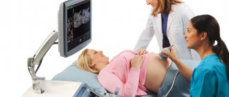
The third scheduled mandatory ultrasound is very important to prepare for childbirth. With an ultrasound at 34 weeks of pregnancy, the normal weight for the fetus is 2100-2400 g, height - 43-44 cm. The size of the fetus is almost equal to the size of a full-term baby (for him, the norm is weight from 2500 g, height - from 45 cm). But, of course, the birth of such a baby will be somewhat premature, because his skin has not yet fully formed, and it will be difficult for the lungs to cope with breathing and atmospheric air on their own.
However, there is very little time left before the birth, and this week very important metamorphoses occur with the baby: the surface of the skin is leveled, becomes smooth, and the embryonic fluff disappears.
The color of the skin loses its redness and gradually becomes pink, and a layer of cheese-like lubricant thickens on the skin. Starting with an ultrasound scan at the 33rd week of pregnancy, the indicators of which are undoubtedly important for assessing the health of the baby, the mother has a pleasant opportunity to take a photo of the baby’s face; modern technologies make it possible to do this even in 3D format. But the main thing, of course, remains the medical examination of the parameters of the fetus and its position in the uterus, because this is the last planned ultrasound before childbirth.
As in previous ultrasound studies, the doctor assesses the number of fetuses, their location in the uterus, determines the dimensions: circumference of the head, abdomen, length of bones. In order to prevent complications during childbirth, a specialist carefully examines the placenta, its thickness, position, degree of maturation, and controls the amount and transparency of amniotic fluid.
Based on the results of the study, a conclusion is drawn up. It records the period in weeks with which pregnancy is correlated, and the size of the fetus relative to developmental norms.
Carrying out all three screening (mandatory) examinations using ultrasound is harmless to the child and provides doctors with valuable information about his condition and the readiness of the mother’s body for natural childbirth.
In total, up to ten ultrasound scans can be performed during pregnancy for medical reasons. The Ministry of Health considers this amount safe.
Interesting Facts
| Options | Indications |
| Time from conception | 32 weeks |
| Period by month | 34 weeks |
| What month | 8 |
| Dimensions and weight of the fetus | 450 mm, 2150 g |
| Uterus dimensions | VDM - 32-34 cm |
| Pregnant weight | Gain from the beginning of pregnancy is 10-14 kg; over the last week 400-500 g |
Your baby is the size of
Dwarf watermelon
450mm Size
2150 g Weight
The thirty-fourth week of pregnancy is a period when you may be surprised to find that your usual actions are no longer available to you. It becomes more and more difficult to climb the stairs, fasten your shoes, and stay on your feet for a long time. Don’t refuse help, rest often and take care of yourself! Right now your body is under enormous stress. Let's find out what happens to the mother and baby, but first let's say a few words about calculating the gestational age.
Feelings of the expectant mother
At 34 weeks of pregnancy, you could feel that breathing became a little easier, heartburn began to occur less frequently or disappeared completely, but all the baby’s movements became much more noticeable. This is due to the fact that the stomach may drop at this stage: the fetus moves lower, pressing its head against the bones of the small pelvis, while the pressure on the stomach and diaphragm decreases.
Training contractions are a common occurrence during the third trimester. There is no need to be afraid of them, they do not in any way speed up the onset of labor. You can alleviate your condition by using measured breathing with an extended exhalation. Rocking on a fitball will also help.
Vaginal discharge, as before, should be clear or slightly whitish, with a neutral, slightly sour odor. Any deviations - mucus, pus, color changes - are a reason to urgently contact a gynecologist.
Fetal development
The height of your unborn child can already reach 45 cm, and weight – 2.1 kg. It is increasingly difficult for him to fit inside the uterus. At this time, most likely, the fetus is already in a cephalic presentation, preparing for birth.
Baby's success
- The immune system has matured: for the first six months of a child’s life, it is protected by antibodies received from the mother, and its own immunity is gradually formed.
- The bones of the body have become dense, but the skull remains soft and mobile. Thanks to this, the baby passes through the birth canal.
- The body of the fetus is increasingly covered with vernix lubrication, which will also help it to be born more easily.
34 weeks pregnant, baby hiccups - is this normal?
The baby, swallowing too much amniotic fluid, actually hiccups later. And at this moment the mother feels rhythmic shudders in her stomach. Many experts believe that this happens when a woman has eaten too much sweets. After all, the taste of amniotic fluid is influenced by what you had for lunch today. What to do in this case? Wait for the hiccups to go away and adjust your diet.
Another cause of hiccups is uncomfortable body position. You can help your child roll over by breathing deeply and evenly, or by sitting down or turning over to the other side.
Useful advice for future parents
If it were possible to stock up on laughter and humor for future use, it would be possible to avoid many worries and fears both during the period of waiting for the Baby and in the first year after his birth.
It is especially important that funny situations are experienced simultaneously by both the baby’s father and mother. The psychotherapeutic value of laughter is difficult to overestimate, so take advantage of every opportunity to laugh: watch a funny movie or show with comedians, download funny videos from the Internet, read funny episodes from books aloud or quote jokes, find the funny and absurd in ordinary things.
Laughter allows you to:
- relieve emotional stress, relax, rest, switch attention, look at a disturbing situation from an unusual perspective;
- strengthen emotional contact with your partner;
- make peace (one of the best family habits);
- to amuse the Baby - so many positive emotions at once!
Of course, you can laugh alone, but laughing together with your loved one increases the effect many times over. By the way, it is the Baby’s father who can take care of the fun in the house, because, according to British scientists, the male hormone testosterone, which is many times more in a man’s body, is responsible for a sense of humor.
What does the Kid experience at the moments when his mother laughs heartily and her body shakes with laughter? Most likely, the hormones of happiness and joy, endorphins, reach the Baby’s body, and pleasant waves of pleasure run through his body, and a smile appears on his face. Only human beings can laugh, and your Baby will learn this very soon - by the end of the fourth month after birth. Or maybe if he often hears your laughter now, and earlier.
Tests and ultrasound
If you haven't had your third scheduled ultrasound yet, now is the time. Its results largely determine the tactics of childbirth. The doctor is interested, first of all, in the growth rate of the baby, his position in the womb, the degree of maturity of the placenta and the volume of amniotic fluid. They can also determine or confirm the sex of your unborn child if this question was previously in doubt.
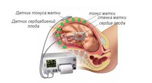
The fetal heartbeat is an important indicator of its condition. To assess it, the cardiotocography method is used. Sensors are placed on the stomach, and you independently note all fetal movements using a special device. The gynecologist performs the interpretation in your presence.
Indicators of the child’s body
Using a small sensor from which ultrasonic waves emanate, you can examine at least 16 parameters characterizing the general condition of the baby.
Weight category
The average weight varies from 1850 g to 2300. However, deviations from the generally accepted value are not always a cause for concern. It is worth noting that the indicator under consideration directly depends on the BMI (body mass index) of both parents: if we are talking about a fairly large physique, the mark can exceed 2500–3000 g, and if it is fragile, then the parameter will be in the range of 1300–1600 g.
Height
Based on the ultrasound results, growth is also determined (not to be confused with the coccygeal-parietal size of the fetus). So, 40–43.5 cm is quite the optimal option. A slight error of 3–5 cm is allowed towards both the minimum and maximum.
Heart rate
Carrying out CTG (cardiotocography) in parallel with the ultrasound process allows you to calculate the exact digital index of heart rate or the number of heart muscle beats in 1 minute.
It is generally accepted that normal values equate to a period from 120 to 160 beats/min. ± 5 units. When the rate drops below 100, they speak of bradycardia; an increase from 170–175 is called tachycardia. Both cases require additional types of examination that will fully clarify the situation.
Head and abdominal circumference
In order to verify the proportional structure of a small body, a specialist uses ultrasound to identify a number of measurements, which include the circumference of the tummy and head. The first parameter should be 258–336 mm, the second – 285–335 mm. In the third trimester, the baby's head has already reached its normal harmonious size.
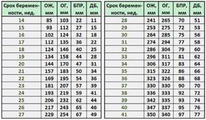
Results of measurements of coolant and exhaust gas during ultrasound
Chest diameter
Another classic indicator is DHA. The special comparative table shows a norm of 83–88 mm.
LZR and BPR
Two values related to each other determine the general structure of the child’s head. Thus, the LZR (fronto-occipital size) is a segment connecting the middle of the occipital bone with the frontal bone. BDP (bipariental size) is the distance between the parietal bones or, to put it simply, the width of the head.
| Indicator name | Norm (mm) | Allowable fluctuation (mm) |
| BPR | 80–85 | 75–91 |
| LZR | 102–109 | 95–115,5 |
Position in the womb
An extremely important factor influencing the tactics of childbirth is the location of the fetus in the mother's womb. If there is a so-called cephalic presentation detected on an ultrasound, then the birth is carried out in the classical way.
Indicators of fetal condition on CTG
Sometimes the baby takes less suitable positions:
- Transverse (perpendicular to the spine).
- Mixed (hips and knees bent at the same time).
- Gluteal (buttocks rest on the mother’s pelvic bones).
- Foot (legs pointing down).
There is an opinion that each of the listed cases requires only a cesarean section, but this is not entirely true. There are known medical facts that indicate the natural normalization of the child’s position even at such a late stage without any intervention.
Absence/presence of umbilical cord entanglement
The norm characteristic of 33-34 weeks of pregnancy practically does not allow the child to be entangled in the umbilical cord. The reason for this is the increased likelihood of fetal hypoxia, which is observed with numerous intrauterine loops. The presence of a single half-loop without tension is the only exception and is not a cause for concern.

Acceptable loop encircling the right leg
Nasal bone
This facial bone must be measured during an ultrasound scan, since in some cases it is an indicator of the presence or absence of Down syndrome. The average value is equivalent to 11.3–11.5 mm. If the detected figure reaches 9 mm or increases to 13.8 mm, then do not panic. Such variations are only a manifestation of individuality, not pathology.
Length of tubular bones
Elongated round bones include the hips, forearms, shoulders, and shins, consisting of the tibia and fibula. The combination of these 4 values reflects the condition of the musculoskeletal system:
| Name of bones | Allowable length (mm) |
| Shin | 83–85 |
| Hip | 63–65 |
| Forearm | 48–50 |
| Shoulder | 56–58 |
Anatomical features
In the form with ultrasound indicators, you can often find data reflecting the development of certain organs, facial structures, as well as bone elements. These include:
- nasolabial triangle;
- lungs;
- kidneys;
- spine;
- cerebellomedullary cistern;
- bladder;
- eye sockets;
- lateral ventricles of the brain;
- stomach;
- cerebellum;
- 4-chamber section of the heart.
The absence of violations is recorded using various entries in the corresponding columns - “no pathologies”, “b/p”, “examined”, “norm” or “+”. If the sonologist identified some abnormality during the ultrasound session, he will describe it in detail on the form.
What to discuss with your doctor
- Normally, you should average 10 episodes of fetal movement within a 12-hour period. If it seems to you that he is moving little or, on the contrary, a lot, tell your doctor about this. This is not always a symptom of pathology, but it is better to keep your finger on the pulse.
- Tone at 34 weeks of pregnancy may be a symptom of labor beginning. Discuss with your gynecologist how to act in case of cramping pain in the lower abdomen, how to distinguish them from training contractions, and where you can go for help.
Risk factors
At the 33rd week of pregnancy, there is a high risk of placental abruption. Normally, this should occur at the end of labor, but in some cases this process begins much earlier. The placenta may detach partially or completely. In this case, severe abdominal pain occurs and bloody discharge from the vagina appears. This condition is dangerous due to large blood loss for the mother and suffocation for the baby, since without the placenta, oxygen ceases to flow to the fetus.
Another problem of 33 weeks of pregnancy is late toxicosis, which also causes oxygen starvation of the child. To avoid such problems, monitor your health and be sure to inform your doctor about any changes.
Possible complications
In the later stages of pregnancy, you should be especially attentive to your well-being. Do not miss visits to the gynecologist to prevent and promptly eliminate various complications of pregnancy.
Premature birth
Observed against the background of infections, stress, shock or trauma, with insufficient cervical length. A woman may suddenly feel a sharp tug in her lower abdomen, her water or mucus plug may break, and regular contractions may occur. However, a child born at this time is quite viable. But he will need special care for a few weeks.
Swelling at 34 weeks of pregnancy
Many pregnant women complain of increased swelling in the last trimester. This is due to increasing water metabolism in the body. The volume of blood and amniotic fluid increases. Reduce your intake of salt, which retains excess water. And don’t forget to take a urine test on time. The appearance of protein in the urine against the background of severe swelling is an alarming symptom of gestosis, which requires immediate treatment in a hospital setting.
How you feel
At 32–33 weeks of pregnancy, you occasionally feel Braxton Hicks training contractions, which prepare your uterus for labor. There should be no painful sensations, and if they do appear, they usually go away while lying down. At such moments, be attentive to your condition and, if contractions do not stop for a long time, and even become regular, call an ambulance immediately! This may be a sign of premature labor.
At 33 weeks of pregnancy, the urge to urinate becomes more frequent, and you have to constantly go to the toilet day and night. Try not to drink a lot of fluids before bed.
Half of expectant mothers develop a runny nose. Women with allergies are most at risk of developing rhinitis. You can combat this problem by rinsing your nose with saline, using special drops (be sure to consult your doctor before using them) and humidifying the premises during the heating season.
Also, at 33 weeks of pregnancy, many women are concerned about inflammation and bleeding of the gums. Gingivitis can be a consequence of both hormonal changes in the body and insufficient oral hygiene. Brush your teeth with toothpaste at least twice a day and simply brush after every meal. Don't ignore dental floss, and the best way to relieve inflammation is to rinse with a saline solution.
At 32–33 weeks of pregnancy, you continue to suffer from pain in the back, sacrum and lower back. Try to maintain correct posture, regularly unload your spine by taking a lying or semi-lying position, wear a bandage (only with the approval of a doctor), and for a comfortable sleep, use an orthopedic pillow and mattress.
A lot of stress is now placed on my legs. Massages and cool foot baths will help reduce pain. And to prevent the occurrence of cramps, you need to introduce foods containing calcium into your diet, and from time to time raise your legs to an elevated position from a lying position.
In addition to the legs, at 33 weeks of pregnancy, the arms, namely the wrists, often suffer. Expectant mothers, especially those whose work involves a computer, may experience carpal tunnel syndrome. Its symptoms are numbness in the fingers, tingling sensation and pain in the hand. Massage and special hand exercises help to cope with the problem.
Lifestyle
In the last weeks of pregnancy, it is important to maintain your weight within normal limits. A fetus that is too large will only complicate the birth. Therefore, limit yourself to simple carbohydrates, junk food and weigh yourself regularly. Eat small meals, do not limit fluid intake.
Is intimacy possible?
If the pregnancy proceeds without complications, sex is not prohibited. However, many experts recommend abstaining from it after the 34th week of pregnancy, when the birth canal begins to prepare for the birth of the baby. Physical intimacy increases the risk of infection, so if you decide to have it, you should use a condom.
Position of the baby in the uterus
A very important indicator that the specialist will pay special attention to is the position of the fetus. After all, the presentation that is observed now during ultrasound will remain until the birth itself. Normally, the baby's position should be in the head position. With such a presentation, natural childbirth is prescribed.
If the baby's position is transverse or pelvic, the obstetrician-gynecologist advises performing a cesarean section in order to avoid the possibility of injury to the fetus. But there is no need to be afraid in advance. Sometimes the baby changes position literally a few days before birth. Also, in order for the fetus to acquire the necessary presentation, the doctor prescribes special exercises.
Also, during ultrasound diagnostics, the doctor examines the location of the umbilical cord. Danger during childbirth is caused by the presence of entanglement of the fetal cervix with this organ. It is difficult for a child to get rid of the entwined umbilical cord, because by the 34th week the size of the child has increased significantly. He can no longer spin freely or tumble inside the amniotic fluid.
Checklist for 34 weeks of pregnancy
- Do Kegel exercises. This is the best prevention of genital prolapse and rupture of perineal tissue after childbirth.
- If you experience itching on the skin of the abdomen or around the navel, be sure to apply moisturizing creams and oils against stretch marks to this area. Daily care is the best prevention of these skin defects.
- When leaving home, do not forget to take with you a pregnant woman’s exchange card, which contains all the necessary information about your health and the course of your pregnancy.
- If, after the start of contractions, you plan to get to the maternity hospital by your own transport, it is worth studying the route in advance, choosing a main route and a spare one in case of difficult traffic.
Prepare for childbirth with the specialists of the Women's Medical Center. We will talk about how to behave correctly during labor, how to breathe and how to move to relieve pain.
Good to know
Why does the baby lie incorrectly in the stomach and how to turn it over?
Placental abruption: delay is unacceptable!
Premature babies: care in the maternity hospital
How to massage the perineum against tears
Placenta - during pregnancy and after childbirth: what you need to know
How does female sexuality change during pregnancy?
All texts for pages about mother and baby were kindly provided by RAMA Publishing - these are chapters from the book by Svetlana Klaas “Your Favorite Little Man from Conception to Birth”, reviewer Irina Nikolaevna Kononova, Candidate of Medical Sciences, Associate Professor of the Department of Obstetrics and Gynecology of the Ural State Medical Academy (Ekaterinburg).

