Ultrasound of the mammary glands during feeding allows timely diagnosis of inflammatory and congestive complications, which often worry women who are breastfeeding. Such pathologies, if left untreated, can lead to irreversible consequences in the future. Therefore, if a woman experiences discomfort or chest pain during breastfeeding, there is no need to postpone a visit to the doctor.
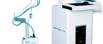
Ultrasound waves are absolutely safe for the health of a woman who has recently given birth, so they do not have any effect on the condition of the gland and the quality of milk. At the International Clinic “Hemostasis” you can sign up for an ultrasound of the mammary glands during breastfeeding at any time. Diagnostics are carried out by doctors of the highest category using highly sensitive equipment of the new generation. We guarantee the quality and most reliable research results.
Indications for ultrasound of the mammary glands
Indications for breast ultrasound include:
- pain in the breast area not associated with menstruation;
- retraction or wrinkling of the breast skin;
- changes in the color of the nipples and breast shape;
- bloody and other discharge from the nipples;
- the appearance of lumps in the breast area;
- traumatic chest injuries;
- enlarged axillary lymph nodes.
For preventive purposes, ultrasound of the mammary glands is performed after completion of breastfeeding (BF). Ultrasound examination of the mammary glands during breastfeeding is not contraindicated.
Indications and contraindications
Ultrasound of the mammary glands during breastfeeding can detect even minimal changes in the structure of the organ under study. During the procedure, the breast tissue is exposed to ultrasonic waves, which are reflected from different structures with different strengths. Diagnostic results in the form of dynamic high-resolution images are visualized on the monitor screen.
This type of diagnosis is not included in the list of mandatory procedures that must be completed during breastfeeding. An examination is prescribed by a doctor if during lactation a woman begins to be bothered by the following pathological signs:
- painful lumps detected during palpation;
- pain, discomfort in the chest that does not go away for a long time;
- pathological discharge from the nipples with purulent, bloody, serous inclusions;
- inflammation of regional lymph nodes;
- low-grade fever of unknown origin;
- breast skin changes;
- pronounced asymmetry of the glands.
Other indications for ultrasound examination:
- neoplasms in the organs of the reproductive system;
- previously suffered mastopathy;
- successfully treated breast cancer;
- previously diagnosed benign neoplasms.
Women who have undergone surgical treatment of tumors in the mammary glands should visit a mammologist in the first three months after the birth of the child. During the postpartum period, serious hormonal changes occur in the body of a young mother, which can become a trigger for the re-development of tumors.
Indications for ultrasound of the mammary glands in men
Ultrasound of the mammary glands in men is prescribed if the development of a benign breast tumor (gynecomastia) is suspected. The price for ultrasound of the mammary glands in men and mammary glands in women is no different. When pathology appears, the size of the mammary gland in men can increase by 1-10 cm. Treatment of gynecomastia can be conservative and surgical, depending on the symptoms of the pathology and the stage at which it was diagnosed.
Symptoms of gynecomastia and indications for breast surgery in men include:
- pain in the chest area;
- nipple discharge;
- increased sensitivity of the nipples;
- the appearance of lumps in the mammary glands;
- increase in size of the mammary glands.
Interpretation of examination results
Normally, the results of an ultrasound examination of the breast during breastfeeding look like this:
- The lobes have an elliptoid shape, the structure is low echogenic.
- The skin of the gland has uniform echogenicity.
- The middle mammary layer is moderately echogenic.
- The width of the ducts is 1 - 2 mm, there are no pathological inclusions, irregularities or compactions.
Any deviations from the norm indicate the development of a pathological process that requires urgent treatment. Common diseases diagnosed in women during breastfeeding:
- Mastopathy. A disease of the mammary gland of a non-inflammatory nature, characterized by changes in the ratio in the structure of the organ of connective and epithelial tissue. The progression of the pathology leads to the formation of cysts and fibrous tumors. With mastopathy, the echogenicity of the glandular structure is increased and looks like light spots.
- Leaf-shaped tumor. May be of benign or malignant etiology. The structure of the neoplasm is heterogeneous, hypoechoic.
- Mastitis. Inflammation of the mammary gland, which mainly affects women who have given birth to their first child and have not fully learned the correct method of latching a child. Ultrasound visualizes focal formations near the areolar segment. With the development of a purulent complication, the alveoli and ducts expand, and the infiltrate surrounding them looks like a honeycomb.
- Cyst. It is a benign cavity neoplasm, the capsule of which is filled with serous fluid. On ultrasound, the formation looks like a clear, smooth capsule with liquid contents.
- Malignant tumor. The structure of the neoplasm is heterogeneous, the boundaries are unclear and blurred.
- Lactostasis. A common complication during breastfeeding. The reason for its development is a disruption in the process of milk outflow from any part of the mammary gland, caused by blockage or obstruction of the milk ducts. Lack of timely treatment of lactostasis leads to the development of mastopathy.
- Fibroadenoma. Often formed against the background of hormonal disorders. Fibroadenoma is an intermediate step between benign and cancerous neoplasms, and therefore requires timely diagnosis and surgical treatment.
How is breast ultrasound performed?
No special preparation is required for breast ultrasound. To carry out ultrasound diagnostics, you need to undress to the waist, lie down on the couch and put your hands behind your head. This position of the body makes it possible to study not only the mammary glands, but also the lymph nodes adjacent to them.
During a breast ultrasound, the doctor moves a sensor along the mammary glands. An image of the organ from different angles is displayed on the screen. Diagnostics takes up to 30 minutes. The results of the study can be obtained immediately after it is carried out.
Only a doctor can interpret the results of a breast ultrasound. Therefore, after diagnosis, you need to make an appointment with a mammologist. Based on the results of the study, the doctor can refer the patient for a consultation with a gynecologist, ultrasound of the pelvic organs and other diagnostic tests.
How is ultrasound performed for breastfeeding?
The diagnostic procedure lasts on average 15 – 20 minutes. All manipulations are painless and do not cause any discomfort. The procedure is as follows:
- The doctor asks the woman to remove clothing from her breasts, lie down on the couch and relax.
- To improve the conductivity of ultrasonic waves, the breast skin is lubricated with gel.
- Using an ultrasound sensor, the diagnostician examines the internal structure of the organ, assesses the ratio of tissues, their echogenicity and other important indicators.
- Directly during the diagnosis, the doctor voices the identified pathologies, so a preliminary diagnosis can be found out immediately.
Upon completion of the diagnosis, the results are recorded in the ultrasound protocol, with which the woman goes to the doctor who gave the referral for the ultrasound. The doctor, based on the protocol data, establishes an accurate diagnosis and, if necessary, prescribes additional research and a treatment regimen.
Application area
Breast doctors work with patients’ concerns about the mammary glands. Sometimes women turn to gynecologists with complaints of pain, a palpable tumor, or discharge from the nipples. Any complaints from this body may be an indication for the use of ultrasound diagnostics:
- change in breast shape and size,
- soreness,
- nipple discharge,
- the presence of compactions, neoplasms,
- previously identified cysts,
- benign tumors.
Ultrasound is also used after surgery to install implants, to monitor ongoing treatment or examine the breasts of pregnant and lactating women. However, not only women can suffer from breast diseases, but also men. In case of gynecomastia, a suspected neoplasm, men are sent for an ultrasound of the mammary glands.
Preparation for ultrasound examination
There is no need to prepare specially for the diagnostic procedure. But for the accuracy of breast ultrasound results, the day of the cycle matters. It is recommended to come for the study between 5 and 10 days after the start of your period.
This is due to the fact that, under the influence of hormonal fluctuations, the structure of the breast changes throughout the cycle. It is in its first half that the study is most informative. But if necessary, the mammologist can send the patient for an ultrasound on another day.
This recommendation does not apply to postmenopausal women - they can diagnose the condition of the mammary glands at any time.
What can a doctor find on a breast ultrasound?
Ultrasound breast screening allows early detection of a number of pathological changes and diseases.
- Abscesses are secondary pathologies that develop in the soft tissues of the mammary glands against the background of inflammation, hormonal disorders, mastitis, injuries, etc.
- Mastitis is an infectious and inflammatory process. Mastitis can be determined without ultrasound (by visual examination and palpation). Sonography is necessary to determine the location and extent of pathology and the presence of complications.
- Mastopathy is a pathology that develops due to hormonal imbalance and is manifested by fibrocystic tissue changes.
- Benign neoplasms (adenomas, intraductal papillomas, breast cysts, lipomas, fibromas, fibrolipomas).
Ultrasound screening of the mammary glands is an important tool for the early diagnosis of breast cancer pathologies. To differentiate benign neoplasms detected on ultrasound from malignant tumors, the mammologist may prescribe additional studies - magnetic resonance imaging, biopsy, etc.
If you need to make a reliable diagnosis of breast pathologies, come to us for an ultrasound of the mammary glands. You can sign up for an ultrasound screening by phone, by writing an online chat, or by filling out an application on this page.
Ultrasound of the mammary glands: possibilities of the method
In modern medicine, ultrasound diagnostics is used everywhere. The method is based on the ability of human body tissues to absorb and reflect ultrasound radiation differently. Ultrasound waves are directed by a radiator to the area under study, and a special sensor reads their reflection. After digital processing of the received data, an image is displayed on the monitor screen, which makes it possible to literally look inside the body and detect any deviations.
Ultrasound of the mammary glands is one of the main methods for diagnosing diseases, including breast cancer. Ultrasound can detect lumps, neoplasms and tumors. The good thing about this method is that it allows you to identify abnormalities at the earliest asymptomatic stages, which makes it possible to carry out timely treatment, preventing the development of serious and life-threatening diseases.
Along with ultrasound, mammography is used to examine the mammary glands. These methods often complement each other, giving the doctor the most complete clinical picture. However, mammography uses ionizing radiation, which is unsafe and contraindicated for some categories of patients. Thus, without special indications, mammography is not prescribed to women under 30 years of age due to the increased sensitivity of tissues to radiation. Ultrasound, unlike X-rays, is harmless, so ultrasound of the mammary glands can be performed even on adolescents, pregnant women and women during lactation.
Advantages of the method
In addition to being harmless, breast ultrasound has many other advantages. In particular, this is high information content and the ability to detect violations at very early stages. The harmlessness of the method allows it to be used as often as necessary, which is important in the treatment of diseases to assess the effectiveness of the therapy or monitor the dynamics of postoperative recovery.
Ultrasound is suitable for examining breasts of any size, while mammography cannot be done on small breasts. Also, mammography is not suitable for examining the breast after injury or surgery. Using an ultrasound, you can examine not only the breast itself, but also nearby tissues and lymph nodes.
Another important advantage of the method is accessibility. Ultrasound machines are used everywhere, and the cost of the examination is low.
How is ultrasound diagnostics carried out?
Sonography is a comfortable study for the patient. If there are no painful areas in the chest, ultrasound does not cause discomfort. To carry out the procedure, the doctor asks the woman to lie on the couch and put her hands behind her head.
Then the specialist applies a transparent gel to the patient’s chest (it can be easily removed with a disposable towel) and applies a special sensor to the treated area. The sensor sends high-frequency waves into the tissue, then receives them back and displays the image on the monitor. When the doctor has seen everything that is needed, the gel is removed from the skin with a disposable paper towel. The patient gets up from the couch, gets dressed and waits for the ultrasound report for a few minutes.