1
What are urates?
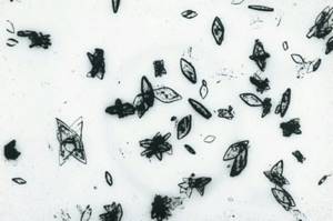
Urates in the urine of women are salts of uric acid in an insoluble form, which can appear in both healthy and sick people. Salts are an unorganized sediment of urine, the composition of which varies greatly and depends on the nature of nutrition, drinking, acidity of the environment, water-salt balance in the body and many other factors. During microscopic examination, these substances are determined in the form of amorphous crystals in the form of barrels, crosses with pointed ends, rhombuses, hexagonal prisms, and less often - in the form of rosettes and bunches.
The formation of urate is mainly associated with the metabolism of uric acid. It is synthesized as a result of the interaction of purine nitrogenous compounds adenine and guanine in all human tissues, but mainly in the liver. Uric acid is present in blood serum, its normal values in adults are
In the body of a healthy person, self-regulation of the processes of formation and release of uric acid occurs. When purine metabolism deteriorates, this balance is disturbed and its hyperproduction occurs; salts in the urine fall out in large quantities in the form of sand. Increased urate excretion is the main factor in urate nephrolithiasis (formation of kidney stones). Urates are in 3rd place in terms of prevalence after oxalates and phosphates in the composition of stones in urolithiasis (19%). A necessary condition for their formation is sharply acidic urine with a pH of 5.0-5.8.
In medicine, a condition in which there is a sharp increase in the acidity of urine and the loss of uric acid salts is called uraturia, or uric acid diathesis. It is often associated with gout (up to 60% of all patients), a metabolic disease characterized by the deposition of urate crystals in the body tissues. Increased secretion of uric acid also increases with inflammation in the kidneys, so chronic pyelonephritis, widespread among women, often causes the formation of urate stones.
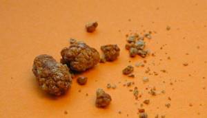
Urate stones can have different shapes and sizes (the average size is about 10 mm, the maximum is 60 mm), their color is from grayish-yellow to red-brown, the surface is rough, less often smooth. They are very hard stones, are clearly visualized by ultrasound and x-ray examination, and in the kidney they can fill the entire renal pelvis. Urates and oxalates are more common in men than in women; people aged 55-65 years are at maximum risk. The mechanism of occurrence of urate stones in the kidneys has 3 forms:
- 1. Idiopathic - patients do not have abnormalities in the amount of uric acid in the blood and urine, but the acidity of urine is constantly reduced. This condition can occur with chronic diarrhea, treatment with drugs that acidify urine, or with ileostomy - removal of the small intestine to the surface of the abdomen.
- 2. Hyperuricemic - in patients with gout (about a quarter of patients), with disorders of purine metabolism, with blood diseases (chronic myeloid leukemia, erythremia, monocytic leukemia and others), with Lesch-Nyhan syndrome - a genetic pathology in boys; after chemotherapy for malignant tumors.
- 3. Dehydration – with insufficient fluid intake, diarrhea, intestinal diseases, increased sweating and other conditions in which concentrated urine is formed.
Damage to kidney tissue as a result of the formation of urate stones leads to the following complications:
- progressive chronic renal failure (causes death in 17-40% of patients with gout);
- acute renal failure due to blockage of the renal tubules by uric acid crystals;
- development of secondary pyelonephritis;
- damage to the blood vessels of the kidneys;
- pyonephrosis (the final stage of purulent pyelonephritis), requiring surgical removal of the kidney.
Although it is believed that the presence of urates in the urine does not have much diagnostic value, there is a certain risk of renal colic, since outside of an attack there may be no significant changes in the tests. Acute uric acid nephropathy is accompanied by the following symptoms:
- a decrease in the amount of urine excreted, and then a complete cessation of its excretion;
- nausea, vomiting;
- dull pain in the lower back;
- the appearance of blood clots in the urine;
- increased blood pressure;
- urate crystals, red blood cells, and a decrease in relative density appear in urine tests;
- increase in ESR in blood serum.
Why do urates appear in urine?
The basis of urates, uric acid, is a product of the metabolism of purine nucleosides. These substances are an integral part of the DNA helix.
Urates begin to precipitate in urine when the amount of uric acid in the body exceeds normal levels. Sometimes this occurs due to physiological reasons (changes in diet or climate, pregnancy). The main pathological causes of uric acid crystal loss include gout, urinary tract infections, and hematological diseases.

Often, small amounts of salts are found in the urine of absolutely healthy people. The main reason for this phenomenon is the characteristics of metabolism and the body as a whole.
How to properly prepare for the test and collect urine
On the eve of urine collection, it is undesirable to consume alcohol, spicy and salty foods, as well as foods that can change the color of the sample (carrots, turmeric, beets). Immediately before collecting the material, hygienic washing is carried out. Women are not recommended to donate urine during their periods. For children, the test is collected in special urinals.
It is important to collect urine in sterile pharmacy containers to avoid the ingress of microorganisms and inorganic elements.
To analyze a single portion, a minimum of 10 ml of sample is required.

If a urine test reveals an increased urate content, the diagnosis is repeated 7-10 days after the diet is normalized.
What does it mean if urates are found in large quantities?
Urate in urine is a nonspecific marker of disease, that is, it can equally indicate a disease and a physiological feature. Therefore, uric acid salts are not the basis for making a diagnosis.
Diseases
A number of diseases are accompanied by increased precipitation of urate. Urine saturated with uric acid salts has a brownish-pink hue. This color is given by amorphous urates in urine.
Pathologies that are accompanied by an increase in the concentration of urate in the urine:
- glomerulonephritis;
- pyelonephritis;
- cystitis;
- urethritis;
- leukemia;
- vascular pathologies of the kidneys and other parts of the urinary system (atherosclerosis of the renal vessels, nephroptosis, hydronephrosis).
What all these diseases have in common is damage to the glomerulus.
Therefore, the filter segment begins to allow substances to pass through that normally cannot pass through its pores. Another serious disease leading to uraturia is gout. Gout refers to metabolic pathologies. It develops when uric acid metabolism is disrupted. First of all, hyperuricemia begins (increased concentration of uric acid in the blood plasma), and then other signs of the disease appear:
- pain in the area of the “bone” of the first toe;
- the appearance of tophi (specific subcutaneous formations that look like wen and consist of uric acid salts);
- uraturia.
The disease requires careful attention because urate crystals damage the kidney filters and tubules. This can lead to a dangerous complication - kidney failure.
Diet
Uraturia is most often a consequence of a gross diet violation. It is the nature of nutrition that causes many diseases.
Large amounts of urate cannot be present in the urine as a suspension. They precipitate. This happens when you abuse the following products:
- meat and meat products;
- offal;
- cheese;
- tomatoes;
- chocolate;
- spinach;
- strong meat broths;
- legumes;
- mushrooms;
- food products rich in salicylates (viburnum, raspberry, linden);
- alcohol.
Uraturia is especially pronounced in people who regularly consume “forbidden” foods. In addition, with such a diet, vegetables, fruits and ordinary clean water are often forgotten.
Pregnancy
Despite the fact that pregnancy is a physiological process, it still represents serious stress for the body. Not only hormonal changes occur, but also changes in metabolism in general. Therefore, pregnant women may experience transient uraturia. Most often, she does not talk about any pathology.

Certain drugs
If an increased amount of urate was detected in the urine, you need to remember your actions before and during the test.
If the biomaterial was collected after taking antipyretic, antiplatelet and painkillers, the result will be unreliable. Acetylsalicylic acid (Aspirin) and all drugs that contain it (Citramon, Coplavix, Excedrin, Migrenol) can cause uraturia. Therefore, three days before the study, it is necessary to avoid taking these medications. If this is not possible, you should consult your doctor.
Other reasons
If the urine is completely strewn with urates, then this may indicate dehydration. Serious fluid loss is caused by:
- fever;
- vomiting and diarrhea;
- heavy physical activity;
- overheating.
To avoid loss of electrolytes and fluids, it is necessary to constantly maintain fluid balance. If your general condition allows, then drink a lot; if not, give droppers with crystalloid solutions.
What are the dangers of excess urate salts?
Urate crystals themselves are not dangerous. They are non-toxic and do not have sharp corners that could injure the epithelium. But these salts are a harbinger of serious diseases.
For example, persistent uraturia in the future will certainly lead to the formation of kidney stones. The urine sediment crystallizes and is retained in the renal pelvis. The pH of urine changes due to metabolic disorders. This creates ideal conditions for stone formation.
Gout, which also causes uraturia, is often complicated by chronic renal failure (CRF). If the terminal phase of chronic renal failure occurs, the person is prescribed lifelong replacement therapy (hemodialysis) or referred for organ transplantation. The first symptoms of gout are pain in the interphalangeal joint of the first toe. The disease requires immediate treatment.
Microscopy of urine sediment
Microscopy of urine components is carried out in the sediment formed after centrifugation of 10 ml of urine. Sediment consists of solid particles suspended in urine: cells, protein-formed casts (with or without inclusions), crystals, or amorphous deposits of chemicals.
2.1. Red blood cells in urine Red blood cells (blood cells) enter the urine from the blood. Physiological erythrocyturia is up to 2 red blood cells/μl of urine. It does not affect the color of urine. During the study, it is necessary to exclude contamination of urine with blood as a result of menstruation! Hematuria (the appearance of red blood cells, other formed elements, as well as hemoglobin and other blood components in the urine) can be caused by bleeding anywhere in the urinary system. The main reason for the increase in the content of red blood cells in the urine is renal or urological diseases and hemorrhagic diathesis.
Reference values: none; with microscopy - up to 2 in the field of view. Red blood cells in urine - exceeding reference values:
- urinary tract stones;
- tumors of the genitourinary system;
- glomerulonephritis;
- pyelonephritis;
- hemorrhagic diathesis (with intolerance to anticoagulant therapy, hemophilia, coagulation disorders, thrombocytopenia, thrombocytopathies);
- urinary tract infections (cystitis, urogenital tuberculosis);
- kidney injury;
- arterial hypertension with involvement of the renal vessels;
- systemic lupus erythematosus (lupus nephritis);
- poisoning with benzene derivatives, aniline, snake venom, poisonous mushrooms;
- inadequate anticoagulant therapy.
2.2. White blood cells in the urine An increased number of white blood cells in the urine (leukocyturia) is a symptom of inflammation of the kidneys and/or lower urinary tract. In chronic inflammation, leukocyturia is a more reliable test than bacteriuria, which is often not detected. With a very large number of leukocytes, pus in the urine is determined macroscopically - this is the so-called pyuria. The presence of leukocytes in the urine may be due to the presence of secretions from the external genitalia in the urine due to vulvovaginitis, or insufficiently thorough toileting of the external genitalia when collecting urine for analysis.
Reference values: none; by microscopy: boys - 0 - 3 in the field of view girls < 14 years old - 0 - 5 in the field of view
An increase in leukocytes in the urine is observed in almost all diseases of the kidneys and genitourinary system:
- acute and chronic pyelonephritis, glomerulonephritis;
- cystitis, urethritis, prostatitis;
- stones in the ureter;
- tubulointerstitial nephritis;
- lupus nephritis;
- kidney transplant rejection.
2.3. Epithelial cells in urine Epithelial cells are almost always present in urine sediment. Epithelial cells originating from different parts of the genitourinary system vary (usually squamous, transitional and renal epithelium are distinguished).
Squamous epithelial cells, characteristic of the lower parts of the genitourinary system, are found in the urine of healthy people and their presence usually has little diagnostic value. The amount of squamous epithelium in the urine increases with urinary tract infection.
An increased number of transitional epithelial cells can be observed in cystitis, pyelonephritis, and kidney stones.
The presence of renal epithelium in the urine indicates damage to the kidney parenchyma (observed in glomerulonephritis, pyelonephritis, some infectious diseases, intoxication, circulatory disorders). The presence of more than 15 renal epithelial cells in the field of view 3 days after transplantation is an early sign of the threat of allograft rejection.
Reference values: none; under microscopy: squamous epithelial cells: other epithelial cells - absent
Detection of renal epithelial cells:
- pyelonephritis;
- intoxication, intake of salicylates, cortisol, phenacetin, bismuth preparations, poisoning with salts of heavy metals, ethylene glycol);
- tubular necrosis;
- kidney transplant rejection;
- nephrosclerosis.
2.4. Cylinders in urine Cylinders are elements of cylindrical sediment (a kind of cast of renal tubules), consisting of protein or cells, and may also contain various inclusions (hemoglobin, bilirubin, pigments, sulfonamides). Based on their composition and appearance, there are several types of cylinders (hyaline, granular, erythrocyte, waxy, etc.).
Normally, renal epithelial cells secrete the so-called Tamm-Horsfall protein (absent in blood plasma), which is the basis of hyaline casts. Hyaline casts can be found in the urine in all kidney diseases. Sometimes hyaline casts can be found in healthy people. As a pathological symptom, they acquire significance when they are constantly detected and in significant quantities, especially when erythrocytes and renal epithelium are superimposed on them.
Granular casts are formed as a result of the destruction of tubular epithelial cells. Their detection in a patient at rest and without fever indicates renal pathology.
Waxy casts are formed from compacted hyaline and granular casts in wide-lumen tubules. They occur in severe kidney diseases with predominant damage and degeneration of the tubular epithelium, more often in chronic than in acute processes.
Erythrocyte casts are formed when red blood cells are layered on hyaline casts, and leukocyte casts are formed by leukocytes. The presence of red blood cell casts confirms the renal origin of hematuria.
Epithelial casts (rarely) are formed when the tubular epithelium is detached. Occurs with severe degenerative changes in the tubules at the onset of acute diffuse glomerulonephritis, chronic glomerulonephritis. Their presence in a urine test a few days after surgery is a sign of rejection of the transplanted kidney.
Pigment (hemoglobin) cylinders are formed when pigments are included in the composition of the cylinder and are observed with myoglobinuria and hemoglobinuria.
Cylinders are long structures consisting of mucus. Single cylindroids are found in urine under normal conditions. A significant number of them occur during inflammatory processes of the mucous membrane of the urinary tract. They are often observed when the nephritic process subsides.
Reference values: hyaline cylinders – single, others – absent
Hyaline casts in urine:
- renal pathology (acute and chronic glomerulonephritis, pyelonephritis, kidney stones, renal tuberculosis, tumors);
- hyperthermic conditions;
- heavy physical activity,
- high blood pressure;
- taking diuretics.
Granular casts (nonspecific pathological symptom):
- glomerulonephoritis, pyelonephritis;
- diabetic nephropathy;
- viral infections;
- lead poisoning;
- fever.
Waxy cylinders:
- chronic renal failure;
- kidney amyloidosis;
- nephrotic syndrome.
Red blood cell casts (hematuria of renal origin):
- acute glomerulonephritis;
- kidney infarction;
- renal vein thrombosis;
- malignant hypertension.
Leukocyte casts (leukocyturia of renal origin):
- pyelonephritis;
- Lupus nephritis in systemic lupus erythematosus.
Epithelial casts (most rare):
- acute tubular necrosis;
- viral infection (for example, cytomegalovirus);
- poisoning with salts of heavy metals, ethylene glycol;
- overdose of salicylates;
- amyloidosis;
- kidney transplant rejection reaction.
2.5. Bacteria in urine
Isolation of bacteria in urine has significant diagnostic value. Bacteria remain in the urine for no more than 1-2 days after the start of antibiotic therapy. The first morning urine sample is preferable for testing. The type of bacteria can be determined and the level of bacteriuria can be assessed, as well as the sensitivity of microorganisms to antibiotics can be determined using bacteriological urine culture.
Reference values: negative