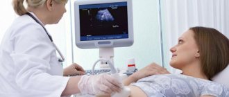
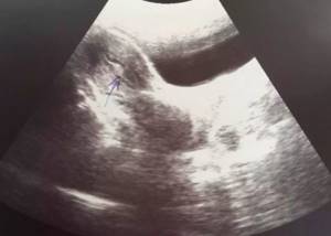
Among modern methods of examining the internal state of the body, the leading place is occupied by ultrasound of the pelvic organs (Ultrasound of the pelvic organs). The study allows, within a few minutes, to obtain a reliable picture of the state and activity of the studied organs and systems in men and women of any age.
PRICE OF PELVIC ULTRASOUND IN OUR CLINIC—RUB 1,000. DOCTOR CONSULTATION ON ULTRASOUND RESULTS – 500 RUR. CLICK TO MAKE AN APPOINTMENT, TEST OR ULTRASOUND
This procedure with the highest reliability allows you to examine the condition of the internal genital organs, urinary tract and determine pregnancy in the early stages. You can undergo a pelvic ultrasound using excellent equipment, inexpensively and without a queue, at the Diana Clinic.
Who prescribes a pelvic ultrasound?
A referral for an ultrasound examination can be given by a gynecologist, urologist, surgeon, and andrologist. Indications for use most often include problems in the functioning of the genitourinary system (bladder, prostate gland, ovaries, uterus, fallopian tubes, etc.). As well as the need to examine the rectum and biliary tract, suspicion of urolithiasis, etc.
In addition, with the help of pelvic ultrasound diagnostics, it is possible to determine pregnancy in its early stages. In the future, ultrasound allows you to monitor the condition and development of the fetus, and promptly identify pathological processes if they occur. The method is also successfully used in determining the causes of infertility.
How often can you undergo a pelvic ultrasound?
The content of the article
The ultrasound procedure itself is absolutely painless and harmless for people of any age, including children. There are no restrictions regarding the number of procedures. Pelvic ultrasound is performed as often as necessary, including as a preventive measure. The following schedule is recommended:
- in the absence of pathologies - once a year, especially after 40 years
- for chronic pelvic diseases - once every 3-6 months;
- when diagnosing the causes of infertility - several times during the menstrual cycle;
- during treatment - several times, as necessary to monitor the results of treatment.
Ultrasound, which has no contraindications and does not require lengthy preparation, can be performed in conjunction with all other diagnostic methods without interrupting treatment that has already begun.
If inflammatory processes in the pelvis are suspected based on ultrasound results, additional diagnostic methods are prescribed: taking tests, endoscopic (internal) examination of the bladder, ultrasound of the bladder.
What diseases are diagnosed using pelvic ultrasound
Thanks to ultrasound diagnostics, it is possible to accurately diagnose numerous inflammatory processes, the formation of tumors of various natures, changes in the location of the pelvic organs, their deformations, the presence of pathological inclusions and other changes. Therefore, based on the results of the study, the following can be diagnosed:
- malformations and deviations in the structure of the uterus, ovaries, fallopian tubes, vagina, in particular bicornuate, childish, saddle-shaped uterus, doubled, the formation of septa in the body of the uterus, etc.;
- cysts in the uterus and ovaries, including polycystic disease, fibroids of various types, cystomas, rupture and torsion of the pedicle of the cyst, fibroids, cystadenomas, teratomas;
- malignant tumors of the reproductive organs, polyps;
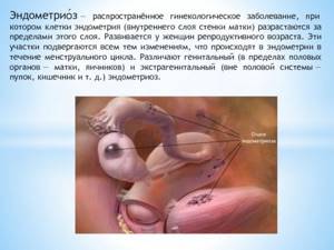
- endometriosis;
- inflammatory diseases, including adnexitis, cervicitis, endometritis, salpingitis, oophoritis, myometritis, parametritis, etc.;
- pathological changes in the fallopian tubes, including hydro- and pyosalpinx, obstruction;
- isthmic-cervical insufficiency, etc.
Ultrasound of the pelvic organs is widely used to diagnose pregnancy, determine its duration, as well as the location of the fetus, i.e., detect an ectopic pregnancy that is dangerous for a woman. Subsequently, 3 screenings are required to promptly detect possible pathologies of intrauterine development and take measures appropriate to the situation. Also, performing an ultrasound during pregnancy allows you to diagnose pathologies of the placenta, premature shortening of the cervix and other disorders fraught with termination of pregnancy or the development of complications.
The method is used to monitor pregnancy when using ICSI, IVF, etc.
The study is widely used to assess the functionality of a woman’s reproductive system, namely:
- the nature of follicle growth and the onset of ovulation, which is important if there are problems with conception;
- size and condition of the corpus luteum after ovulation;
- correct installation of the intrauterine device.
It is also used to monitor a woman’s condition after diagnostic invasive interventions, abortions, and various surgical interventions, including after treatment of cervical erosion. In addition, the procedure can be carried out for preventive purposes even in the absence of complaints about the condition of the genitourinary system. It is worth undergoing it once a year as part of a routine examination.
Methods for performing pelvic ultrasound
In our clinic, ultrasound diagnostics of the pelvic organs is carried out in 3 ways, which are selected depending on the gender and age of the patient, as well as existing problems and expected results. .
Transabdominal ultrasound - through the abdominal wall
Transabdominal examination (through the wall of the abdominal cavity) diagnosis is the most familiar to ordinary people. Before the procedure, you need to drink water so that the filled bladder displaces the intestines from the examined area.
The patient exposes the part of the body being examined and lies down on the couch, usually on his back. The doctor applies a special gel to his skin, which ensures a tight fit of the sensor to the skin and improves the passage of ultrasound, after which he begins to move the sensor over the part of the body being examined. All that the patient feels at this moment is light pressure and smooth movements of the tip. In fact, sound waves, as a result of close contact of the sensor with the skin, penetrate deep into the body, and a clear picture appears on the device’s screen, allowing the specialist to almost accurately determine the condition of the organ being examined.
This method of pelvic ultrasound is suitable for all patients, with the possible exception of very obese and overweight people - their large body weight and fat deposits can make it difficult for waves to pass through tissue.
Transabdominal ultrasound diagnostics is universal, and difficulties during its implementation can arise only if the intestines are filled with gases that prevent obtaining a clear picture on the screen. This problem is completely solvable: on the eve of the examination, it is enough to follow a few simple recommendations from the doctor to eliminate increased gas formation in the digestive system.
Transvaginal ultrasound – through the vagina
This procedure allows you to clearly examine the pelvic organs, since the intestinal loops do not fall into the field of view of the device; or rather, the doctor sees them, but they do not interfere with examining other organs. It is advisable to conduct the examination a week after the start of menstruation.
The examination is prescribed to women by a gynecologist as the most appropriate for diagnosing and treating gynecological diseases, as it allows a detailed examination of the condition of all internal genital organs from different viewing angles.
The exceptions are virgins and women with serious neoplasms in the pelvic organs or with a long period of pregnancy - the latter undergo only a transabdominal ultrasound.
The procedure itself is a bit like a gynecological examination: the woman lies on her back and spreads her legs, and the doctor inserts a sensor into the vagina, on which a special condom is put on (you can also use a regular disposable one, but without lubricant and not grooved). To ensure the necessary contact with the surface of the organs being examined, the same harmless water-based gel is applied to the condom.
The procedure, as a rule, does not cause discomfort, and minor painful sensations may appear only if the tip of the sensor is pressed on the diseased organ. Since transvaginal ultrasound allows you to examine the uterus, fallopian tubes, and ovaries from different sides, this helps to obtain a more objective picture of their condition and timely prescription of treatment.
Transrectal ultrasound - through the rectum
This examination is carried out after taking carminatives and performing a cleansing enema.
The patient lies on his side and slightly tucks his legs. The doctor inserts a sensor with a condom on it into the rectum. The patient may experience only some psychological discomfort, which cannot be compared with the final result - accurate and without the feeling of pain.
Sonohysterography and combined methods of ultrasound of the pelvic organs
Sometimes, in parallel with transvaginal ultrasound, the gynecologist performs sonohysterography, the essence of which is as follows. First, an ultrasound machine diagnoses the woman’s internal organs (transvaginally), then the vaginal cavity and uterine wall are treated with an antiseptic and a saline solution is slowly injected into the cervix through a thin tube connected to a syringe. It enters the uterine cavity, straightens its walls and then flows into the pelvic cavity through the fallopian tubes. This method allows you to clearly examine the uterine cavity during a transvaginal ultrasound performed at this moment and determine the patency of the fallopian tubes.
Sometimes the doctor prescribes 2 types of ultrasound - this allows you to see the same organ from different sides and significantly complement the existing medical history. The image is displayed on the screen, and the doctor sees what is happening in the patient’s body.
Any ultrasound procedure lasts no more than half an hour, and the results obtained are interpreted immediately. In addition, all ultrasound data can be recorded on disk, which will allow you to compare records in the future and monitor the dynamics of treatment.
Stages of work during ultrasound examination
Conversation with an ultrasound diagnostic doctor.
Comfortable position on the couch.
Applying media gel to the study area or to the sensor (a special sterile condom is placed on the vaginal sensor).
With smooth movements, the ultrasound sensor moves in the study area, receiving images of the organs being examined through a piezoceramic transducer.
At the end of the study, the remaining media gel is removed with a disposable napkin.
Drawing up and issuing an ultrasound examination protocol to the patient.
Which ultrasound method to choose: transvaginal, transabdominal, rectal
Indications for the procedure for women
Many patients, noticing obvious signs of urological or gynecological diseases, want to undergo an ultrasound scan before seeing a doctor. In such cases, you need to study the information on when a pelvic ultrasound is prescribed.
If you have visited a doctor, he will prescribe such an examination if there is:
- menstrual irregularities, unknown vaginal discharge, uterine bleeding;
- pain in the lower abdomen;
- problems with urination;
- suspected tumors of internal organs located in the pelvis;
- suspicion of any formations (from cysts and fibroids to cancerous tumors) in the genital organs;
- infertility;
- congenital anomalies and inflammatory processes;
- suspicion of stones in the urinary tract;
- pregnancy - in this case, an ultrasound is performed several times and helps determine the presence of genetic abnormalities in the development of the child, the gender of the unborn baby, its size, the expected date of birth, etc.;
- monitoring the patient after an abortion;
- less often - monitoring the intrauterine device if it causes discomfort or causes discharge and pain.
In this case, it is necessary to take into account the optimal time for the procedure. Usually this is the first 7-10 days from the start of the menstrual cycle, although it all depends on the expected or already established diagnosis. For example, if endometriosis is suspected, the examination is carried out before the start of menstruation, and the detection of uterine fibroids is carried out immediately after their end.
Special requirements relate to monitoring folliculogenesis (primarily, this applies to problems with conception) - ultrasound is prescribed several times per cycle, depending on its duration and the characteristics of each woman.
Also, a pelvic examination is indicated for representatives of both sexes if there is a suspicion of urolithiasis or problems with the urinary and gall bladder, kidneys, liver, or rectum. It is often given to young children for diseases of the genitourinary system.
You can undergo a pelvic ultrasound at the Diana private clinic on your own initiative, without a referral.
Features of pelvic ultrasound in men
Ultrasound examination of the pelvic organs in men is the most reliable and easiest way to assess the general condition and functioning of the pelvic organs.
Indications for examination of men
Men over the age of 40 should undergo an ultrasound examination of the pelvis for prevention every year, since this age is characterized by a high risk of developing prostate diseases, including cancer. Also, the doctor may insist on performing this type of ultrasound when diagnosing a patient with sexually transmitted infections, in order to identify disorders of the reproductive system caused by this infection.
The following complaints may be the reason for conducting a study:
- pain in the lower abdomen, localized in the perineum, scrotum or pubis;
- disturbance in the frequency of urination or a feeling of discomfort when emptying the bladder;
- difficulty or complete inability to urinate, too weak a stream of urine;
- the presence of abnormal discharge from the urinary tract;
- infertility;
- violation of potency;
- changes in biological fluids, for example, the presence of blood in urine, semen.
Pelvic ultrasound is the most important stage of the mandatory annual examination of the male body, which is recommended by doctors for men of all age groups in order to prevent urological and andrological diseases. Men are unconditionally referred for a pelvic ultrasound for any pathology of the prostate gland and seminal vesicles.
Ultrasound of the pelvic organs in men allows you to visualize and examine the following organs:
- bladder;
- prostate (prostate gland);
- seminal vesicles.
Despite the fact that the kidneys are not included in the list of organs examined for this type of diagnosis, a specialist can detect and visualize the presence of stones in this paired organ. If this happens, additional ultrasound and laboratory tests are prescribed.
How is an ultrasound of the pelvic organs performed in men?
The procedure can be performed in two ways, the appropriateness of which is determined by the attending physician, depending on the expected results.
- Abdominal examination (transabdominal)
- Rectal TRUS examination (transrectal)
During an abdominal ultrasound examination, a specialist applies a specific water-based gel to the surface of the anterior abdominal wall, which fills the air voids between the body and the device’s sensor. Moving the sensor along the surface of the abdomen, pressing lightly on it, the doctor monitors the condition and functioning of the internal organs and systems on the monitor. The procedure is absolutely painless, and the patient may feel discomfort only in the case of severe inflammatory processes in the organs being examined with severe pain.
The transrectal method of performing the procedure (TRUS) is much more informative and is usually the most preferable for the doctor, however, many patients are concerned about the possibility of discomfort and pain. In fact, the technique is painless, since the sensor is inserted into the rectum only 4-5 centimeters and is not felt even in the presence of small hemorrhoids. A contraindication for TRUS can only be an exacerbation of severe forms of hemorrhoids.
Preparing for a pelvic ultrasound
Ultrasound of the pelvic organs in men does not require complex special training. The main condition is the release of gas from the intestines, which is achieved by a diet that must be followed a few days before the procedure. You will have to exclude milk, fresh fruits and vegetables, legumes and bread. Ultrasound diagnostics of the pelvis using the TRUS method should also completely empty the intestines.
Our clinic offers all types of ultrasound of the pelvic organs in men at affordable prices by highly qualified specialists.
Benefits of the study
Among the main advantages of ultrasound technology are the following:
- Non-invasive. The procedure does not require injections, incisions or other tissue damage.
- Painless. During the ultrasound, the patient does not experience any discomfort.
- No ionizing radiation. This allows you to prescribe ultrasound for adults, children, the elderly and even pregnant women. The procedure does not affect the growth and development of the fetus.
- Good visualization. Highly sensitive sensors allow you to visualize on the screen the slightest changes in the organ that are not visible during x-ray examination.
- Ability to conduct research in emergency situations. In the event of the development of an acute pathology, the doctor will be able to identify in real time the cause of the disorder and the extent of damage to the diseased organ.
- An ideal method for monitoring the development of pregnancy. Transabdominal ultrasound is prescribed several times during pregnancy. With its help, the doctor will confirm a successful conception, will be able to monitor the intrauterine development of the fetus or promptly diagnose congenital anomalies.
Disadvantages of transabdominal ultrasound
If you ignore the preparation rules, the results of the study may be unreliable, and some organs may not be examined at all. Imaging problems often occur in people diagnosed with obesity. The fact is that excess fat deposits become a serious obstacle to ultrasonic waves.
In some cases, it is not possible to detail the organ being examined in detail, so the specialist may not notice the problem at the initial stage of development. Often, to confirm the diagnosis, the patient is referred to other high-precision diagnostic procedures - CT or MRI, which are more expensive.
Contraindications to pelvic ultrasound
Ultrasound is difficult to perform in patients with severe abdominal obesity. Fat slows down the passage of ultrasound waves. Also, the ultrasound method cannot be used to examine organs containing large amounts of gas, for example, the intestines.
For girls who are not sexually active, only transrectal and transabdominal pelvic ultrasound is performed. In late pregnancy, women's pelvis is examined using a sensor placed on the skin of the abdomen.
Advantages of the ultrasound method in diagnosing the pelvis
Ultrasound has no disadvantages, but the advantages of the method are obvious:
- painlessness, ease, accessibility and safety;
- absence of harmful radiation affecting sexual function. Such examination can be carried out an unlimited number of times, even during pregnancy;
- no special preparation is needed for the examination (except for transrectal ultrasound);
- It is possible to obtain detailed images of tissues, which show how organs work.
How to prepare for the examination?
Except in emergency cases, the doctor gives a referral for an ultrasound scan a few days before the procedure. This allows the patient to prepare for the diagnosis, which will eliminate the occurrence of any hitches and troubles during its implementation. Moreover, each type of ultrasound requires a special preparatory stage - not difficult, but very necessary.
- Transabdominal, transrectal ultrasound. 3-4 days before transabdominal and transrectal ultrasound of the pelvis, foods that promote gas formation should be excluded from the diet - a list of them is usually given in the referral for ultrasound diagnostics or given by the doctor during a consultation or appointment. These days you can additionally take enzyme preparations, for example, Mezim. Also, 3-4 hours before the procedure you should not visit the toilet, since a full bladder raises the intestines. This will allow the specialist performing the ultrasound to clearly examine all the internal organs of the pelvis and determine their condition. If you cannot endure for such a long time, you can drink about a liter of water - plain, still - 0.5-1 hour before the ultrasound.
- Transrectal diagnosis, for obvious reasons, requires a complete cleansing of the intestines, so on the eve of the procedure it is necessary to give several enemas. The recommended amount is at least 2 in the evening and 1 in the morning, before visiting the doctor. As a last resort, you can resort to special medications that can solve this problem - a doctor should give recommendations on their use.
- Transvaginal ultrasound. For transvaginal ultrasound, no special preparations are required, except, perhaps, hygienic procedures. This also applies to filling the bladder: you can visit the toilet before the procedure.
IMPORTANT!
- Transvaginal and transrectal ultrasounds require the use of a condom. Therefore, the doctor must know in advance that the patient has an allergic reaction to latex.
- If, in parallel with a transabdominal examination, a gastroscopy or other similar procedure is prescribed that promotes the injection of gases into the intestines, it must be performed after an ultrasound of the pelvis.
- For convenience, it is recommended to take a diaper to the ultrasound - they cover the couch - and a towel in order to remove the gel from the skin immediately after the procedure. Other recommendations are usually given by the doctor individually.
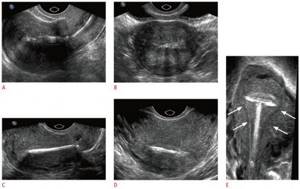
Decoding pelvic ultrasound: what the results say
The decoding of the received data - they are reflected in the diagnostic protocol, drawn up in a special form - is carried out by the doctor who issued the referral for the ultrasound. Only he can say with a high degree of confidence whether there are pathologies in the examined organ, and draw a conclusion about the patient’s health status. Here are examples of what a specialist can see when conducting a pelvic ultrasound and interpreting its results in a woman.
- The echogenicity is homogeneous. Traditionally, it decreases somewhat in size in older women during the postmenopausal period.
- Leaned back - the position is abnormal. During pregnancy can lead to complications.
- The contours are uneven - fibroids or a tumor is developing, blurred - there is an inflammatory process (parametritis, endometritis, endometrial hyperplasia).
- The size is reduced - underdevelopment (infantilism) of the uterus, increased - a sign of pregnancy, fibroids, cancer, adenomyosis.
Hypoechogenicity - occurs with polyps, fibroids, and cancerous tumors. In women who have reached menopause, a change in the thickness of the endometrium may indicate the onset of hyperplasia and cancer. At other times, it indicates the presence of inflammation or a tumor.
Pelvic ultrasound interpretation table: norms and deviations
| Organ being examined | Norm | Signs of pathology |
| Uterus | Deflected forward, the contours are smooth and clear, the shape is pear-shaped with a length of about 40-50 mm, a width of about 40 mm. | |
| Endometrium | The structure of the mucous membrane and its thickness change at different periods of the cycle, so the data in each specific case correlate with the norm characteristic of a certain phase of the menstrual cycle. | |
| Cervix | Length – 35-40 mm, canal diameter – 2-3 mm and filled with liquid, anteroposterior size – 25-30 mm. The echogenicity is homogeneous. | The structure is changed, the cervix is expanded - signs of inflammation, endometriosis, cancer. |
| Ovaries | Dimensions (in mm): width – up to 25, length – up to 30, thickness – up to 15, volume – 2-8 cubic meters. cm. The contour is clear, lumpy due to the presence of growing follicles up to 6 mm in size. And in the middle of the cycle and with a dominant follicle, it is about 15-25 mm. The echostructure is mostly homogeneous, with small areas (several mm) of fibrosis. | Increased size and volume, altered contours, impaired echostructure and fibrosis are a sign of inflammatory processes (usually oophoritis), polycystic disease. The presence of a fluid-filled cavity is a cyst. |
| Uterine (fallopian) tubes | Almost invisible and invisible during ultrasound. | Visualization of the tubes during diagnosis - salpingitis (inflammation) or intrauterine pregnancy. The expansion of the tubes and the accumulation of fluid in them is hydrosalpinx. |
| Presence of free fluid | Present in small quantities (no more than a few mm) only in the period after ovulation, i.e. approximately 2 weeks after the last menstrual period. |
Thus, ultrasound diagnostics of the pelvic organs in women makes it possible to detect the presence and development of such serious diseases as uterine fibroids in the early stages; endometriosis; various inflammations (adnexitis - appendages, endometritis - uterine mucosa, etc.); dysfunctions in the functioning of the ovaries, including those leading to infertility; cysts and tumors, as well as detect early pregnancy, monitor the development of the fetus and the condition of the genital organs after an abortion, etc.
What the study won't show
The only drawback of ultrasound is that with just one such diagnosis it will be difficult for a doctor to distinguish a fibroid from a neoplasm. That is why, identifying any pathology of the pelvic organs during an ultrasound is the primary reason for prescribing an additional comprehensive examination. It may include all kinds of tests and diagnostics.
How is a transabdominal examination performed?
An ultrasound examination is carried out in an ultrasound room, which has all the necessary equipment for modern, high-precision diagnostics. First, the doctor will ask the patient to free the upper body from clothing, then ask him to lie down on the couch and relax.
The anterior wall of the abdominal cavity is lubricated with a special gel, which enhances the conductivity of ultrasonic waves. Next, using a sensor, the specialist slowly moves it along the surface of the abdomen, examining the pelvic organs. During diagnosis, the patient does not feel any pain or discomfort. The procedure is safe for children and the elderly because it does not cause complications and does not cause radiation exposure. The examination takes on average 15 – 25 minutes. After this, the patient can return home and do their usual activities.
Where to do a pelvic ultrasound in St. Petersburg
Both municipal clinics and antenatal clinics, as well as private medical centers, carry out ultrasound diagnostics. The advantage of the former is often only that the procedure is free – although this is also not always the case. Often, ultrasound is performed only according to a pre-planned schedule, which is difficult to get into, and on old equipment.
The private clinic Diana - St. Petersburg is free of these shortcomings, equipped with the latest modern ultrasound machines and constantly working to create comfortable conditions for patients. Here you won’t have to sit in lines or wait for your turn for several weeks.
You can sign up for an ultrasound of the pelvic organs in St. Petersburg right now by making a free call to 8-800-707-15-60
Features of ultrasound for women and on what day of the cycle is ultrasound done

Transvaginal ultrasound makes it possible to diagnose almost all pathologies of the reproductive system in women, ranging from endometriosis and uterine fibroids to obstruction of the fallopian tubes and pregnancy pathologies.
In addition, almost all gynecological manipulations, be it placing an intrauterine device or assessing the ongoing treatment over time, at some stage may require an ultrasound of the pelvic organs.
That is why it is so difficult to overestimate the importance of this research method, and that is why we tell you how to do a pelvic ultrasound so that you have no doubts about the painlessness and safety of the method.
As for the question of what day of the cycle an ultrasound is performed, experts recommend that women contact an uzist on the 5-10th day of the menstrual cycle (the first day of the menstrual cycle is the first day of the last menstruation).
During this period, due to the physiological processes occurring in the body, the visualization of most pelvic organs becomes even clearer and more informative.
How much does a pelvic examination cost?
The price of the study depends on the device and the pricing policy of the clinic. Our pelvic ultrasound is quite inexpensive. The procedure is performed using a new expert-class ultrasound machine.
| 57 | Consultative consultation with a doctor based on the results of an ultrasound examination | 500 |
| 58 | Foliculometry, determination of endometrial thickness, IUD control using ultrasound | 900 |
| 59 | Ultrasound examination of the pelvis | 1000 |
| 60 | Ultrasound examination of the pelvic organs in girls (transabdominal) | 1000 |
At the Diana clinic, a pelvic ultrasound in St. Petersburg can be done any day - we work seven days a week.
Do not be afraid of such a useful procedure. After all, it allows you to timely identify and completely cure many serious diseases, without causing any harm or pain to the patient.
<
p style=”text-align: justify;”>
CLICK TO MAKE AN APPOINTMENT, TEST OR ULTRASOUND
If you find an error, please select a piece of text and press Ctrl+Enter