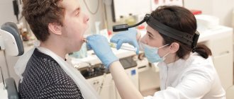
Ultrasound of the scrotum is recommended for all men for the purpose of prevention and timely diagnosis of diseases of the reproductive system. The procedure is prescribed to patients who have consulted a doctor with certain complaints, based on which the doctor may suspect damage to the testicles, epididymis, or spermatic cords.

Ultrasound of the scrotal organs1,400 rub.
All prices Make an appointment
No special preparation is required for the diagnostic study. The results will be deciphered by the doctor who gave the direction for the ultrasound. With the help of ultrasound diagnostics, the doctor will be able to diagnose almost all pathologies affecting the organs of the scrotum: cysts of the testicles and appendages, acute and chronic inflammatory processes, neoplasms, abscesses, testicular torsion, traumatic lesions.
The essence of the ultrasound procedure of the scrotal organs
Ultrasound of the scrotum is a safe, painless, non-invasive, highly informative procedure with which the doctor can assess the condition of the organs of the male reproductive system.
The scrotal organs are examined through the skin of the scrotum, over the surface of which an ultrasound probe moves. The studied organs are visualized on the monitor screen in volume. The essence of the procedure is that the ultrasonic waves generated by the sensor are reflected from soft tissues and transformed into electrical impulses, which, in turn, visualize the picture on the screen. Modern software allows you to measure all important parameters as accurately as possible and identify even minor deviations from the norm.
To conduct an ultrasound examination of the scrotum, sensors with a shallow scanning depth are used - up to 10 cm. Such sensors are intended for examining superficially located organs. Using ultrasound, you can evaluate important parameters with millimeter accuracy. Thanks to highly sensitive equipment, the doctor will detect even minor changes and diagnose pathology at an early stage of development.
Expert opinions and different approaches
In modern medicine, ultrasound is considered the best way to detect and prevent diseases in the early stages. But the number of patients is growing every year, so doctors have to introduce new technologies. This is necessary to obtain a more accurate picture of the disease. Color Doppler mapping, abbreviated as color doppler, helped to greatly expand the capabilities of conventional ultrasound. Although, using this technology in practice, some experts evaluate it ambiguously.
There are three main approaches:
- According to a large number of doctors, the method is of great value.
- The technology has no value and does not provide new insights into the disease.
- CDC provides useful information only when combining ultrasound with needle biopsy.
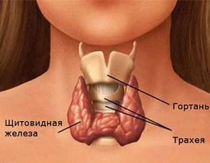
The main value of the method is not only in identifying defects in the blood supply to the thyroid gland, but also in the ability to detect tumors in the initial stages of development. CDC allows you to obtain information from the very depths of what is happening. Thanks to this, a qualified specialist can differentiate deviations.
He will be able to recognize where the tumor is benign and where it is malignant. The equipment allows you to see the structure and structure of tissue on several scales.
The patient is prescribed a thyroid ultrasound with colorectal dosage if he experiences the following symptoms:
- Adenoma;
- Various oncological diseases;
- Chronic thyroiditis;
- Suspicion of benign nodular formations.

Ultrasound of the scrotum with color circulation
Ultrasound of the scrotum with color Doppler mapping is used for the differential diagnosis of tumors of the scrotum. The essence of the method is the ability to visualize all moving fluids in real time and analyze their movement. The fact is that the blood flow of newly formed tumor vessels has its own characteristics. The vascularization of the neoplasm is represented by many small, abnormally located vessels, which are randomly scattered within the pathological tissues. The blood flow in such vessels is characterized by extremely low resistance, high speed and multidirectionality.
Ultrasound with Color Doppler is characterized by high sensitivity and accuracy in diagnosing tumors of a malignant nature. In addition, with the help of ultrasound with color doppler, it will be possible to predict the growth rate of the diagnosed tumor.
Pelvic ultrasound in men
Ultrasound of the ureters
Ultrasound of the bladder
Information obtained using the CDC
This method can now be considered the most used in the world. After all, information obtained with the help of modern technology helps to understand the degree of damage to the endocrine gland, as well as the entire system as a whole. If you use DK in color, the doctor can examine not only the thyroid gland, but also the fluid around it. No other equipment has this capability. The specialist will check the condition of the patient’s blood vessels.
Color Doppler mapping makes it possible to analyze information “live”. On the display, the endocrinologist sees red and blue colors, which transmit data about blood movement. It should be noted that colors are not the type of vessels and arteries. To do this, the doctor uses a special list that describes in detail the problem and type of disease.
Basic data received by the equipment:
- Blood flow speed.
- Color information about the vascular system.
- The structure and type of tissue of a certain vessel.
- The rhythm of the thyroid gland during operation.
- Presence or absence of tumors.
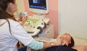
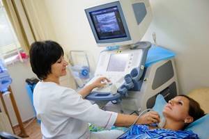
How the research is carried out
How is an ultrasound of the scrotum done with color doppler or dopplerography? The procedure is carried out in a separate ultrasound room, equipped with modern, highly sensitive, new generation equipment.
The doctor will ask the patient to remove the private parts from clothing, including underwear. The procedure is carried out in a supine position, while the man must still spread his legs to the sides. The genital organ is located on the stomach so that during the examination it does not interfere with the doctor’s manipulation of the sensor.
The skin of the scrotum is pre-treated with a special gel, which enhances the conductivity of ultrasonic waves. Next, using ultrasound sensors, the specialist alternately examines the right and left testicle in all projections. At the end of the procedure, Doppler scanning of the vessels is performed.
An ultrasound examination can last 15–30 minutes. If this is a routine examination, the examination will take on average 10–15 minutes. If the patient comes to the doctor with certain complaints, then the diagnosis will be carried out until the cause of the deterioration in health can be identified.
Decoding the results
The ultrasound results are recorded in a special report form. It describes in detail the parameters of the studied organs, identified abnormalities and pathologies.
Normally, an ultrasound of the scrotum shows the following results:
- Testicles. The structure is homogeneous, the outlines are even. Length 25 – 60 mm, width 15 – 30 mm.
- Scrotum. The wall thickness is no more than 8 mm; up to 2 ml of liquid without pathological inclusions can be determined between the wall and the appendages.
- Epididymis. The head is 10–15 mm long; the body and tail are not normally visualized.
- Vessels. The veins and arteries are not dilated, the blood flow is not impaired.
Signs of pathology
The progression of inflammatory processes in the scrotal organs will be indicated by the following signs:
- increase in organ size;
- heterogeneous structure;
- accumulation of excess fluid;
- pain when touched by the sensor.
Pathology is the detection of neoplasms of both benign and malignant etiology. Common diseases diagnosed using ultrasound:
- Congenital developmental anomalies. These include pathologies such as cryptorchidism, in which the testicle is located in an uncharacteristic location, as well as the absence of testicles.
- Cysts of the testicles and appendages. Neoplasms of a benign nature with liquid serous contents. Cyst-shaped tumors have clear contours, a dense capsule filled with fluid. Sometimes such tumors compress the appendages, causing infertility.
- Epididymitis. A disease of inflammatory nature, in which the epididymis is involved in the pathological process.
- Hydrocele or hydrocele of the testicle.
- Pyocele. It develops against the background of infectious processes, causing suppuration of testicular tissue.
- Varicocele. Vascular pathology, characterized by dilation of the vessels of the spermatic cord by more than 3 mm.
- Malignant or benign neoplasms. It is almost impossible to determine the nature of the tumor using ultrasound alone. The doctor can only see signs of a space-occupying formation: irregular shape, reduced echogenicity, heterogeneous structure, the presence of calcifications.
- Testicular torsion. It is detected by ultrasound and is characterized by a lack of blood flow in the vessels.
- Abscess of the appendage. A pathological neoplasm with purulent contents, which can be damaged at any time, spreading to neighboring tissues.
Indications for Doppler ultrasound
The procedure can be prescribed if there is a discrepancy between the parameters of the fetal body when measured during ultrasound screening, to predict the development of complications during screening studies. These are the main indications for this study. In addition, Doppler testing is recommended for pregnant patients with:
- preeclampsia;
- too little or too much;
- amniotic fluid;
- umbilical cord pathologies;
- pregnancy due to diabetes, hypertension and other chronic diseases;
- Rh factor conflict;
- growth retardation of one of the twins;
- for pathologies of the placenta;
- systemic collagenosis in the expectant mother;
- the patient has a history of miscarriage and premature birth;
- unsatisfactory results of cardiotocography.
Among other things, Doppler measurements are performed at the patient’s request. This can be done at any of the branches of the Medok clinic network.
Cost of ultrasound procedure of the scrotum organs
The price of an ultrasound scan of the scrotum depends on the scope of the study and ranges from 300 to 1000 rubles. Advantages of performing an ultrasound of the scrotal organs in our clinic:
- Rapidity. The primary diagnosis can be obtained within 20 minutes.
- Availability. Prices for ultrasound diagnostics in our clinic are affordable for every patient in need of professional medical care.
- High information content. The ultrasound room is equipped with highly sensitive diagnostic equipment that allows diagnosing any pathology in the early stages of development.
- Dynamic image. The diagnosis provides a clear picture in real time, which allows it to be used during surgery or biopsy.
Registration at our clinic
Ultrasound of the scrotum is a fairly intimate procedure, so it is not possible to undergo it comfortably in every medical center. If the confidentiality of the examination is important to you, then contact the International Hemostasis Clinic in Moscow. The price of such a service starts from 1,400 rubles.
All questions about the nuances of the procedure can be asked through the online form on the website or by phone.
Question answer
- How to prepare for an ultrasound of the scrotum and testicles? The patient will not require any preparation other than careful hygiene of the genitals. To avoid discomfort after applying the conductive gel, you can shave the hair in the groin area.
- Is it possible to eat and drink before undergoing diagnostics? There are no restrictions on food and water before the examination. However, alcohol should be avoided a few days before the event.
- Does ultrasound affect potency? The procedure is absolutely safe and does not affect male potency. Unlike X-rays, ultrasound does not contain radioactive radiation.
Description of the procedure, preparation for it
Before the procedure, the patient must familiarize himself with it and prepare mentally. Visiting a doctor in a state of shock or stress is strictly prohibited. This state will not allow you to understand the exact picture of what is happening, because all organs will be very tense. It is recommended to limit the consumption of meat products before the procedure.
When visiting the ultrasound room, the patient takes with him some kind of towel or diaper, on which the person lies during the examination. Use a towel to remove excess gel, which is used to lubricate the examination site. This procedure does not affect the body in any way.

There are some contraindications that prohibit thyroid examination:
- Presence of open wounds on the neck.
- Presence of burns in the area of examination.
Scientists highlight several advantages of ultrasound with color Doppler mapping:
- Wide information content of the results.
- No harm to the body.
- High accuracy of the information obtained.
- Little time investment for research.
The most important advantage of CDC is the safety of the procedure for pregnant women. This method does not cause any harm to the developing fetus. It is actively beginning to be introduced into clinics and hospitals around the world.