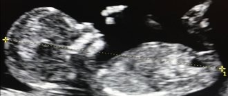
There comes a touching, important moment in every woman’s life when she realizes that she will become a mother. Much in the child’s future life will depend on her compliance with medical recommendations. During the entire period of gestation, the woman is under the close attention of specialists, and the first main study in which the mother sees her baby is an ultrasound scan in the first trimester of pregnancy.
Appointment with a gynecologist - 1000 rubles. Comprehensive pelvic ultrasound - 1000 rubles. Ultrasound during pregnancy - from 1300 rubles. Appointment based on ultrasound or test results - 500 rubles (optional)!
Timing of planned and unscheduled ultrasound in the 1st trimester, indications
The content of the article
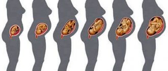
Pregnancy is a special period in the life of the expectant mother and in the intrauterine development of the child. The woman’s body gets used to new stress, and the baby, meanwhile, grows and develops. A normal pregnancy lasts for 9 months or 38-42 weeks. This period is conventionally divided into three parts, each of which is called a trimester. The first three months of pregnancy take place in the first trimester. It is during this period that all the child’s organs are formed.

Ultrasound in the first trimester corresponds to the period from 9 weeks to 12 weeks of gestation (in some cases it is prescribed from 11 to 14 weeks). The scheduled inspection does not cause alarm and proceeds as normal. There are medical indications that are alarming with dangerous consequences. Such unscheduled examinations must be carried out in consultation with the attending physician.
It is important!
“An ultrasound examination of the fetus in the first trimester of pregnancy makes it possible to verify the viability of the fetus, determine the number of fetuses, accurately determine the gestational age, detect gross developmental anomalies and measure the cervical fold, which, in combination with the age of the mother, is an important component of screening, allowing up to 75% confidence in early detection of some chromosomal abnormalities.”
The data obtained during many years of research and observation of fetal development and the course of pregnancy allowed us to draw several conclusions that are used today in ultrasound diagnostics.
- Fetal development occurs unevenly: some organs and parts of the body are visualized already in the first weeks of pregnancy, while others are visualized only in the second or third trimester. But with normal fetal formation, the sequence of development is the same. Thanks to this, it became possible to detect any abnormalities at an early stage, when many of them can be corrected, and the pregnancy can be preserved.
- During an ultrasound, pregnancy complications can also be detected - anomalies in the attachment or thickness of the chorion (the membrane that will subsequently be responsible for the nutrition and breathing of the fetus until its birth) or other disorders. Based on the detected problems, the woman may be prescribed measures to reduce the risk to the health of the fetus and the likelihood of spontaneous abortion (drug therapy, physiotherapy, spa treatment, restrictive physical activity, etc.).
At what weeks the first ultrasound is performed during pregnancy is decided by the attending physician. Modern standards require ultrasound diagnostics to be carried out at 11–13 gestational weeks, since this is the optimal time at which important features of the course of pregnancy can already be noticed. Contrary to popular belief, the principle “the sooner the better” does not apply to ultrasound, since at too early stages the examination readings will be uninformative and have a high level of error.
“An ultrasound in the early stages of pregnancy is very uninformative, so many false negative and false positive conclusions can be made. Determining the gestational age before 11–12 weeks has a wide range of errors, which means it can be very inaccurate. This is a completely false idea that the earlier an ultrasound is done, the more accurately the gestational age will be determined.”
Objectives of ultrasound in the first trimester during routine examination
Ultrasound examination helps to identify possible abnormalities in fetal development. This includes chromosomal pathologies, which are accompanied by thickening of the folds in the neck area.
The collar space or fold on the neck usually resolves in the second trimester; if this does not happen, swelling or a cystic formation develops. This is a symptom of Down syndrome, Edwards syndrome, Patau syndrome, etc. The tasks of the first trimester include early detection of disorders. During this period, it is still possible to safely terminate the pregnancy, while in later stages it will be difficult or impossible.
It is important!
Today, ultrasound examination is often resorted to in the earliest stages in order to confirm the actual fact of pregnancy and clarify its duration. So, a woman, noticing a delay in her period or other signs of a possible pregnancy, can independently contact a medical center for an ultrasound scan. This usually occurs between the 3rd and 5th week. An ultrasound performed at this stage only serves to confirm the fact of pregnancy! It is not possible to conduct an adequate diagnosis of the fetus at this stage, since many organs and systems of the fetus are not yet formed to the extent that any conclusion about normality or pathology can be made. An ultrasound to confirm pregnancy does not replace the need to conduct the first ultrasound diagnosis within the period indicated above: 10–14 weeks.
What does an ultrasound show in the 1st trimester?
Ultrasound in the first trimester is of particular importance for resolving doubts about an ectopic pregnancy, the possible absence of an embryo, and provides accurate results regarding the timing of pregnancy.
If the pregnancy is uterine, then the study includes many stages:
- the size of the collar thickening is studied,
- the presence or absence of nasal bones in the embryo is determined;
- the size is taken from the coccyx to the parietal part of the head (KTR);
- head circumference (BPR) is measured;
- the size is taken from the forehead to the occipital bone;
- the symmetry of the cerebral hemispheres is checked;
- the frequency of contractions of the heart muscle is checked;
- the length of the humerus and forearms, shins and femurs is measured;
- the location and size of the heart and stomach are calculated;
- the placenta with its characteristic features is studied;
- The condition of the uterus is checked.
The results are compared with tabular standards corresponding to the gestational age.
Extraembryonic structures and their characteristics

There are also some extraembryonic structures that must be studied using ultrasound in the first trimester of any pregnancy. These include the yolk sac, chorion and amnion.
Throughout pregnancy, nutritional and hematopoietic functions will be performed by the yolk sac. It begins to be visualized at 5 weeks, at 10 weeks it begins to reach a size of 7 millimeters, but after the 12th week it becomes impossible to normally identify or assess the condition and size of the yolk sac.
At the same time, there is a direct relationship between the size of the yolk sac, its shape and walls with the development of the embryo - if the parameters of the yolk sac are violated, specialists can draw conclusions about disturbances in the course of pregnancy as a whole.
The fleecy membrane of the fertilized egg is called the chorion. In the first trimester of pregnancy, the thickness of the chorion is usually equal to the gestation period itself in weeks. With changes in the structure of the chorion, specialists can predict dangers to the viability of the embryo. When the structure of this membrane changes, the villi, with the help of which the chorion is attached to the walls of the uterus, cease to fix the fertilized egg, and the process of miscarriage begins.
The water sac where the embryo is located in the fertilized egg is called the amnion. The amnion must be studied in the early stages of pregnancy to determine the absence or presence of pathologies in the diameter of the amniotic cavity.
If the size of the amnion is small, pregnancy will have difficulties in its own development, and if it is large, experts will conclude that intrauterine infectious processes have occurred.
Placenta and yolk sac on ultrasound
Life support for the developing fetus during intrauterine development occurs through the placenta (baby place). This is how the fetus is supplied with blood and nutrition. The study shows whether there are certain abnormalities or threats of pregnancy complications based on the position of the placenta. A low position of the place in relation to the fundus of the uterus is not a good sign. The condition requires constant monitoring. During pregnancy, the position of the placenta may change in the right direction. An ultrasound in the second trimester will bring certainty.
By the end of the first and beginning of the second trimester, the yolk sac is formed. This temporary organ indicates the viability of the embryo. If the shape of the sac on the side of the peritoneum is curved, enlarged or reduced, this is an alarming signal. The sac contains the required amount of vital yolk, which replaces the functions of the liver, spleen and even supplies primary germ cells that form immunity and metabolism.
Features of preparation
No special preparation is required before an ultrasound examination. A couple of days before the procedure, you should exclude from your diet foods that lead to increased gas formation. Otherwise, the survey results may be distorted. But changing the diet also depends on what kind of ultrasound is performed - transabdominal or transvaginal. If TVUS is performed, then you don’t have to limit yourself in food.
During transabdominal ultrasound, in addition to excluding foods that provoke increased gas formation, you should take carminatives 2-3 days before the procedure in case of intestinal bloating. With transvaginal it is not necessary to take anti-flatulence tablets.
Recommendations regarding the drinking regime also differ. With the standard method of conducting ultrasound diagnostics, one hour before the procedure you should drink 1 liter of liquid, i.e. the bladder should be full. You will be able to go to the toilet only after the ultrasound is completed. With TVUS, on the contrary, the bladder should be empty, so you need to go to the toilet before the procedure. In addition, on the day of the examination you need to take a shower and perform hygiene of the external genitalia.
What to do if pregnancy pathologies are detected
Pathological deviations from the norm are detected at different periods of gestation. Deviations from the normal state can be congenital (genetic) or may appear during pregnancy (depending on the external environment). Genetic disorders occur at the chromosomal level and are detected in newborns in the form of syndromes, expressed by signs of dementia, growth retardation or appearance features, sexual problems, deafness,
What to do if the examination shows fetal abnormalities? The first and most important condition is not to lose composure, listen to the gynecologist and follow his recommendations.
The doctor may suggest the following options:
- A mutation at the gene level will lead to the inevitable death of the fetus, so the doctor will recommend a medical abortion, available at 6 weeks.
- The few possible external problems are being treated now, so the doctor will suggest continuing the pregnancy and getting ready for further plastic surgery.
Making a fateful decision regarding an abortion is not easy; a woman must weigh the pros and cons, after which it will be clear what to do.
The modern level of medical diagnostic equipment helps expectant mothers safely bear healthy children.
How is ultrasound diagnostics performed?
The first ultrasound examination of pregnant women is carried out according to the same scheme as a regular ultrasound of the pelvic organs. In the early stages, it is better to perform a transvaginal ultrasound, because it is more informative than a transabdominal one. In the second case, the results may be distorted by the subcutaneous fat layer.
Transvaginal ultrasound is performed according to the following scheme:
- Preparation of the procedure site. An oilcloth is placed on the couch, the woman undresses, as during a regular gynecological examination.
- The patient lies on her back. The legs are spread apart, bending at the knees, or placed in a support device.
- A condom is placed on the sensor of the device. Next, it is lubricated with a special hypoallergenic gel.
- The sensor is carefully inserted into the vagina. If a woman experiences any discomfort, she should immediately inform the doctor.
- The diagnostician moves the sensor in different directions, scanning from different angles.
- After the procedure is completed, the examination protocol is printed.
A regular ultrasound through the abdominal wall is carried out according to the traditional scheme: the woman lies on the couch, exposes her stomach, then the doctor lubricates the area in the area of the ovaries and uterus with gel. After the procedure is completed, the woman wipes herself with a towel.
Second ultrasound: 20-24 weeks
During the second ultrasound, the doctor measures the abdominal circumference, the length of the femur, and the interparietal size of the fetal head.
Based on these indicators, one can judge whether there is a delay in the development of the unborn baby. In addition, the ultrasound specialist evaluates indicators such as blood flow in the vessels of the placenta, its location, degree of maturity, and structure. This is very important, since premature placental abruption is very dangerous and can be a reason for hospitalization. And thickening of the placenta is often a sign of infection, diabetes and other diseases that can harm the fetus. Boy or girl Theoretically, the sex of the fetus can be determined as early as the 12th week of pregnancy. However, it is best seen at 16-20 weeks. By this time, the fetal genitals have noticeably increased in size and are easier to see. So it’s worth asking the question of determining the sex of the child during the second ultrasound. However, they will not give you a 100% guarantee. The child's posture may simply not allow the doctor to discern his gender.
Examination of amniotic fluid can provide additional information about fetal kidney development. Polyhydramnios may indicate Rh conflict or some kind of infection. In both cases, special therapy and observation by a doctor will be required. Examining the umbilical cord, the doctor looks to see if it is entangled. However, at this stage it is not as important as during the subsequent examination.
Examination of the cervix allows you to clarify whether there is isthmic-cervical insufficiency. This is a pathology in which the cervix begins to dilate before 37 weeks, which can lead to the threat of premature birth.
The condition of which organs is monitored by ultrasound during pregnancy?
The uterus is where the embryo develops and the fetus is born; it is thanks to the peculiarities of its muscles that the fetus is expelled from the mother’s womb. Therefore, diagnosing her condition is very important. Particular attention is paid to:
- Uterus size
- The condition of its walls (tone, thickness, presence/absence of myomatous nodes)
The placenta not only protects the fetus from the harmful effects of the external environment, but is also a direct supplier of oxygen and nutrients to the developing body of the child. That is why it is so important to monitor its condition, degree of maturity and normal functioning. Umbilical cord – connects the placenta to the fetus. And a lot depends on the quality of this connection. A more specific target of the study is the umbilical cord vessels. Amniotic fluid - by its condition, quantity, structure, transparency, you can determine the condition of the placenta. Cervix - during pregnancy, its length changes, the internal and external pharynx should be closed, and closer to childbirth the surface of the cervix begins to smooth out. If any of these processes slows down or, on the contrary, accelerates, this requires special attention from the doctor leading the pregnancy.