When a woman is carrying a baby, a lot of changes occur in the body: chronic diseases and inflammatory processes worsen. Cystitis is no exception. But bladder pain is not necessarily an indicator of illness. Discomfort occurs due to an enlarged uterus, which puts pressure on the bladder. Sometimes pain in the bladder indicates renal pathology. Pregnant women who experience symptoms of inflammation are prescribed an ultrasound of the kidneys and bladder.
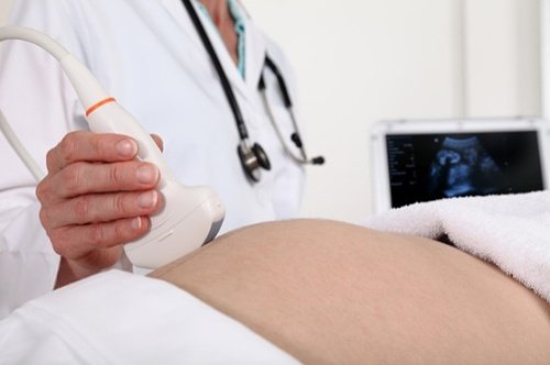
Each diagnostic procedure must be motivated. The ultrasound diagnostic method is the safest and most accessible way to examine internal organs. During the ultrasound procedure, the condition of the organs of the genitourinary system is visually assessed.
Many doctors are confident that ultrasound of the kidneys and bladder should be prescribed to all expectant mothers. This screening will help identify changes in the urinary system in their infancy. In addition, ultrasound can examine the bladder and kidneys of the fetus.
The fetus in the womb gradually grows, oppressing the organs. If a pregnant woman has been diagnosed with kidney or bladder diseases in the past, the diseases will certainly make themselves felt. Kidney pathologies cause dangerous complications that negatively affect the health of the child and mother. Therefore, it is advisable to perform ultrasound of the bladder and kidneys systematically, for medical reasons.
Many pregnant women are interested in the question: how harmful is ultrasound of the kidneys and bladder? The diagnosis uses standard ultrasound frequency, which is harmless. However, before you do an ultrasound of the urinary and kidneys, you need a written referral from the gynecologist who is monitoring the pregnancy. It is the doctor who determines the need for an ultrasound of the bladder and kidneys.
When is it necessary to make an appointment with a urologist?
The appearance of symptoms of urological diseases is a direct indication to see a urologist, and regular preventive examinations are the way to maintaining health.
A consultation with a urologist is necessary if an adult (male or female) has:
- painful sensations that occur when urinating;
- frequent feeling of fullness of the bladder even with a small amount of accumulated urine;
- recurring urinary retention;
- cloudiness of urine or change in color not associated with eating certain foods (beets, etc.);
- foreign discharge when urinating;
- pain localized in the lower abdomen and lumbar region.
Indications
When is an ultrasound of the kidneys prescribed during pregnancy? Ultrasound examination is prescribed for pregnant women in the following situations:
- If protein, bacteria, leukocytes and other abnormalities are detected in the OAM (general urinalysis).
- Frequent pulling sensations in the lower back.
- Painful urination.
- Blood in the urine or unusual coloring.
- Diabetes mellitus and other diseases of the endocrine system
- The presence of chronic diseases of the genitourinary system.
List of necessary things:
- Diaper
- Wet wipes
- Disposable dry wipes
- A 0.5 liter bottle of distilled water without gas (up to 12 weeks).
- Exchange card and direction
In what cases is it necessary to see a urologist for men?
A visit to a urologist is necessary for men if one or more signs are present:
- Discharge from the urethra . The urethra is designed to remove urine and ejaculate; normally, this may also be a secretion produced by the urethral glands, but its quantity is so small that it is problematic to notice it. In other cases, we may be talking about the presence of diseases transmitted during intercourse, inflammation of the urethra, or prostate as a result of hypothermia. Discharge may indicate complicated pathology of other organs.
- Cutting in the urethra, burning and itching . Sometimes similar phenomena can be observed in a completely healthy man, which is caused by synthetic underwear, the use of coated condoms and lubricants, and the consumption of spiced dishes. However, such symptoms, occurring regularly and increasing over time, indicate the presence of pathology.
- Complete lack of libido or decreased libido.
- Pain in the testicles and perineum, near the anus and in the lumbar region . If there was no increased physical activity on the eve of the onset of symptoms, we can talk about urological problems.
- Impaired urination . It is evidenced by a weakening of the stream, the inability to relieve oneself at will, although it occurs pathologically often.
- Rashes and neoplasms on the penis . These signs most often occur against the background of pathologies transmitted through sexual contact without barrier contraceptives.
- Erectile dysfunction . This can manifest itself as ordinary fatigue, stress and lack of sleep, or the problem lies in hormonal imbalance, diseases of the vascular and genitourinary systems, and internal organs.
- Asthenovegetative syndrome . With chronic prostatitis, general weakness, deterioration in performance, irritability, insomnia, increased sweating and increased heart rate often develop.
- Changed color of urine , presence of purulent and bloody inclusions in it or ejaculate.
- Lack of children in marriage with a full sex life.
Rules for preparing for visiting a urologist for men
- Before visiting a doctor, you need to take a hygienic shower and put on clean underwear . At the same time, for more reliable research results, it is not recommended to wash thoroughly; simply washing the external genitalia is sufficient.
- To perform a smear in the urethra for possible infections, it is necessary: sexual abstinence for 1-2 days before seeing a doctor; do not urinate for 2-3 hours before taking it; Do not drink alcohol for 1 day before your appointment.
- To perform an analysis of ejaculate (spermogram) for the problem of infertility in marriage or before planning pregnancy, it is necessary: sexual abstinence before the examination for 3-5 days; abstaining from taking alcoholic beverages (including beer), medications (hypnotics and sedatives, antibiotics) for 14 days; abstaining from visiting the sauna, bathhouse, as well as taking hot baths for 14 days; exclusion of X-ray examinations within 14 days; On the eve of the analysis, it is advisable to get enough sleep and not be overtired.
- When referring and performing an ultrasound of the bladder: do not urinate for 2 hours before the examination; 1-1.5 hours before your appointment, drink 1 liter of liquid (still water, tea or juice). When applying for surgical pathology (varicocele, dropsy, phimosis), no special preparation is required.
- When examining sexual disorders, preliminary preparation for special research methods is discussed individually with the doctor.
Normal ultrasound indicators of the fetal kidneys
A picture of a healthy fetal urinary system looks like this:
- Shape: with longitudinal scanning – bean-shaped or oval, with transverse scanning – round.
- Location: on both sides of the spine.
- The calyces are one of the structural units of the kidney. When they merge, one common cavity is formed - the renal pelvis. It gradually narrows and continues into the ureter, which flows into the bladder.
- The diameter of the renal pelvis: in the 2nd trimester – 4-5 mm, in the 3rd trimester – 7 mm.
- Normally, the ureters are not visualized.
Normal kidney size by week of pregnancy. The maximum size of the kidney is determined by longitudinal scanning.
| Duration (weeks) | Length(cm) | Duration (weeks) | Length(cm) |
| 18 | 1,6 – 2,8 | 30 | 2,9 – 4,6 |
| 19 | 1,5 – 3,1 | 31 | 2,8 – 4,6 |
| 20 | 1,8 – 3,4 | 32 | 3,1 – 5,1 |
| 21 | 2,1 – 3,2 | 33 | 3,1 – 4,7 |
| 22 | 2 – 3,4 | 34 | 3,3 – 5 |
| 23 | 2,2 – 3,7 | 35 | 3,2 – 5,2 |
| 24 | 1,9 – 4,4 | 36 | 3,3 – 5 |
| 25 | 2,5 – 4,2 | 37 | 3,3 – 5,1 |
| 26 | 2,4 – 4,4 | 38 | 3,2 – 5,6 |
| 27 | 2,7 – 4,4 | 39 | 3,5 – 4,8 |
| 28 | 2,6 – 4,2 | 40 | 3,2 – 5,3 |
| 29 | 2,3 – 4,8 | 41 | 3,9 – 5,1 |
Examination of the internal structure of a child’s kidneys is possible only from 20-24 weeks. It will identify a number of anomalies in the development of the paired organ, such as the absence of one or both kidneys, atypical localization, excess size, expansion of the calyces and/or pelvis, and the presence of cysts.
Rules for preparing for visiting a urologist for women
The preparation procedure for representatives of the fair sex is not much different from that before visiting a gynecologist. Moreover, the woman will even be examined in a gynecological chair.
Before use, toilet the genitals with plain running water. It is prohibited to treat the genitals with Miramistin, Chlorhexidine and other antibacterial agents. Since the analysis results in this case will not be reliable.
Before going to the doctor, avoid sexual intercourse for at least 24 , and preferably 48 hours. It is recommended to have a diaper or a special towel with you.
The influence of kidney ultrasound on fetal development
Most pregnant women are always wary of being referred for additional examination methods, and ultrasound is no exception. In modern medicine, there are studies about the possible negative impact of ultrasound waves on the development of the fetus, but over the years of practice, not a single case of a negative impact of ultrasound on the course of pregnancy has been identified.
For a woman in a position who is unsure whether to agree to an ultrasound, it is extremely important to understand that the presence of an undiagnosed disease of the urinary system poses a much greater danger than the influence of ultrasonic waves. You should not ignore the referral of a gynecologist for further examination, because you can miss the onset of the development of the disease, and this will lead to serious complications during pregnancy, among which even the worst thing for a woman is the loss of the desired baby.
If you find an error, please select a piece of text and press Ctrl+Enter
What questions can a urologist ask during a consultation?
During the consultation, the doctor may ask the patient about the circumstances of the disease, its manifestations, and so on.
At the first consultation, the urologist may ask:
- How long ago did the disease begin?
- How does the disease manifest?
- Do you have problems with urination?
- What causes/increases symptoms?
- Does the patient suffer from any known diseases of the genitourinary system?
- Did your parents or close relatives (siblings) have similar diseases?
- Does the patient have chronic diseases of other organs and systems (heart, liver, etc.)?
- Does the patient have a regular sexual partner?
- What methods of contraception (protection) does the patient use?
- Has the patient suffered from sexually transmitted diseases?
- Does the patient have children?
- Is the patient taking narcotic drugs?
- Does the patient abuse alcohol?
- Does the patient smoke?
It is worth noting that the list of questions may differ significantly depending on which organ is affected and how severely it is affected.
How does an appointment with a urologist work for men?
After interviewing the patient, the doctor must examine the external genitalia.
During the examination, the doctor assesses:
- the shape of the penis - its excessive curvature can cause infertility, and also indicate a high probability of other developmental anomalies;
- size of the penis - its underdevelopment is possible with a reduced concentration of the male sex hormone in the blood;
- the condition of the skin in the genital area - in order to identify foci of inflammation, ulcers, cracks or other deformations;
- the condition of the head of the penis (for this the doctor exposes it) - to identify phimosis or inflammatory processes in this area;
- condition of the testicles - the doctor palpates (feels) the testicles and epididymis, assessing their shape, size and consistency;
- condition of the scrotum - in order to identify varicocele or an infectious-inflammatory process;
- condition of the bladder - for this, the doctor may ask the patient to lie down, and then begin to lightly press on the bladder area (just above the pubis);
- kidney condition - the urologist can lightly tap the patient’s lumbar region (to which the kidneys are projected) with the edge of his palm, assessing his reaction (the occurrence of pain may indicate the presence of an inflammatory process);
- Also, a mandatory stage of the examination is a digital rectal examination of the prostate: the patient lies on his side and tries to press his knees to his chest, the doctor puts on a sterile glove, lubricates it with special oil and inserts the index finger into the patient’s anus; at a depth of several centimeters, he identifies the prostate, which is located between the bladder and intestines (the doctor palpates it through the wall of the rectum); Next, the doctor evaluates the size, consistency and shape of the prostate. If during the examination the patient feels sharp stabbing pain, he should inform the doctor (this symptom may indicate the presence of prostatitis).
Ultrasound of fetal kidneys
Assessment of the condition of the fetal kidneys is carried out in the second half of pregnancy. The study is necessary to identify the expansion of the pelvis in the unborn baby.
The formation of a child’s urinary system is a complex multi-stage process that continues from the beginning of embryogenesis until the prenatal period. The development of fetal kidneys occurs in 4 stages:
- 5-15 weeks – formation of the collecting ducts of the kidneys and their pelvis, formation of urine (from 11 weeks);
- from the 16th week - the appearance of renal arcades - a special localization of nephrons, in which they communicate with the ampoules of the tubules;
- 23-26 weeks - attachment of nephrons to the terminal sections of the collecting duct;
- Weeks 33-36 – growth and maturation of the interstitial component of the kidneys.
In the 2nd and 3rd trimester, ultrasound statistically reveals about 85% of fetal kidney diseases that arise as a result of:
- early cessation of kidney development;
- disturbances in the development of kidney tubules;
- disturbances in the movement of the kidney from the pelvic area to the abdominal cavity;
- decreased urinary tract patency.
Often, abnormal kidney development is combined with other pathologies, often caused by oligohydramnios.
How does an appointment with a urologist work for women?
Survey. The appointment begins with the woman being asked about any complaints. The doctor clarifies how long the woman has been bothered by certain symptoms and how much they affect the quality of life. Under some circumstances their presence is felt more strongly, and under what circumstances their severity, on the contrary, is less. After the interview, the woman is asked to lie down on a gynecological chair.
Inspection. During the examination, the doctor pays attention to the structure of the visible genital organs. Notes the presence or absence of redness, irritation, and various skin defects. A simple vaginal examination is often then performed to assess the condition of the genitals that can be seen. However, the urologist may not conduct such an examination, limiting himself to an external examination and taking samples.
Also, the doctor must palpate (feel) the area of the bladder and kidneys, assessing the patient’s reaction. The occurrence of pain in the pelvic area or in the lumbar region may indicate the presence of inflammatory processes and usually requires additional instrumental studies.
What are the dangers of kidney disease during pregnancy?
Untreated kidney disease during pregnancy can lead to serious complications, such as:
- Reddening of the kidneys, which can lead to high blood pressure and kidney failure;
- Blood poisoning, also called septicemia. The kidneys filter waste from the blood and return the filtered blood to the body. However, a kidney infection allows bacteria to enter the bloodstream, causing blood poisoning;
- A kidney infection causes bacteria to enter the amniotic sac and fluid. This can cause the sac to rupture and lead to premature birth;
- Premature birth can result in a low birth weight baby;
- Pyelonephritis with high fever is a severe case of kidney infection that can lead to miscarriage.
How does an appointment with a urologist end for both men and women?
The final stage of the first visit to the urologist is drawing up a plan for laboratory tests and additional instrumental examinations necessary to confirm the disease suspected during examination and conversation and determine the severity of possible changes.
Depending on the specific situation, these could be:
- general blood and urine tests;
- blood chemistry;
- bacteriological culture of urine for flora;
- urinary tract swab;
- enzyme immunoassays;
- DNA diagnostics (PCR);
- Ultrasound of the urinary system
- X-ray examinations, etc.
Men should be examined by a urologist, both elderly and young, at least once every 1 year. Annual examinations for women are not mandatory, but require an individual approach, as well as the presence of relevant complaints. As a rule, if a particular urological pathology is suspected, the referral is made by a gynecologist.

Embryonic aspects of congenital anomalies of the kidneys and urinary tract (cakut syndrome)
Vasiliev A.O., Govorov A.V., Pushkar D.Yu.
Department of Urology, State Budgetary Educational Institution of Higher Professional Education, Moscow State Medical and Dental University named after A.I. Evdokimov Ministry of Health of Russia, Russia, Moscow
Address: 127206, Moscow, st. Vucheticha 21, bldg. 2, tel. 8(495)6113129 Email, [email protected] ,
Introduction. The spectrum of congenital anomalies of the kidneys and urinary tract is extremely wide and varies from mild and asymptomatic defects, such as duplication of the ureter, to severe, sometimes incompatible with life, such as bilateral renal agenesis or dysplasia. As a result of the close embryogenetic connection of the human urinary and reproductive systems, anomalies in the development of the organs of the urinary system (US) in 33% of cases are associated with malformations of the genital organs, which in the future can lead to the development of infertility. In the total number of lesions, developmental anomalies of CMS account for an average of 25% of the total number of all genetic defects diagnosed in utero [1]. Congenital malformations of the genitourinary system are one of the largest groups of congenital anomalies, including damage to the kidneys, ureters, bladder, urethra, as well as male and female genitalia.
The possibility of simultaneous development of congenital anomalies of the check and lower urinary tract was reported in the manual of A. Ya. Pytel. and Goligorsky S.D. [2]. In 1998, E. Yerkes and H. Nishimura proposed the term CAKUT (congenital anomalies of the kidney and urinary tract) - congenital anomalies of the kidney and urinary tract. In their study, the authors described the influence of angiotensin in the formation of congenital anomalies of the kidneys and urinary tract in mice and humans [3]. Currently, CAKUT syndrome accounts for about 30% of all developmental anomalies of CMS and up to 50% of all cases of palpable abdominal mass in newborns [4, 5]. The incidence of this syndrome is 1 case per 500 newborns [6].
CAKUT can be either a component of various multiorgan genetic syndromes or an independent lesion. As part of other genetic syndromes, CAKUT has been described in approximately 500 multiorgan syndromes, such as renal cystic syndrome and diabetes mellitus [7]. According to Renkema KY et al. up to 10% of isolated forms of CAKUT syndrome can be associated with a hereditary factor. In most cases of familial forms, the condition is asymptomatic in close relatives [8]. CAKUT syndrome includes renal dysplasia with or without hypoplasia, urinary tract obstruction, and vesicoureteral reflux (VUR) (Table 1).
The main causes of the development of CAKUT syndrome include simultaneous mutation of the PAX2 and EMX2 genes. Studies have shown the absence of such gene mutations in healthy mouse embryos and in humans; in humans, both genes are located on chromosome 10q and their mutation is accompanied by complete destruction of the chromosome [9]. Having studied the role of PAX2 in the development of CMS, Chuary Ya. came to the conclusion that the relationship of the gene under study with transcription factors such as Gdnt, Ret, SHH, Wnt4, Fgt has a negative effect on nephrogenesis [10].
Table 1. Structure of CAKUT syndrome
| Bud | Urinary tract |
| Kidney agenesis | Agenesis – absence of the triangle of the bladder |
| Kidney dysplasia (including cystic and multicystic) | Stenosis of the pelvic ureter |
| Kidney hypoplasia | Megaureter |
| Duplication of the collecting system | Posterior urethral valve |
| Horseshoe kidney | Vesicoureteral reflux |
To fully understand the causes of congenital developmental anomalies, we consider it appropriate to briefly outline the main stages of CMS embryogenesis [11].
Intrauterine development of a person lasts on average 280 days, during which it is customary to distinguish three periods: initial (1st week), embryonic (2-8 weeks), fetal (from the 9th week of development to birth). By the end of the embryonic period, the laying of the main embryonic rudiments of tissues and organs is completed. During human embryogenesis, three paired excretory organs are sequentially formed: the anterior kidney or prebud (pronephros), the primary kidney (mesonephros) and the permanent (definitive) kidney (metanephros).
The kidney is formed from the anterior 8-10 segmental legs (nephrotomes) of the mesoderm and consists of epithelial tubes, one end of which is blindly closed, and the other end faces the somites, where the tubules unite to form the mesonephric (Wolffian) duct. In the human embryo, the kidney does not function as a urinary organ and soon after development undergoes reverse development. The mesonephric duct persists and grows in a caudal direction.
The primary kidney is formed from a larger number of nephrotomes (up to 25) located in the area of the embryo’s body. Over time, the segmental legs are separated from the somites and splanchnotome and turn into blind tubules of the primary kidney. The tubules grow towards the mesonephric duct and merge with it at one end. Towards the other end of the tubule of the primary kidney, vessels grow from the aorta, which break up into capillary glomeruli. The tubule with its blind end grows over the capillary glomerulus, forming the glomerular capsule. Capillary glomeruli and capsules together form the renal corpuscles. The mesonephric duct, which appears during the development of the kidney, opens into the hindgut.
The formation of the final bud occurs at the 2nd month. intrauterine development, but its final development is completed only after the birth of the child. This kidney is formed from two sources, the mesonephric duct and nephrogenic tissue. The latter represents areas of mesoderm not divided into segmental stalks in the caudal part of the embryo. The mesonephric duct grows towards the nephrogenic primordium, and from it the ureter, renal pelvis with renal calyces are subsequently formed, and from the latter outgrowths arise that turn into collecting ducts and tubules. These tubes play the role of an inducer during the development of tubules in the nephrogenic primordium. From the latter, clusters of cells are formed, which turn into closed vesicles. Growing in length, the vesicles turn into blind renal tubules, which bend in an S-shape as they grow. When the wall of the tubule adjacent to the blind outgrowth of the collecting duct interacts, their lumens are united. The opposite blind end of the renal tubule takes the form of a two-layer cup, into the recess of which a glomerulus of arterial capillaries grows. Here the vascular glomerulus of the kidney is formed, which together with the capsule forms the renal corpuscle. Once formed, the final bud begins to grow rapidly and from the 3rd month. turns out to lie above the primary kidney, which atrophies in the second half of pregnancy.
The impact of unfavorable factors on the body of a pregnant woman during the embryonic period (from 4 to 8 weeks) can lead to agenesis of the kidneys and ureters, ectopia of the ureteral orifice, the formation of an additional ureter with a blind end or a poorly developed kidney.
Exposure to various adverse factors for 9-12 weeks. development can lead to the formation of a retrocaval, retroiliac ureter, heterolocal dystopia of the kidneys and ureters, ectopia of the ureteric orifice, the formation of narrowings or valves of various parts of the ureters, insufficiency of the folds of the mucous membrane of the ureteral orifice, congenital underdevelopment or absence of the muscular layer, high discharge of the ureter from the renal pelvis.
During the fetal period (from the 13th week), it is possible to develop tortuosity of the ureter, its bends, shortening, lengthening, imbalance of contractile function, congenital insufficiency of the innervation apparatus and ureteral dysplasia [12].
The ontogenetic mechanisms underlying the etiology of developmental anomalies and CAKUT syndrome are multifactorial. Many of them were obtained by dissecting human embryos, others - in animal experiments. Their detailed characteristics are presented in Figure 1 [5].
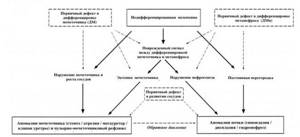
Fig.1. Ontogenetic basis of the formation of CAKUT syndrome. The dotted arrow indicates a crossover anomaly.
Materials and methods. Sources in the English-language literature were analyzed, the search was carried out in the Cochrane Library's, Medline (Pubmed, BioMedNet), Scopus and Biosis databases, using the keywords “developmental anomalies”, “congenital malformations”, “CAKUT”, “embryogenesis of the urinary system organs” "
Discussion. The manifestation of congenital malformations of CMS, including CAKUT syndrome, is quite fully reflected in the world literature, however, these anomalies are characterized by features that have not been fully studied. For example, such anomalies as stenosis of the ureteropelvic segment (UPS), atresia and multicystic dysplasia of the kidney are most often unilateral; Prenatal hydronephrosis of newborns associated with VUR and primary megaureter is overwhelmingly found in men [13, 14]. Some of the described anomalies often develop simultaneously [15]. For example, hypoplasia and dysplasia are often accompanied by VUR or LMJ stenosis, atresia of the ipsilateral or contralateral kidney.
The most common congenital anomaly of UMC is duplication of the ureter, which can be complete or incomplete. Incomplete duplication often occurs together with a bifurcation of the pyelocaliceal system (PSS). Most cases are asymptomatic, but the risk of upper urinary tract infection increases 20-fold [16]. Atwell JD et al. when examining 30 children with incomplete duplication of the ureter, they came to the conclusion that this anomaly has an autosomal dominant mode of inheritance, since up to 66% of first-degree relatives had the same lesion [17].
PUJ obstruction is the most common cause of antenatal hydronephrosis and occurs with a frequency of 1 in 1000-1500 newborns [18]. Of all the known reasons for the development of this anomaly, most authors suggest that incomplete muscularization of the ureter and excessive apoptosis of smooth muscle cells are the main ones [19, 20].
In the case of fusion of the lower poles of the kidneys, a horseshoe-shaped kidney is formed, located in most cases lower than usual. In rare cases, the presence of a horseshoe kidney is associated with PUJ obstruction and urinary tract infection, hematuria, and a palpable abdominal mass. One of the best treatment options for this anomaly is transperitoneal laparoscopic pyeloplasty, which allows a detailed examination of the renal vascular system and the presence of renal vascular anomalies [21].
Posterior urethral valve is considered the most common cause of lower urinary tract obstruction in neonates and male fetuses [22]. This malformation is often associated with plum belly syndrome, which has an incidence of 3.8 per 100,000 male newborns and is characterized by congenital absence, insufficiency or hypoplasia of the abdominal wall muscles in combination with megacystis, dilatation of the ureters and prostatic urethra, as well as bilateral cryptorchidism. Some of the possible treatment methods in this situation are to perform a vesico-amniotic shunt or perform a vesicocentesis after birth. A distinctive feature is the characteristic wrinkled appearance of the anterior abdominal wall after surgery, which gives the name to this syndrome [23, 24].
In most cases, the manifestation of obstructive renal dysplasia (in particular, megacystis) can be detected by fetal ultrasound, while dilatation of the ureter, calyces or pelvis can only be suspected with careful examination. The histological appearance may vary depending on the duration of intrauterine obstruction and includes compressed medulla as a result of excess hydrostatic pressure created by accumulated urine in the pelvic area [25]. The most common and studied syndromes of renal dysplasia (a group of ciliopathies) include Meckel-Gruber syndrome, Joubert syndrome, short rib syndrome, Bardet-Biedl syndrome, asplenia or polysplenia syndrome and VACTER-L.
Meckel-Gruber syndrome was first described by the German biologist F. Meckel in 1822. The frequency of manifestation in newborns is 1:9000. The syndrome is usually fatal in early childhood due to severe pathology of the kidneys and central nervous system and is characterized by a combination of polycystic kidney disease, abnormal development of the central nervous system (occipital encephalocele), fibrotic changes in the liver and polydactyly [26]. The syndrome is genetically heterogeneous , an autosomal recessive disease. Currently, about 10 genes are known whose mutations can lead to the development of the syndrome, including MKS1, TMEM216, TMEM67, etc.
Joubert syndrome is a rare autosomal recessive pathology characterized by the complete absence or underdevelopment of the cerebellar vermis, mental retardation, retinal dystrophy, ocular coloboma, nystagmus, polydactyly, cystic renal dysplasia and congenital liver fibrosis. The syndrome was first described by M. Joubert in 1969 during an examination of four newborns in one French-Canadian family. The prevalence of kidney disease (cystic renal dysplasia and tubulointerstitial nephritis) in Joubert syndrome and related disorders averages 30% [27].
Short rib syndrome is a genetic pathology that also belongs to the group of ciliopathies and is characterized by the presence of short ribs, shortened limbs, polydactyly and early mortality due to respiratory failure. Traditionally, four subtypes of this syndrome are distinguished: Saldino-Noonan syndrome, Majewski syndrome, Verma-Naumoff syndrome and Beemer-Langer syndrome [28]. The histological picture of the kidneys is similar to that of Meckel-Gruber and Joubert syndromes. Most of the genes associated with short rib syndrome control the process of intraflagellar transport. The most studied mutations are in the NEK1 and TTC21B genes [29, 30].
Bardet-Biedl syndrome is a genetically heterogeneous disease and occurs with a frequency of 1:120,000 newborns. Characterized by the presence of at least four of the six primary symptoms - obesity, retinal degradation, polydactyly, polycystic kidney disease, hypogonadism and mental retardation. Secondary symptoms may include diabetes, liver fibrosis, ataxia, various speech disorders, asymmetry of visceral organs, dental pathology, anosmia and hearing loss. There are 18 known genes whose mutations can lead to the development of the syndrome [31].
In 1959, Ivemark BL et al. a variant of renal-hepatic-pancreas dysplasia in combination with asplenia was described [32]. Currently, the literature contains no more than 10 reports describing children with this autosomal recessive syndrome, characterized by multicystic dysplasia of the kidney, liver and pancreas, underdevelopment of the lungs, heart defects and in most cases leading to early infant mortality. At the molecular level, two genes whose mutation leads to the development of this condition have been studied: NPNP3 and NEK8. Macroscopically, multicystic renal dysplasia is clearly visible [33].
The VACTER-L association is a group of combined developmental anomalies, the name of which is made up of the first letters of the defects that are part of the syndrome [34]:
- V (Vertebral anomalies) - spinal anomalies, A (Anal atresia) - anal atresia,
- C (Cardiovascular anomalies) - septal defects and other heart defects,
- TE (Tracheo-esophageal fistula) - tracheoesophageal fistula with esophageal atresia,
- R (Renal defects) - kidney anomalies, such as agenesis, dysplasia, hydronephrosis; the only umbilical artery.
- L (Limb defects) - radial bone defects - hypoplasia of the first finger or radial bone, preaxial polydactyly and syndactyly. Most authors note that a combination of at least three of the listed symptoms is necessary to make a diagnosis. The described lesion occurs with a frequency of 1 in 10,000–40,000 newborns [14]
Conclusions. Congenital anomalies of the kidneys and urinary tract are a group of conditions with varying degrees of severity, many of which require a multidisciplinary approach to accurately diagnose and improve subsequent treatment. Given that many of the reported congenital anomalies are hereditary, advances in prenatal diagnosis, fetal surgery, and targeted therapy will improve the prognosis and quality of life of newborns.
Literature
1. dos Santos Junior, AC Congenital anomalies of the kidney and urinary tract: an embryogenetic review / AC dos Santos Junior, DM de Miranda, Silva AC e Simões // Birth Defects Res C Embryo Today. – 2014, Vol. 102, No. 4 – P. 374-381.
2. Pytel, A.Ya. Selected chapters of urology and nephrology. Part 1 / A.Ya. Pytel, S.D. Goligorsky. L.: Medicine, 1968. 312 pp..
3. Yerkes, E. Role of angiotensin in the congenital anomalies of the kidney and urinary tract in the mouse and the human / E. Yerkes, H. Nishimura, Y. Miyazaki // Kidney Int. – 1998; Vol. 67, pp. 75–77.
4. Toka, HR Congenital Anomalies of Kidney and Urinary Tract / HR Toka, O. Toka, A. Hariri, HT Nguyen // Samin Nephril. – 2010, Vol. 30, No. 4 – P. 374–386.
5. Pope, JC IV. How they begin and how they end classic and new theories for the development and deterioration of congenital anomalies of the kidney and urinary tract, CAKUT / JC Pope IV, JW Brock III, MC Adams // JASN. – 1999, Vol. 10, No. 9 – P. 2018–2028.
6. Loane, M. EUROCAT statistical monitoring: identification and investigation of ten year trends of congenital anomalies in Europe / M. Loane, H. Dolk, A. Kelly // Birth defects Res A Clin Mol Teratol. – 2011, Vol. 91, No. 1 – P. 31-43.
7. Erman, M.V. Nephrology of childhood / M.V. Erman. St. Petersburg: SpetsLit, 2010. 683 p.
8. Renkema, KY EUCAKUT consortium/Novel perspectives for investigating congenital anomalies of the kidney and urinary tract (CAKUT) / KY Renkema, PJ Winyard, IN Skovorodkin // Nephrol. Dial. Transplant. – 2011, Vol. 26, No. 12 – P. 3843–3851.
9. Boualia, SK. Vesicoureter Reflux and Other Urinary Tract Malformation in Mice Compound / SK Boualia, Y. Gartan, I. Murawski // PRoS One. – 2011, Vol. 6, No. 6 –P. 215–224.
10. Chuary, Ya. The role of PAX2 is regulation of kidney development and kidney diseases / Ya Chuary, // Ya Chion. – 2011, Vol. 33, No. 3 – P. 231–238.
11. Afanasyev, Yu.I. Histology, embryology, cytology. 6th edition / Yu.I. Afanasyev, N.A. Yurina. – M.: GEOTAR-Media, 2012. – 800 p.
12. Zwolińska, D. Genetics of congenital anomalies of the kidney and urinary tract / D. Zwolińska, D. Polak-Jonkisz, I. Makulska // Postepy Hig Med Dosw. – 2011, Vol. 15, No. 65 – P. 829-837.
13. Uehling, DT Urologic implications of the VATER association / DT Uehling, E. Gilbert, R. Chesney // J Urol. – 1983, Vol. 129, pp. 352–354.
14. Solomon, BD VACTERL/VATER association / BD Solomon // Orphanet J Rare Dis. – 2011, Vol. 6, p. 6.
15. Atiyeh, B. Contralateral renal abnormalities in multicyctic-dysplastic kidney disease / B. Atiyeh, D. Husmann, M. Baum // J Pediatr. – 1992, Vol. 121, p. 6567.
16. Decter, R. Renal duplication and fusion anomalies / R. Decter // Pediatr Clin North Am. – 1997, Vol. 44, pp. 1323–1341.
17. Atwell, JD Familial incidence of bifid and double ureters / JD Atwell, PL Cook, C. Howell // Arch Dis Child. – 1974, Vol. 49, pp. 390–393.
18. Williams, B. Pathophysiology and treatment of ureteropelvic junction obstruction / B. Williams, B. Tareen, MI Resnick // Curr Urol Rep. – 2007, Vol. 8, pp. 111–117.
19. Aoki, Y. Id2 haploinsufficiency in mice leads to congenital hydronephrosis resembling that in humans / Y. Aoki, S. Mori, K. Kitajima // Genes Cells. – 2004, Vol. 9, pp. 1287–1296.
20. Kajbafzadeh, A.M. Smooth muscle cell apoptosis and defective neural development in congenital ureteropelvic junction obstruction / A.M. Kajbafzadeh, S. Payabvash, A.H. Salmasi // J Urol. – 2006, Vol. 176, pp. 718–723.
21. Blanc, T. Laparoscopic pyeloplasty in children with horseshoe kidney / T. Blanc, E. Koulouris, N. Botto // J Urol. – 2014, Vol. 191, pp. 1097–1103.
22. Quintero, RA In utero management of fetal lower urinary tract obstruction with a novel shunt: a landmark development in fetal therapy / RA Quintero, LA Gomez, LA Castro, C. Bermudez // J Matern Fetal Neonatal Med. – 2010, Vol. 23, pp. 806–812.
23. Tonni, G. Prune-belly syndrome: case series and review of the literature regarding early prenatal diagnosis, epidemiology, genetic factors, treatment, and prognosis / G. Tonni, V. Ida, V. Alessandro, MP Bonasoni // Fetal Pediatr Pathol. – 2013, Vol. 31, pp. 13–24.
24. Granberg, CF Genetic basis of prune belly syndrome: Screening for the HNF1β gene / CF Granberg, SM Harrison, D. Dajusta // J Urol. – 2012, Vol. 187, pp. 272–278.
25. Yosypiv, IV Congenital anomalies of the kidney and urinary tract: a genetic disorder? / IV Yosypiv // Int J Nephrol. – 2012, Vol. 90, pp. 83-90.
26. Opitz, J. M. Annals of morphology. Meckel on developmental pathology / JM Opitz, R. Schultka, L. Göbbel // Am J Med Genet. – 2006, Vol. 140A, pp. 115–128.
27. Joubert, M. Familial agenesis of the cerebellar vermis. A syndrome of episodic apnea, abnormal eye movements, ataxia, and retardation / M. Joubert, E. JeanJacques, JP Robb, F. Anderman // Neurology. – 1969, Vol. 19, pp. 813–825.
28. Dagoneau, N. DYNC2H1 mutations cause asphyxiating thoracic dystrophy and short rib-polydactyly syndrome, type III / N. Dagoneau, M. Goulet, D. Geneviève, Y. Sznajer, J. Martinovic, S. Smithson, C. Huber, G. Baujat, E. Flori, L. Tecco, D. Cavalcanti, A. L. Delezoide, V. Serre, M. Le Merrer, A. Munnich, V. CormierDaire // Am J Hum Genet. – 2009, Vol. 84, No. 5 – P. 706-711
29. Thiel, C. NEK1 mutations cause short-rib polydactyly syndrome type Majewski / Thiel, A. Giessl // Am J Hum Genet. – 2011, Vol. 88, pp. 106–114.
30. Davis, EE TTC21B contributes both causal and modifying alleles across the ciliopathy spectrum / EE Davis, Q. Zhang, Q. Liu // Nat Genet. – 2011, Vol. 43, pp. 189–196.
31. Green, JS The cardinal manifestations of Bardet–Biedl syndrome, a form of Laurence–Moon–Biedl syndrome / JS Green, PS Parfrey, JD Harnett // N Eng J Med. – 1989, Vol. 321, pp. 1002–1009.
32. Ivemark, BL Familial dysplasia of kidneys, liver and pancreas: a probably genetically determined syndrome / BL Ivemark, V. Oldfelt, R. Zetterstrom // Acta Paediatr. – 1959, Vol. 48, pp. 1–11.
33. Frank, V. Mutations in NEK8 link multiple organ dysplasia with altered Hippo signaling and increased c-MYC expression / V. Frank, S. Habbig, MP Bartram // Hum Molec Genet. – 2013, Vol. 22, pp. 2177–2185.
34. Uehling, DT Urologic implications of the VATER association / DT Uehling, E. Gilbert, R. Chesney // J Urol. – 1983, Vol. 129, pp. 352–354.
Magazine
Bulletin of Urology No. 2/2015
Comments
To post comments you must log in or register