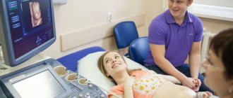
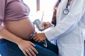
Ultrasound examination of pregnancy is an important and mandatory method of examining pregnant patients, gynecologists note. “Usually during pregnancy we perform an ultrasound examination of the fetus, otherwise called a scan, 2-3 times. Sometimes an ultrasound scan of a pregnant woman needs to be done more times, for example, if some problems stand out, or the doctor wants to reconsider something, or there are doubts,” says obstetrician-gynecologist Angelina Brainina .
Using ultrasound, you can not only study the state of pregnancy, but also determine existing defects based on the results of an ultrasound examination of the fetus.
Article on the topic Multiple pregnancy and ultrasound: features of the procedure
When can defects appear?
As Angelina Brainina notes, there are several reasons for the appearance of fetal malformations. Among them, says the expert:
- heredity;
- mother's age is over 35 years, father's age is over 40 years;
- unfavorable environmental conditions, for example, deficiency of iodine or some other important microelements and substances;
- working with chemicals at parents’ place, for example in hazardous industries;
- bad habits - smoking, alcohol abuse, drug addiction;
- viral or bacterial infections suffered by the mother, especially if they occurred in the first trimester (dangerous ones include influenza, rubella, toxoplasmosis, etc.);
- taking a number of medications;
- strong psycho-emotional stress.
Therefore, says Angelina Brainina, it is extremely important to plan the child so that most of the problems can be eliminated at this stage.
Doppler ultrasound during pregnancy: features, indications, results Read more
What are the main syndromes identified by NIPT?
- Down syndrome (trisomy 21)
The frequency of occurrence is 1:600 births. Moreover, the risk of trisomy 21 in the fetus increases with the woman’s age. One of the forms of genomic pathology, in which most often the karyotype is represented by 47 chromosomes instead of the normal 46, since the chromosomes of the 21st pair, instead of the normal two, are represented by three copies. There are two more forms of this syndrome: translocation of chromosome 21 to other chromosomes (usually on 15, less often on 14, even less often on 21, 22 and Y chromosome) - 4% of cases, and a mosaic variant of the syndrome - 5%. Characteristic features associated with Down syndrome: “flat face”, abnormal shortening of the skull, skin folds on the neck in newborns, short limbs, open mouth and other signs that can be accurately diagnosed by a geneticist.
- Edwards syndrome (trisomy 18)
The chromosomal disease is characterized by a complex of multiple malformations and trisomy 18 chromosomes. Described in 1960 by John Edwards. The worldwide population frequency is 1:5000 as of 2021. Children with trisomy 18 are more often born to older mothers, and the relationship with maternal age is less pronounced than in cases of trisomy 21. The main signs of Edwards syndrome: low weight, abnormalities in the shape of the skull, heart defects, ventricular septal defects, mental retardation.
- Patau syndrome (trisomy 13)
Frequency – 1:16000 births. Characterized by developmental disorders of the nervous system, eyes, severe facial deformation, malformations of the heart and other internal organs. Characterized by profound mental retardation. 90% of children die before the age of 1 year.
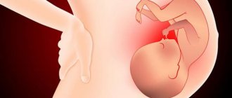
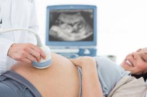
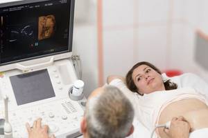
Ultrasound examination of the fetus: what defects can be seen?
Fetal development on ultrasound is an important point in studying the state of pregnancy. A description of various anomalies may appear in the pregnancy ultrasound report. Angelina Brainina identifies several groups of child developmental defects that can be seen in utero.
- Anomalies in the development of the fetal neural tube , that is, disruptions in the development of nervous tissue, the brain and spinal cord. Here we are talking about diseases such as absence of cerebral hemispheres, hydrocephalus, brain hernia, microcephaly.
- Malformations of the spine.
- Heart pathologies.
- Anomalies in the development of the gastrointestinal tract in children, for example, problems with the stomach, duodenum, anterior abdominal wall, when as a result of the formation of the fetus it simply does not exist, and the internal organs, the same intestines, exit into the amniotic fluid.
- Congenital kidney anomalies - underdevelopment of the organ, the presence of only one kidney, polycystic disease. Structural changes in the urinary tract may also be noted.
- Much and little water.
Ultrasound also shows a frozen pregnancy or intrauterine fetal death.
Baby in 3D. Will ultrasound be prohibited during pregnancy? More details
Neural tube malformations, notes Angelina Brainina, are most often diagnosed by ultrasound in the first trimester of pregnancy - at 10-12 weeks. “Sometimes they ask to redo the ultrasound at a later date - most often we are talking about 18 weeks. Here you can already see quite well the pathologies of the development of the neural tube,” says the obstetrician-gynecologist. As for spinal anomalies, they are more often detected by ultrasound in the second trimester of pregnancy - at 15 weeks, on average at 15-20 weeks.
Congenital heart and gastrointestinal defects can also be seen on ultrasound in the second trimester of pregnancy - at 20 weeks. As for problems with the abdominal wall, they can be examined quite early - with an ultrasound in the first trimester of pregnancy, at 12 weeks. Problems with the urinary system are noted at the study at 18-20 weeks.
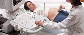
Many people are also concerned about genetic abnormalities in fetal development. “The conclusion of an ultrasound during pregnancy cannot 100% diagnose genetic pathologies. Usually, doctors may suspect the presence of some abnormalities based on a number of signs. Therefore, now there are so-called screenings - comprehensive studies that include the study of blood parameters and ultrasound screening of the fetus,” explains Angelina Brainina. The result of ultrasound screening of pregnancy, together with blood tests, is calculated using special programs depending on one and another parameter, as a result of which the risk of having a child with chromosomal pathologies is calculated. Ultrasound screening of pregnant women and blood tests are performed twice. One - in the first trimester (9-13th week), the other - in the second (13-24th week). “At the same time, the fetus is examined completely, as well as the amniotic fluid to ensure its sufficiency,” says the specialist.
How are you doing there? Why do pregnant women need ultrasound Read more
Prenatal diagnosis is divided into invasive and non-invasive.
- Invasive diagnostic methods - such as chorionic villus biopsy, amniocentesis (sampling of amniotic fluid), cordocentesis (sampling of blood from the umbilical cord).
- Non-invasive diagnostic methods are more gentle. These include non-invasive prenatal testing for common chromosomal abnormalities that are compatible with life, but such chromosomal abnormalities as Down, Edwards and Patau syndromes are accompanied by multiple malformations and cannot be cured. NIPT is more reliable than biochemical screening, as it is a direct method for studying fetal DNA. At the same time, the expectant mother does not have the risk of unnecessary anxiety during pregnancy if she falls into a risk group for chromosomal abnormalities, and there is no risk of spontaneous abortion. NIPT is approved in many countries and is included in the voluntary health insurance system (developed countries such as Switzerland, USA, Canada, the Netherlands, Germany).
Types of prenatal screening by timing of research
During pregnancy, a woman must undergo prenatal screening three times. Each examination has its own characteristics and is aimed at identifying diseases and pathologies of the embryo.
First
Early screening is carried out at 11–13 weeks. There is no point in undergoing an examination before the 11th week, since it will not be possible to undergo a thorough, comprehensive examination of the fetus. The first screening takes place in two stages: ultrasound and biochemical examination (blood test).
Early ultrasound is of great importance for the expectant mother. Using it, you can set the date of birth, assess the level of physical development of the embryo, determine the number of fetuses in multiple pregnancies, and also identify possible pathologies and genetic abnormalities.
Indicators that are taken into account during ultrasound:
- the size of the embryo from the parietal part to the coccyx;
- biparietal size - the distance from the temple to the temple of the fetus;
- collar space thickness;
- heart rate;
- nasal bone size;
- the size of the chorion, amnion, yolk sac;
- T-shirt neck size;
- volume of amniotic fluid.
Any deviations in indicators may indicate possible pathologies and defects of the embryo. However, for a final diagnosis, additional examinations will be required.
A biochemical blood test takes place on the same day as the ultrasound. Blood must be donated on an empty stomach. A blood test helps determine the development of possible pathologies in the embryo. If abnormalities are detected in the fetus, additional genetic testing will be required.
Second
The second prenatal screening is carried out at 18-22 weeks. The examination takes place in the form of an ultrasound. The second screening is also of great importance and helps to establish the following indicators:
- distance between temples;
- Head circumference;
- size of the femur;
- humerus length;
- cervical size;
- volume of amniotic fluid;
- localization of the placenta;
- the site of attachment of the umbilical cord.
A biochemical analysis is performed along with an ultrasound scan only if, for some reason, the pregnant woman did not pass the test during the first screening.
Third
The third screening is carried out shortly before birth at 30-34 weeks. The purpose of diagnosis is to establish the development of the embryo and determine possible risks of delivery. The results of the study determine whether the birth will take place naturally or by caesarean section.
Fetal health
Today, every expectant mother can obtain comprehensive information about fetal malformations and intrauterine infections. The Internet, magazines and books describe in detail the causes and consequences of such diseases. Laboratory diagnostics, in turn, offers a range of examinations “for any case.” But is it necessary to take them all? At what time are they most informative? And how to understand the abundance of tests for “fetal health”?
Intrauterine infections
Intrauterine infections (IUI) occupy a leading place among the causes of developmental defects. And the most common pathogens of dangerous infections are representatives of the “TORCH” group:
- T - toxoplasmosis - toxoplasmosis;
- Other – other infections (viral hepatitis, chlamydia, ureaplasmosis, mycoplasmosis and other infections that provoke adverse pregnancy outcomes);
- R – rubella – rubella;
- C – cytomegalovirus – cytomegalovirus infection;
- H – herpes – herpes type I and II.
The danger of TORCH infections is due to their widespread distribution, high contagiousness (infectiousness) and often hidden course (except for rubella). Infection (first encounter with infection) at:
- up to 12-16 weeks may result in spontaneous miscarriage;
- from 16 to 28 weeks – the formation of severe malformations;
- more than 28 weeks – development of infection and inflammatory processes in the fetus
The expectant mother may not be aware of the infection, while for the fetus this is a serious threat.
The risks of infections can be assessed both at the stage of preparation for pregnancy and already in pregnancy. Blood testing for antibodies to pathogens of TORCH infections is a simple diagnostic test that allows you to identify both current and past infectious processes.
IgM antibodies appear in the blood approximately 2 weeks after infection and “speak” of a high risk of complications for the fetus.
IgG antibodies are a marker of a previous infectious process, the presence of immunity to the pathogen or its carriage. And its identification requires clarification of the “prescription” of infection.
A blood test for the avidity of IgG antibodies allows you to calculate “how long ago” antibodies appeared in the blood. The higher the percentage of avidity, the “older” the infection, and therefore safer for the fetus.
Each of these indicators can be passed separately, as well as as part of complexes:
"TORCH-complex screening"
It includes the determination of only IgG to the pathogens presented (toxoplasma, herpes viruses, rubella, cytomegalovirus), and is used during the preparation for pregnancy for both partners.
"TORCH-complex basic"
Suitable for examining pregnant women and includes the determination of IgG and IgM to pathogens of TORCH infections (toxoplasmosis, rubella, herpes and CMV - cytomegalovirus).
"TORCH-complex with avidity"
Allows you to determine the presence of acute or previous TORCH infections in a pregnant woman, as well as the duration of infection and the risk to the fetus.
"TORCH-complex extended"
Includes determination of IgG and IgM for all of the above infections, including chlamydia, which allows you to obtain the most complete information.
Genetic pathology of the fetus
Until recently, the ability to detect genetic “damages” in the fetus was limited to prenatal screening or analysis of fetal biomaterial obtained by invasive methods (blood from the umbilical cord, amniotic fetuses, and others).
At the same time, chromosomal abnormalities and microdeletion syndromes threaten severe developmental defects associated with the death or severe retardation of the child.
In most cases, chromosome number pathologies (mono-, di-, trisomy) are related to the age of the mother. Whereas microdeletion syndromes are in no way related to age. The incidence of both types of “breakdowns” is on average 1 in 1000 newborns, which causes the same risks for pregnant women both before and after 30 years.
Today, it is possible to exclude up to 18 syndromes at a time using just one study - NIPS (Genomed).
Non-invasive prenatal tests are based on fetal DNA analysis from the mother's blood and are highly accurate (99.8%).
Unlike the PRISCA prenatal screening program (“double” and “triple” tests), the conclusion on NIPT is based on the quantity and quality of the baby’s chromosomes, and not on indirect signs of pathology (fetal size and hormone levels). This result allows us to make the most accurate prediction of the risk of genetic abnormality
Among the chromosomal disorders diagnosed using NIPT Panorama, basic panel (Natera):
- Down syndrome (trisomy 21),
- Edwards syndrome (trisomy 18),
- Patau syndrome (trisomy 13),
- absence of one chromosome (X chromosome monosomy = Shereshevsky-Turner syndrome),
- “extra” X chromosome in a male fetus (Klinefelter syndrome),
- triploidy.
Microdeletion syndromes can be further seen using the Panorama NIPT test, extended panel (Natera) (chromosome screening: 13, 18, 21, X, Y, triploidies and microdeletion syndromes:
- Prader-Willi syndrome,
- Angelman syndrome,
- Cri-du-sha syndrome (“cat cry”),
- syndrome 1p36,
- syndrome 222.
In addition, some tests in the NIPT line, for example, the NIPS test (Genomed), in addition to the main chromosomal pathology, allow us to identify carriage of the most common mutations in the mother, leading to genetic diseases of the fetus:
- Cystic fibrosis (autosomal recessive inheritance, search for CFTR gene mutation)
- Neuronal ceroid lipofuscinosis (autosomal recessive type of inheritance, search for TPP1 gene mutation)
- Phenylketonuria (autosomal recessive type of inheritance, search for PAH gene mutation)
- Galactosemia (autosomal recessive type of inheritance, search for GALT gene mutation)
- Hearing loss (various types of inheritance, search for mutations of the SLC26A4, GJB2 genes)
The test does not require special preparation; you just need to donate blood at a full 10 weeks of pregnancy.
There are some limitations to using the test:
- less than 10 weeks of pregnancy,
- number of fruits more than 2,
- there are signs of a frozen pregnancy,
- organs, tissues and stem cells were transplanted,
- have cancer,
- Whole blood transfusions were performed within 6 months before the test.
Analyzes can be carried out both “in the homeland” of the test - in the Natera laboratory (USA), and under a technology transfer company license in Russia (partner - Genomed laboratory). The need for transportation abroad or the lack thereof, as well as the dollar exchange rate, determines the pricing policy for research. Otherwise, the quality of analyzes carried out in the Russian Federation is under the control of the “parent” company.
Thus, the line of NIPT studies (non-invasive prenatal tests) are the most accurate, safe and fast ways to exclude severe genetic abnormalities in the very early stages of pregnancy.