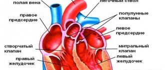
Heart ultrasound (ECHO-CG, echocardiography) is one of the highly informative modern methods of instrumental diagnostics. With the help of the study, the doctor can evaluate the functioning of the heart and its individual structures - valves, atria and ventricles. Typically, echocardiography is prescribed for all children twice: in the first year of life and before starting school. The study allows you to exclude or diagnose various pathologies of the heart and nearby large vessels. As a rule, cardiologists prescribe treatment only after receiving the results of echocardiography and ECG.
Indications for ECHO-CG
An ultrasound of the child’s heart should be performed in the following cases:
- presence of heart murmurs on auscultation;
- ECG changes;
- child's complaints of stabbing, pulling, aching pain in the heart area;
- chest deformation;
- high blood pressure;
- prolonged dry cough;
- sensation of vibration or “trembling” over the heart area or in the subclavian fossa;
- frequent coldness, pallor, blueness of the limbs, nasolabial triangle (in small children when crying, sucking the breast);
- increased fatigue, weakness;
- repeated fainting in the child;
- frequent pneumonia;
- heart disease in close relatives;
- excessive sweating, swelling of the extremities;
- retardation in physical development, etc.
Specialists at the PreAmbula Clinic recommend undergoing a heart ultrasound, regardless of the presence of complaints. Many diseases can be asymptomatic for a long time until decompensation occurs. For example, some congenital heart defects can only be detected by echocardiography. The outcome of the disease largely depends on the timeliness of diagnosis and treatment.
Transesophageal ultrasound
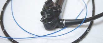
The transesophageal echocardiography procedure is more complex and has a number of limitations. An ultrasound sensor is inserted through the esophagus, which makes it possible to see the condition of the organ from all sides. The examination may be indicated for severe heart defects, chest deformities and lung diseases, inflammation of the aorta after prosthetics, etc. Preparation includes abstaining from food at least 6 hours before the examination, and during the procedure special medications may be required to improve the tolerability of the examination. Thus, it is sometimes advisable to perform intravenous anesthesia if a pronounced gag reflex is observed.
What heart pathologies are diagnosed using ultrasound?
Ultrasound of the heart is a fundamental method for diagnosing the following cardiovascular pathologies:
- various congenital and acquired heart defects (ventricular and atrial septal defects, patent ductus arteriosus, mitral stenosis and mitral insufficiency, tricuspid and aortic valve defects, etc.);
- aortic aneurysm;
- hypertrophy of the myocardium of the ventricles or atria (often develops secondary to defects);
- various types of cardiomyopathy (hypertrophic, dilated);
- blood clots in the chambers of the heart;
- myocarditis (inflammation of the middle layer of the heart muscle);
- endocarditis (inflammation of the inner layer of the heart muscle);
- changes in the myocardium (presence of scars, sclerotic processes);
- additional chords in the chambers of the heart;
- ischemic disease, etc.
Timely diagnosis of these heart diseases is necessary for proper treatment. If any heart defect is detected in a small child, the issue of the need for surgical intervention is decided. Early stages of diseases that can be detected by ultrasound of the heart are more treatable than advanced forms. In addition, the examination is necessary to determine the child’s physical fitness group. If you have any cardiac pathology, some sports may be contraindicated.
Therefore, before enrolling in the sports section, it is better to conduct an ultrasound of the heart.
When performing an ultrasound of the heart, a small child is often diagnosed with a patent foramen ovale. This is a small opening that connects the right and left atria. Normally, it heals after some time, so there is no need to worry if your baby is found to have an atrial septal defect. In this case, you need to consult a cardiologist and come for a repeat echocardiogram after some time. It is believed that the interatrial window closes normally before the age of 5 years. If after this moment it remains, then they already talk about congenital heart disease and decide on the choice of optimal treatment.
Advantages of visiting the Family Doctor clinic
Advantages of research in our clinic:
- experience and professionalism of diagnosticians;
- high-precision equipment from leading manufacturers;
- individual approach to each patient.
The Family Doctor clinic in Moscow offers to conduct an Echo-CG for the child, based on the results of which you will receive a detailed conclusion immediately after the procedure. You can also get advice from a specialized specialist of the highest category.
Make an appointment by calling the contact center +7 (495) 775 75 66, through the on-line registration form and at the clinic reception.
Preparing for echocardiography
No special preparation is required before performing echocardiography. Experts do not recommend engaging in sports training before the procedure. If your child is taking any sedative or stimulant medications, you should consult your doctor. Some medications may need to be stopped for 1-2 days before the ultrasound so that they do not affect the picture of the heart muscle. Immediately before the test, you should try to calm the child so that his pulse is within normal values.
Popular questions
How often can echocardiography be repeated in young children? How safe is the procedure?
Unlike radiographic methods, echocardiography does not pose any potential harm to health; there are no restrictions. You can be examined as often as the situation requires.
How is transesophageal echocardiography performed in infants? A child cannot fast for 6 hours.
In some cases, it is possible to carry out the procedure between feedings. The doctor referring you for diagnosis will tell you more about the preparation and procedure for conducting an examination on an infant.
How is the procedure done?
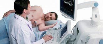
In the cardiac ultrasound room, the child should undress to the waist and lie down on the couch. If the baby is very small, then one of the parents helps the specialist by holding and calming the child. A gel is applied to the chest, which is necessary for better contact of the sensor with the skin. From the ultrasound sensor, the image of the internal structures of the heart is transmitted to the monitor, where the doctor records the parameters being studied. The ultrasound diagnostic procedure usually takes no more than 15-20 minutes.
All indicators obtained using ultrasound are entered into a special form. The results are given to the patient along with the doctor’s report. If necessary, the ultrasound technician may refer you to a cardiologist or other doctor.
Order of conduct
Echo-CG is performed absolutely painlessly and does not pose any risks to the child’s health, so the study can be repeated as often as the clinical case requires. There are also no age restrictions - the procedure can be carried out from the first days of life.
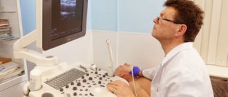
An ultrasound takes no more than 20 minutes. The child sits on the couch, the doctor applies a special gel to the skin of the chest and a special sensor to improve the conductivity of ultrasound. Then he applies the sensor and moves it across the chest, at which time an image of the heart appears on the monitor. The condition of the organ is assessed visually in real time, and with the help of the program, the necessary changes are made and blood flow is assessed. During the procedure, you may need to change your body position. As a rule, you should lie on your left side or back most of the time. The examination does not cause any discomfort, but for the baby’s peace of mind, the parent can be nearby.
Question answer
— Why is it worth choosing cardiac ultrasound as a diagnostic method?
This diagnostic method allows you to identify the presence of pathology at an early stage of development, which allows the doctor to prescribe timely treatment and monitor its progress and results. In addition, ultrasound diagnostics is a safe and absolutely painless research method.
— At what age can a child have an ultrasound of the heart?
An ultrasound of a child’s heart can be done even in the first days of life. Ultrasound does not cause any harm to both adults and children, so the frequency of this procedure is not limited.
— We need a good pediatric cardiologist in Saratov. Is it possible to make an appointment and undergo an examination at your center?
Of course, in our Center, a cardiologist of the highest category, candidate of medical sciences, Elena Nikolaevna Shulgina, conducts consultations. You can make an appointment for a consultation and, if necessary, sign up for an examination. You can make an appointment for a consultation from 8.00 to 20.00 by calling (8452) 244-000. Reception is by appointment only.
Make an appointment with a cardiologist
Choose a doctor
Interpretation of EchoCG results in adults
The diagnostic conclusion is made only by the attending physician. The document in written (sometimes electronic) form is transferred to the patient in the diagnostic room, after which it is recorded in the hospital record. The individual indicators of the person undergoing the examination are entered into a special table, which already contains the normal figures for a specific age and gender.
Trying to decipher an ultrasound of the heart on your own is stupid and unsafe. The combination of several deviations may indicate different pathologies. You can’t do without experience and knowledge here.
| Cardiac parameter | Men's answers | Women's answers |
| LVMM—left ventricular myocardial mass | 135-180 g | 95-142 g |
| LVMI—left ventricular mass index | 71-92 g/m2 | 71-88 g/m2 |
| ESD – LV end-systolic size | 31-42 mm | 31-42 mm |
| ESD – LV end-systolic size | 31-42 mm | 31-42 mm |
| EDV - end diastolic size | 46-58 mm | 45-58 mm |
| Volume of fluid in the pericardial cavity | 10-30 ml | 10-30 ml |
| LV wall thickness in diastole | up to 11 mm | up to 10 mm |
| Blood ejection volume during LV systole | 60-100 ml | 60-100 ml |
| Wall thickness of the RV - right ventricle | 5 mm | 4.8-5 mm |
| LA size - left atrium | 18.5-33 mm | 17.5-33 mm |
| LA end-diastolic volume | 50-82 ml | 38-57 ml |
| End-diastolic volume of the RA – right atrium | 20-100 ml | 20-100 ml |
| Thickness of the IVS - interventricular septum in systole | 5-9.5 mm | 5-9.0 mm |
| IVS thickness in diastole | 7.5-11 mm | 7.5-11 mm |
| Aortic opening area | 20-35 mm2 | 20-35 mm2 |
| Thickness of the outer membrane of the pericardium | 1.2-1.7 mm | 1.2-1.7 mm |
Only a cardiologist can evaluate an ultrasound of the heart; the results are interpreted according to standard values, taking into account the general health of the patient, the medications he is taking and other factors. The size, weight, volume and location of the organ, the condition of the valves and tissues are studied. Parameters for contractions, blood vessels and blood flow, wall thickness and other important nuances are recorded. The speed of blood distribution through the chambers of the heart helps to determine Doppler measurements. It is often performed in conjunction with traditional ultrasound examination.
Deviations from the norm
There are a huge number of indicators indicating deviations from the norm. Eg:
- Stenosis is said to occur when the valve opening is reduced in diameter, making it difficult to pump blood.
- If the valve leaflets interfere with the reverse blood flow, perform their natural function inadequately, insufficiency is suspected.
- Pericarditis is detected when, with a normal fluid level of 20-30 ml, all 500 ml are traced.
The list of deviations from the norm and possible diagnoses associated with this can be endless. But you shouldn’t take deciphering an ultrasound of the heart so lightly. Here it is inappropriate to guess and assume without the help of a doctor. Mistakes lead to irreparable consequences. Therefore, experiments should be abandoned.
Norms of cardiac echocardiography in children
In pediatrics, standard indicators are directly related to the patient’s body area. It can be determined using a formula using the child’s height and weight data. During diagnostics, review and note information on:
- cavities of the right/left ventricle, as well as the left atrium;
- wall density;
- aorta, especially its ascending section;
- hemodynamics;
- the septum between the heart ventricles.
Competent interpretation of cardiac echocardiography in children requires care and responsibility in all situations.
| Parameters for diagnosing myocardium in newborns | Male | Female |
| Left ventricular EDR | From 19 to 25 mm | From 18 to 24 mm |
| Left atrium diameter | From 13 to 18 mm | From 12 to 17 mm |
| Left ventricle diameter | From 6 to 14 mm | From 5 to 13 mm |
| Left ventricular ESR | From 12 to 17 mm | |
| MZhP (thickness) | From 3 to 6 mm | |
| Right ventricular wall thickness | From 2 to 3 mm | |
| Blood flow speed | About 1.3 m per second | |
| Ejection fraction varies in infants | From 65 to 75% | |
Please note that sometimes after cardiac echocardiography has been performed, interpretation is carried out directly in the diagnostician’s office. The fact is that often the hardware examination is carried out by the attending physician himself, which significantly saves time for the patient and helps the doctor quickly make the correct diagnosis.
Sometimes the list of parameters in the response is different. This happens if the research was carried out with devices of different manufacture and power, that is, devices with different technical characteristics and capabilities.
Pediatric cardiology in Saratov: what should be done if a child has heart problems?
If you notice that your child is exhibiting certain symptoms characteristic of heart pathologies, we recommend making an appointment with a pediatric cardiologist. A pediatric cardiologist is a doctor involved in the prevention, diagnosis and treatment of diseases of the cardiovascular system and connective tissue. The First Children's Medical Center employs highly qualified and competent specialists - pediatricians and cardiologists, with sufficient experience, who are able to establish the true cause of the dysfunction of the heart and blood vessels, and who are also able to find an approach to a child of any age - from infants to adolescents.
To timely identify a possible pathology of the cardiovascular system in a child, the Clinic carries out the necessary examinations using modern equipment (such as ECG, ultrasound of the heart, exercise tests, daily ECG Holter monitoring, as well as daily blood pressure monitoring). Also in the arsenal of the First Children's Medical Center there are the necessary methods for laboratory diagnosis of diseases of the cardiovascular system.
The First Children's Medical Center has programs for long-term monitoring of your child, here you can get advice from other specialists. For your convenience, the clinic has a day hospital.