For future fathers and mothers, the anticipation of the birth of a child is a long-awaited and touching event. Women treat this with trepidation, because they feel a new life inside them. At this time, an important issue is determining the sex of the baby. This information will allow you to prepare well for the long-awaited moment. Knowing the gender of the unborn child, you can choose the necessary things for newborns.
To clarify the sex of the fetus, an ultrasound examination is performed. You can learn about which ultrasound to find out the sex of the child in this article.
What is blood taken for during the first screening?
A blood test is done to determine the level of substances such as β-hCG and PAPP-A. Beta-chorionic gonadotropin is important for the fetus in the early stages of development. Normally, the level of this hormonal substance increases and then gradually decreases.
Excessively high levels of β-hCG indicate the risk of Down syndrome. These indicators are also characteristic of early toxicosis, multiple pregnancy, and sugar toxicosis in the mother. If the level of beta-chorionic gonadotropin is below normal, pathologies such as:
- placental insufficiency;
- ectopic pregnancy;
- Edwards syndrome in the fetus.
Protein A is essential for the proper development and function of the placenta. An increased level of PAPP-A does not cause alarm, but a decreased level indicates a threat of spontaneous abortion and the development of de Lange and Down syndromes.
Detailed description of the study
Prenatal (antenatal) screening is a comprehensive medical study (laboratory and instrumental) aimed at identifying a group at risk for the development of chromosomal abnormalities of the fetus during pregnancy. The screening results allow you to decide on the need for a more detailed examination (invasive diagnostics, genetic consultation).
Laboratory Gemotest LLC performs prenatal screening of the first trimester of pregnancy using PRISCA (PrenatalRiskCalculation) software from the manufacturer Siemens Healthcare Global. To calculate the biochemical risk of chromosomal abnormalities, the program uses indicators of biochemical blood markers, anthropometric data of the fetus based on ultrasound results, as well as the pregnant woman’s personal data.
Prenatal screening in the first trimester:
- It is carried out during 11-13 weeks of pregnancy;
- Biochemical (serum) markers: free β-hCG and PAPP (Pregnancy Associated Protein A);
- Ultrasound data of the first trimester indicating the date of execution: coccygeal-parietal size (CTR) for calculating the gestational age, nuchal translucency thickness (NVP), visualization of the nasal bone;
- Personal details of the pregnant woman: age, weight, race, bad habits (smoking);
- History of the pregnant woman: number of previous pregnancies, presence of multiple pregnancies, in vitro fertilization, presence of diseases (insulin-dependent diabetes mellitus).
To correctly calculate the risk of developing chromosomal abnormalities, the laboratory must have accurate data on the gestational age, ultrasound data (CTR, TVP and visualization of the nasal bone for the first trimester) and complete information about all factors necessary for screening (indicated in the referral form). The gestational age can be calculated by the date of the last menstruation (LMP), the date of conception (estimated period).
For prenatal screening, it is recommended to use a more accurate and informative method - calculating the gestational age according to ultrasound data (CTR and BPR). Due to the existence of several statistical methods for determining the gestational age based on fetal ultrasound data, the calculation results performed using the PRISCA software may differ slightly from the gestational age determined by the ultrasound doctor (range up to 5 days in the first trimester).
The computer program for calculating the analysis results PRISCA allows you to:
- calculate the probability of various risks of fetal pathology
- take into account the patient’s individual data
- consider factors influencing the detection of abnormal levels of biochemical markers.
Screening parameters in the 1st trimester (11-14 weeks):
- Ultrasound measurement of CTP + TVP (coccygeal-parietal size should be in the range of 38-84 mm; measurement of the cervical fold is important when diagnosing Down Syndrome and is increased in pathology (normally about 1 mm)
- Immunochemical determination of the free beta subunit of human chorionic gonadotropin and pregnancy-associated protein A (PAPP-A).
Features of risk calculation:
- Risk calculation depends on the accuracy of the data provided for analysis
- risk calculation is the result of statistical data processing
- Results must be confirmed (or excluded) by cytogenetic studies.
1. Pregnancy associated protein A (PAPP-A) is a plasma protein produced in large quantities by placental fibroblasts during pregnancy. Detected in the mother's bloodstream. Its concentration increases throughout pregnancy. This protein ensures the full growth and development of the placenta. With a chromosomal abnormality with fetal malformations, the concentration of PAPP-A in the blood decreases significantly from the 8th to the 14th week of pregnancy. The most dramatic decrease is observed with trisomies on the 21st, 18th and 13th chromosomes. In Down syndrome, the PAPP-A indicator is an order of magnitude lower than in the norm. The level of PAPP-A in the mother's blood serum drops even more sharply if the fetus has a genetic pathology with multiple malformations - Cornelia de Lange syndrome.
2. Free beta subunit of human chorionic gonadotropin is a hormone that is produced in the fetal membrane of the human embryo. It is an important indicator of the development of pregnancy and its deviations. According to its chemical structure, it is a combination of protein and complex carbohydrates. There are several variants, or isoforms, of hCG, including free, or unbound, beta-hCG. An increase in the concentration of free beta-hCG in pregnant women in the first trimester of pregnancy may indicate a risk of developing trisomy 21 (Down syndrome) in the fetus. The risk of developing other fetal aneuploidies also increases - conditions in which the body's cells contain an altered number of chromosomes that is not a multiple of the normal haploid set. A decrease in hormone concentration may indicate the development of other chromosomal abnormalities in the fetus, in particular Edwards syndrome, trisomy 18.
In accordance with the order of the Ministry of Health of the Russian Federation dated November 1, 2012 No. 572n. The procedure for providing medical care in the field of “obstetrics and gynecology (except for the use of assisted reproductive technologies)”: it is necessary to conduct prenatal screening in the first trimester at 10-13+6 weeks of pregnancy.
Attention! Taking medications can affect the results of prenatal screening!
First screening: what preparation is needed for donating blood?
Before donating blood for beta-human chorionic gonadotropin and protein-A, a number of conditions must be met. You should:
- give up intimacy in 2-3 days;
- the day before, exclude fatty meat, chocolate, and seafood from the menu;
- do not eat anything 6 hours before the test.
It is also important to be in a calm state, not to be nervous, and not to speed up your pace when walking. Stress and physical activity can cause an increase in the concentration of certain substances in the body, which will distort the test results.
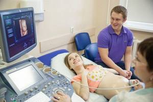
Which ultrasound is used to determine the sex of the child?
Over the course of 9 months, expectant mothers undergo ultrasound 3 times. If there are compelling indications, the gynecologist will refer you for additional examination. The study is carried out to assess the child’s condition, as well as to identify possible developmental pathologies. Using this method you can find out who will appear: a boy or a girl.
The first procedure is carried out at 12 weeks. It allows you to identify developmental defects. It is still impossible to find out the sex of the fetus at this stage. The baby’s development during this period is almost complete and the genitals can already be seen. However, not every device will allow a doctor to determine gender.
The second ultrasound is performed at 20-22 weeks. The genitals are formed, it will be easy to see them. During the 2nd screening, the size of the fetus, heart rate will be determined, and pathologies will be excluded. Ultrasound diagnostics will determine how the fetus is located in the uterus. If it is turned away from the outer wall, it will be difficult to determine the floor.
You can find out the sex of the baby using an ultrasound between 32 and 36 weeks. During the last screening, fetal pathologies are excluded, and the specialist also determines the sex of the baby, if this could not be done earlier. At the last ultrasound, the specialist receives important data before childbirth: weight, height, position of the child.
What should you not eat before your first blood screening?
2-3 days before the double test, you must adhere to a special diet. This is a very important requirement, since failure to comply will result in distorted results.
Before blood screening you need to avoid:
- fried and spiced foods;
- fatty meat;
- meat products (sausage, lard, bacon, smoked meats);
- canned food
Chocolate, nuts, chicken eggs, seafood, cabbage, rye bread, and legumes are also prohibited.
Cereals, butter and vegetable oil, pasta, honey, fresh and boiled vegetables are allowed. For meat dishes, boiled chicken is shown.
Are ultrasounds mistaken about the sex of the baby?
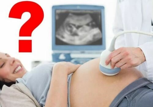
The accuracy of the first ultrasound examination is directly related to the qualifications of the doctor and the class of equipment used for diagnosis. You should not take sex determination data during the first screening as truth. In such a short period of time, the probability of error is too high. Often the doctor does not answer parents’ questions about the sex of the unborn baby and suggests asking them about this question in 4-6 weeks.
Which ultrasound is used to accurately determine the gender of the baby? With a probability of up to 90%, it is possible to determine who will be born no earlier than at 15 weeks. During the second screening, the doctor will be able to answer this question. By this period, girls' labia and penis can be clearly seen. After 20 weeks, an error in determining gender is unlikely.
According to gynecologists, the most favorable time to find out the sex of the child by ultrasound is from 23 to 25 weeks. During this period, babies can pull themselves up and straighten in the womb, allowing the doctor to examine the genitals. After 6-8 weeks, the baby will grow up and will take uncomfortable positions for diagnosis.
Many babies in the 3rd trimester are positioned head down. Due to lack of space in the uterus, the legs are bent. In this position, it is not easy to determine the sex, because the umbilical cord is located between the legs.
Expectant mothers should be aware that it is impossible to determine gender with 100% accuracy. Any doctor, based on the data obtained, can only give from 85 to 90% probability. Therefore, every tenth pregnant woman will give birth to a baby of the wrong gender.
We have found out which ultrasound is used to determine the sex of the child, now let’s talk about whether preparation is needed before conducting the study.
What types of screenings are there?
There are 5 types of screening during pregnancy:
- Ultrasonic;
- Biochemical;
- Immunological (testing a pregnant woman for a number of infections that potentially impair the development of the fetus. These include rubella, herpes, chicken pox, cytomegalovirus, toxoplasmosis, etc. Prescribed to everyone when registering for pregnancy);
- Cytogenetic (optional, performed according to indications);
- Molecular (detection of rare mutations occurring with a frequency of no more than 1%. Prescribed in unique cases)
Separately, invasive diagnostics are distinguished: chorionic villus biopsy (sampling a piece of tissue from the outer membrane around the embryo), amniocentesis (puncture to collect amniotic fluid), placentocentesis (biopsy of the placenta) and cordocentesis (sampling of the fetal umbilical cord blood). These are complex manipulations, they are necessary for cytogenetic research when searching for chromosomal diseases. It is carried out extremely rarely and only after a cascade of other examinations with disappointing results in order to confirm or exclude the diagnosis.
In addition to 5 types of screening, every 2-4 weeks pregnant women donate blood and urine for general clinical analysis.
Let's take a closer look at why each examination is needed.
3D ultrasound: pros and cons

Equipment plays a key role in the fetal examination process. Some pregnant women undergo 3D ultrasound. In three-dimensional photographs, you can easily examine sexual characteristics, as well as identify developmental pathologies. An addition for parents will be photographs that the specialist will provide after the procedure.
Not every clinic has such equipment, and not all doctors advise undergoing 3D ultrasound. This type of examination uses powerful ultrasound. Gender determination using such devices is not recommended in the first trimester. Just at this time, the formation of all vital organs and systems occurs; any negative influences from the outside may not have the best effect on the developing organism.
Why is screening needed?
To a greater extent, prenatal (prenatal) diagnosis is necessary for two purposes: to predict the risks of complications during pregnancy and to identify the preconditions for the development of fetal malformations.
Possible pregnancy complications detected during screenings:
- Preeclampsia is a pathological condition characterized by increased blood pressure, proteinuria (the presence of protein in the urine), possible edema and damage to the kidneys and other organs. More often develops in the second half of pregnancy.
- Eclampsia is a life-threatening condition for the mother and fetus, characterized by increased blood pressure, convulsions, coma and death (of the mother and/or fetus)
- Fetoplacental insufficiency (FPI) is a pathological condition of the mother (the placenta in particular), leading to hypoxia (lack of oxygen) and delayed fetal development with a high risk of intrauterine fetal death.
- Intrauterine growth retardation (IUGR) of the fetus is a pathological condition in which the fetus’s height and body weight lag behind its gestational (expected) age.
- Frozen pregnancy is a pathological condition in which the fetus stops developing and dies before 28 weeks.
Congenital malformations of the fetus recorded during screenings:
- Anencephaly is the complete or partial absence of the cerebral hemispheres, soft tissues and bones of the cranial vault.
- Microcephaly is a pronounced reduction in the size of the skull and brain while maintaining the normal size of other parts of the body.
- A cleft palate (“cleft lip”) is a tear/cleft in the middle part of the palate of the fetus, formed due to non-closure of the halves of the palate.
- Hydrocephalus is an excessive accumulation of cerebrospinal fluid in the ventricles of the brain.
- Congenital heart defects are defects in the structure of the heart and/or great vessels in the fetus.
- Many syndromes: Down, Patau, Shereshvsky-Turner, etc.).
After screening, pregnant women are divided into risk groups for the possible development of complications in the women themselves and their fetuses. Further management of pregnancy (diagnosis, treatment and obstetric care) differs for each group. Thanks to the screening system, the birth rate of healthy children has increased and the mortality rate of fetuses during gestation has decreased.
What determines the sex of the unborn child?
Many people already know well from their school biology course that all female reproductive cells contain an X chromosome, and male reproductive cells contain either an X or a Y chromosome. During conception, only one sperm can penetrate the egg. And the sex of the unborn child depends on which chromosome set it ends up with: if it is a sperm carrying an X chromosome, then it is a boy, if it is a sperm carrying a Y chromosome, then it is a girl .
Different sperm have their own characteristics. So Y-sperm are more mobile, faster, but live no more than 48 hours. X-sperm are larger and therefore, on the contrary, slower, but they remain viable for up to 5 days.
Thus, the possibility of fusion of an egg with sperm carrying a Y chromosome and the birth of a boy is higher during sexual intercourse the day before ovulation or in the next 12 hours after it. If sexual intercourse was several days earlier or later than ovulation, then there is a greater chance of a sperm with an X chromosome penetrating the egg and giving birth to a female child.
What other methods of determining gender exist?
Expectant parents often wonder whether there are more accurate methods for determining sex. Nowadays, such methods exist. First of all, this is a DNA test. It determines the gender of the fetus with a probability of up to 99%. This method can be carried out after 8 weeks of pregnancy. However, the DNA test has a big drawback - high cost.
There are also invasive methods of sex determination. They are carried out using special surgical instruments, which take a piece of skin or a small amount of fetal blood. Such an analysis determines with high accuracy not only gender, but also the presence of pathologies.
This type of diagnosis is carried out only if there are strong indications, as it is very traumatic. Parents' interest should not cause harm to the baby's health.
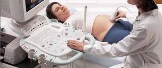
Ultrasound screening
Ultrasound examination (ultrasound) is a highly informative and harmless method with which doctors monitor the development and condition of the fetus from the earliest stages. A pregnant woman does not require special preparation before the procedure.
Expert class ultrasound machine from SAMSUNG
A total of three screening ultrasounds are performed:
- The first screening is at 10-13 weeks of pregnancy;
- The second screening is at 18-20 weeks of pregnancy;
- The third screening is at 30-34 weeks of pregnancy;
On an ultrasound examination, the fertilized egg in the uterus is visible from the 3rd week. The fetal heartbeat is detected by ultrasound from 4-5 weeks, and motor activity - from 7-8 weeks.
Now let's look at what is determined in each mandatory screening performed once every trimester.
Third screening
It is carried out at 30-34 weeks of pregnancy.
The diagnostic complex usually includes:
- General clinical analysis of blood and urine;
- Blood chemistry;
- Ultrasound of the uterus and fetus;
- Dopplerography - examination of fetal vessels using ultrasound techniques;
- CTG – cardiotocography. This is the study of fetal cardiac activity. It is prescribed for the risk of intrauterine hypoxia (oxygen starvation), and from 33 weeks once every 2 weeks until birth for all pregnant women.
- Screening for preeclampsia (measurement of blood pressure, detection of proteinuria).
Objectives of ultrasound in the third trimester:
- Checking for malformations and delayed fetal development;
- Assessment of fetal condition (motor activity, respiration and blood flow);
- Determination of the location, thickness and structure of the placenta.
By 36-37 weeks, the placenta stops growing, after which it remains at the same level or decreases. The condition of the placenta directly affects the health of the fetus.