The importance of the carotid arteries, located in the neck and chest, in the human body is difficult to overestimate. The carotid arteries participate in the general blood flow and play a critical role in supplying the brain with oxygen. Any problems related to the carotid arteries can have a negative impact on your overall health. Therefore, today there are quite a lot of examination methods, including ultrasound of the carotid arteries.
Ultrasound of the carotid arteries
Ultrasound of the carotid arteries reliably shows the condition of the blood vessels and is one of the safest and most painless technologies. Ultrasound waves do not harm the body, and the non-invasiveness of the method negates the risk of vessel damage. Therefore, this type of diagnosis of diseases and pathologies of the carotid arteries is the most common. There are practically no obstacles in the path of ultrasonic waves when examining the carotid arteries using ultrasound, which makes it possible to obtain reliable readings about the condition of the vessel. A pleasant advantage of this research method is its low cost.
MRI of neck vessels with contrast
Sometimes native MR angiography turns out to be uninformative. Vessels (even of small diameter) can be visualized more clearly by contrast-enhanced examination. It involves the injection into a vein of a drug that increases the vibrations of hydrogen atoms in water molecules in tissues and makes the latter more visible in photographs.
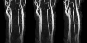
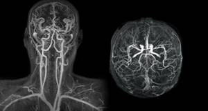
MR angiography of neck vessels
Contrast is required if abnormal vascular development or tumor changes are suspected. With its help, you can assess the condition of the veins and arteries and the speed of blood flow in them. The procedure for MR angiography with contrast is as follows:
- After standard patient preparation, native scanning begins.
- Once the shooting is complete, a contrast agent is injected into a vein in the forearm.
- Repeat the study.
The procedure takes 30-35 minutes. Contrast MR angiography is contraindicated in pregnant women. During lactation, you need to make a supply of breast milk for the next 2 feedings, which you will have to skip.
When is ultrasound scanning of the carotid arteries prescribed?
Duplex ultrasound scanning of the carotid arteries is the general name for ultrasound of the cervical and clavicular regions. Due to vague symptoms, diagnosis of many diseases is difficult, which is why ultrasound of the carotid arteries is prescribed. Indications for ultrasound of the carotid arteries may include headache, dizziness, blurred vision, increased or decreased blood pressure, increased cholesterol levels in the blood, as well as fainting and limb dysfunction.
These alarming symptoms can be signs of many other diseases. Therefore, why to do an ultrasound of the carotid artery is decided by the doctor, based on other signs of a possible disease. Ultrasound of the carotid arteries is also prescribed for vascular damage due to neck injuries, osteochondrosis and spinal injuries.
Indications for ultrasound examination
The carotid arteries are of great importance to human health. Timely detection of problems in the carotid artery will allow a specialist to prescribe treatment and save you from many problems. Carotid artery examination can be performed regularly as a preventive examination. Ultrasound is also used to diagnose the carotid artery in the following cases:
- blindness in one eye that lasts for a short period of time,
- noise in the head, frequent dizziness,
- migraines and headaches, the localization of which is impossible within a clear framework,
- temporary conditions in which paralysis of the body and difficulty speaking can be noted,
- sensation of flashing before the eyes,
- short-term loss of consciousness,
- sudden falls while maintaining consciousness.
If the carotid artery is not functioning properly, it can cause many problems. During an ultrasound examination of the carotid arteries, CityClinic specialists will identify any deviations in their functioning and determine the causes. The condition of our arteries depends on many factors, so as we age, they should be given special attention. Timely diagnosis and treatment of arteries will allow you to avoid serious health problems.
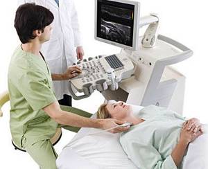
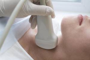
How is the carotid artery ultrasound procedure performed?
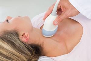
The ultrasound procedure of the carotid arteries does not cause discomfort or pain and is performed with the patient in a supine position. A special pillow is placed under the patient's shoulders so that nothing interferes with the view of the arteries. Throughout the procedure, the sonologist moves the scanner over the skin of the neck, lubricated with gel, and from time to time changes the sensitivity of the device.
There are several methods for ultrasound of the carotid arteries. However, it does not depend on this how ultrasound of the carotid arteries is done - in any case, the sonologist moves the scanner in the neck area, if necessary, telling the patient to turn his head or stretch his neck, examining the subclavian region.
Dopplerography
Using Doppler ultrasound, disturbances in blood flow in the vessels are detected, and ultrasound of the carotid arteries is one of the most effective diagnostic methods. If the blood flow is not visible, the sonologist can apply digital pressure on the artery to obtain more accurate information. In some cases, signals obtained using Doppler ultrasound can be converted into sound, this helps when examining seriously ill patients.
Duplex scanning
Duplex scanning is a combination of Doppler ultrasound, which determines blood flow disturbances, with traditional scanning, which shows the echostructure of the arteries. Duplex scanning of the carotid and vertebral arteries is the most common procedure, since it solves two problems simultaneously.
Triplex scanning
Triplex scanning of the carotid arteries involves research in three modes. The greyscale scanning mode provides information about the structure and shape of the artery, as well as any changes in it. Dopplerography
reveals the quality of blood flow inside the artery. And color Doppler mapping provides a color image that provides comprehensive information about any changes in the structure and blood flow of the artery.
Advantages of performing ultrasound scanning of the carotid and vertebral arteries in CELT
The multidisciplinary clinic CELT invites you to undergo a diagnostic test in accordance with international standards. Our name is well known in the capital, since we have been working in the domestic market of paid services since 1993. During this time, our specialists correctly diagnosed and restored health to thousands of patients, earning positive reviews and a good reputation.
Our diagnostic department is staffed by doctors of the highest category and candidates of medical sciences with at least fifteen years of experience. They know how to scan in such a way as to detect pathological changes even in the initial stages. This is important because timely treatment increases the chances of success, and especially when it comes to vascular diseases and cerebrovascular disorders.
Our specialists have two ultrasound devices in their arsenal: “Electric Medical System” (USA) and “Philips” (Holland), which allow duplex and triplex diagnostics in real time and provide high-quality images.
You can find out prices for services in this section of the website by going to the “Services and Prices” tab. To avoid misunderstandings, we ask you to contact our operators to clarify the cost: +7 (495) 788-33-88.
Decoding the results
The interpretation of the diagnosis of ultrasound of the carotid arteries is carried out by the attending physician based on the results obtained. However, a sonologist can also clarify the situation by recording the examination results at the end of the procedure. Typically, a sonologist comments on the visible echostructure of the vessel during scanning and blood flow.
The norm for ultrasound of the carotid arteries is that the blood flow in the internal and external arteries is equal in power, as well as the location of the carotid artery to the left of the aorta. The thickness of the vessel walls should not exceed 1.2 mm, and the pulsation in a healthy artery should be continuous.
What diseases can be detected by ultrasound of the carotid arteries?
Ultrasound of the carotid arteries allows you to examine not only the brachiocephalic arteries, but also simultaneously assess the condition of the vertebral vessels of the neck. The norm for ultrasound of the carotid arteries implies an internal and external diameter of the walls sufficient for good blood flow. Ultrasound of the carotid arteries most often reveals atherosclerotic plaques and kinks, which cause strokes. Ultrasound of the carotid arteries allows you to see even the structure of plaques, for example, those that have calcium impurities and are especially difficult to treat.
No less dangerous are blood clots and aneurysms, which are clearly visible using ultrasound. Stenosis visible on ultrasound of the carotid arteries may explain ischemic attacks, since blood flow worsened due to narrowing of the artery does not allow the brain to receive enough oxygen. An alarming symptom is a change in the level of the bifurcation of the carotid artery, visible on ultrasound. However, one must understand that ultrasound of the carotid artery can only identify these problems; subsequently, it is recommended to conduct a more in-depth study.
How is an MRI of the cervical spine done?
At the Magnit diagnostic clinic, scanning is carried out by appointment. During its registration, the patient must inform the medical staff about the presence of any metal structures in the body, namely:
- pins;
- knitting needles;
- endoprostheses;
- middle ear implants, etc.
The presence of products and metals with ferromagnetic properties in the body can affect the quality of the images. Clinic staff must be informed of the specific material the structure is made of. You can obtain this information from the medical institution where the operation was performed. You need to ask for an extract from there and take it with you to the procedure to show the radiologist.
MRA of head and neck vessels
You need to arrive for the MRI 5-10 minutes earlier than the appointed time so that you can fill out the documents without rushing. The study is preceded by a consultation at which the patient is asked about contraindications to the procedure, which are:
- first trimester of pregnancy;
- presence of cardiac pacemakers (pacemakers, defibrillators);
- implantation of an insulin pump;
- the presence of metal stents or hemostatic clips in arteries and veins;
- other ferromagnetic implants.
During the interview and introduction, the X-ray technician explains how an MRI of neck vessels is performed and the rules of behavior in the diagnostic room. Patients with claustrophobia are offered to undergo the procedure on an open tomograph. In case of increased anxiety, it is possible to use sedatives, but only as prescribed by a doctor. For patients with acute pain and neuropsychiatric diseases, MRI is performed under sedation or anesthesia in a hospital setting.
Preparing for the examination involves removing all metal objects. The patient must remove jewelry, glasses, hairpins, and items of clothing with such accessories. Electronic devices (phone, watch, hearing aid) remain outside the tomography room. The procedure for performing MRI of neck vessels is as follows:
- The patient is taken to a special room and placed on the tomograph platform. The conveyor moves so that the neck area is in the center of the device frame.
- The laboratory assistant fixes the position of the person’s head, places cushions for convenience, hands the emergency button and leaves the office.
- An x-ray technician watches the procedure from the next room through glass. He checks the functionality of the speakerphone, reminds the patient to remain still until the end of the study, and turns on the device.
During scanning, the tomograph makes sounds (clicking, humming) that may cause discomfort. At the Magnit diagnostic clinic, patients are offered headphones to listen to pleasant music during the examination. You can get regular earplugs instead. Preparation takes 5-10 minutes, scanning lasts 15-20.
Interpretation of photos of MRI images of neck vessels
The scanning results are layer-by-layer monochrome images of the area under study. They are deciphered by a radiologist. The specialist sees and records any deviations in tissue structure from the norm. All pathological changes will be recorded in the conclusion, and the images will be recorded on digital media.
Preparing results takes from 15 to 60 minutes. During this time, the patient can walk or rest in the waiting area. Study protocols are sent by email if you specify it when filling out the documents. You can pick up the original on any other day.
The radiologist describes what MRI shows in the arteries and veins of the neck, he gives explanations when presenting the results and recommendations on which doctor to contact. The specialist does not diagnose, make prognosis or prescribe treatment. If results are received by email, the patient will not be able to consult with a radiologist.