We will select a clinic or diagnostic center for free. Call 8(499) 116-77-86
Find a clinic
Sign up for an MRI
Magnetic resonance imaging (MRI) of the ankle provides high-quality imaging of the structures of the ankle, which helps doctors diagnose a wide range of diseases and conditions. Patients who have ankle pain may be referred for an MRI scan. Most often, an MRI examination of the ankle joint is prescribed to diagnose injuries to the bones, tendons, ligaments and cartilage of the ankle joint. Imaging using MRI can also diagnose tumors, arthritis, and joint infections.
Various tendons and ligaments in the ankle, including the Achilles tendon, provide flexibility and range of motion to the joint. Forceful movement of the ankle in an awkward position, outside of the typical range of motion, can cause damage to the ankle tendons and ligaments.
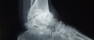
MRI of the ankle can detect both tears and complete ruptures of ligaments and tendons. For most ankle tendon and ligament injuries, MRI is the most informative and reliable diagnostic method. Thinning and changes in the structure of the cartilage tissue of the ankle joint of an involutional degenerative nature are also well visualized using MRI of the ankle. The bones of the ankle (including the talus and calcaneus) and the bones of the foot are clearly visible on MRI of the ankle and allow one to determine the presence of fractures. MRI of the ankle also allows you to determine the presence of bone bruises, the presence of dislocations or manifestations of osteoarthritis. In addition, an MRI of the ankle provides good information about the presence of tumors and blood accumulation in the soft tissues around or inside the ankle joint. MRI of the ankle can also evaluate the condition of the distal tibial or fibular tissue, as well as the muscles of the foot. The introduction of contrast allows for more detailed visualization of the structures of the ankle and identification of small morphological changes.
Main indications for MRI of the ankle:
- Tendon injuries
- Ligament injuries
- Cartilage injuries
- Fractures
- Tumors (soft tissue and bones)
- Infections
- Aseptic necrosis
- Pseudarthrosis or unconsolidated fractures
- Arthritis, arthrosis
- Tendonitis, tendinosis
- Presence of a tumor in the joint area
- Presence of pathology on radiography
- Congenital anomalies
- Pain, swelling, redness in the ankle area
- Decreased range of motion in the joint
- Unclear genesis of pain in the ankle joint
- Preparation for surgical treatment
What does an MRI of the ankle ligaments show?
Magnetic resonance imaging allows us to evaluate some pathological processes. Damage in this area occurs quite often.
MRI can detect arthrosis, inflammatory lesions in rheumatic pathologies, capsule ruptures, developmental anomalies and oncology. Diagnostics allows us to identify the causes of chronic pain, swelling, and stiffness of movement.
After the procedure is completed, the patient receives pictures with a description. When undergoing a procedure on the direction of a doctor, you must show him the conclusion. Performing an MRI allows the doctor to assess the severity of the injury, its consequences, and prescribe the correct treatment, which will quickly and effectively relieve all unpleasant symptoms.
Preparing for an ankle MRI procedure
The patient can use special disposable clothing or wear his own clothing during the procedure if it is loose and does not have metal fittings.
Eating during an MRI of the ankle is not regulated, but it is better to refrain from eating several hours before the examination if an examination with contrast is planned. If contrast administration is planned, the MRI technician will need information about the presence of an allergy to the contrast agent or bronchial asthma.
The contrast agent most commonly used for MRI studies contains a metallic substance (gadolinium). And although gadolinium very rarely leads to complications, unlike iodine contrast (which is used in CT studies), its administration is nevertheless undesirable in the presence of serious somatic diseases, especially chronic kidney diseases. If MRI of the spine is performed on women, then information about the presence of pregnancy is required. And although long-term studies have not shown any harmful effects on the fetus, MRI is not recommended for pregnant women, especially in the first trimester. Carrying out MRI with contrast is possible only in exceptional cases, according to clinical indications. If claustrophobia is present, MRI examinations are recommended to be performed using open-type machines. During an MRI examination, young children require sedation so that the child can lie still during the examination. Sedation is performed by an anesthesiologist.
All objects containing metal must be removed before MRI is performed. These are items such as:
- Jewelry, watches, credit cards and hearing aids that may be damaged
- Pins, hairpins, metal zippers and similar metal objects that may distort the MRI image
- Removable dentures containing metal
- Pens, pocket knives and glasses
- Body piercing
MRI is contraindicated if the patient has implants or implanted electronic devices:
- Cochlear implants
- Some types of clips used on cerebral aneurysms
- Some types of metal coils placed within blood vessels (stents)
- Artificial heart valves
- Implanted infusion pumps
- Implanted electronic devices, including defibrillator, pacemaker
- Joint endoprostheses (with metal content)
- Implanted nerve stimulators
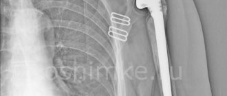
- Metal pins, screws, plates, stents, or surgical staples
- Metal parts in the human body (such as bullets or shrapnel), as a strong magnetic field can cause metal objects to dislodge and damage tissue. And therefore, in such cases, it is necessary to conduct radiography before the MRI examination.
Parents accompanying children must also remove all metal objects and report the presence of objects containing metal in the body.
Anatomy of a stretch
Ligaments are made of strong connective tissue. They usually stretch a certain amount and then return to their normal state. A severe stretch actually tears the ligaments, at which point you may feel or hear something like a pop or crack.
An ankle sprain is a common injury. Inversion-type collateral ligament injuries account for approximately 85% of all cases. According to statistics, at the beginning of 2020, the highest incidence rate was among those aged 15 to 19 years. Half of all ankle sprains occur during sports, such as basketball (41.1%) and soccer (9.3%). And in relation to all sports injuries, it occurs in 77-83% of the total number of cases.
The most common risk factor is a previous sprain. It can compromise the strength and integrity of stabilizers by interrupting sensory nerve fibers. Gender, height, weight, limb dominance, postural sway, and foot anatomy are intrinsic risk factors that may also influence the potential for injury. External causes include how protective systems are attached, type of footwear, duration of competition and intensity of activity.
In addition to injuries in sports, an ankle injury can be caused by simply stepping on your foot incorrectly. For example, when a person stumbled and stood not on the plantar part of the foot, but on its lateral surface. This can happen when walking down stairs or running.
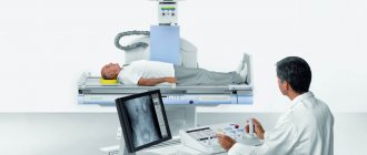
Procedure for performing MRI of the ankle joint
A traditional MRI machine (closed type) is a large cylindrical tube surrounded by a magnet. During the examination, the patient lies on a movable table, which moves to the center of the magnet. There are also open-type MRI machines, where the magnet does not completely surround the patient, but is open on the sides.
Studies on open-type devices (and they are predominantly low-field) are useful for studying patients with claustrophobia or heavy weight. Recently, open-type devices with a high field (1 or more Tesla) have appeared, which allow obtaining high-quality images, in contrast to the main models of open MRI, where the magnetic field is low and the image is of lower quality.
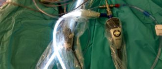
When performing an MRI of the ankle, a coil is placed on the ankle joint. During the procedure, the patient must lie still for a certain time (on average 30-40 minutes). When studying with contrast, the duration of the study will be longer.
The MRI procedure is completely painless and, however, some patients may experience a feeling of warmth in the area where the study is performed, which is a normal tissue reaction to the magnetic field. Typically, the patient is alone in the MRI room during the examination, but there is two-way audio communication between the radiologist and the patient and the doctor can see the patient. After undergoing an MRI procedure, the patient does not need time to adapt.
Recently, it has become possible to perform MRI of the ankle joint using small-sized devices, in which only the joint is examined and the whole body is not in a magnetic field, especially since in such devices the magnetic field is quite powerful and allows one to obtain a high-quality image.
Relationship between MRI and metal
The MRI machine is a huge magnet that, like all magnets, attracts metal elements. What does this mean? Metal elements can be located both inside (catheters/prostheses) and outside (piercings/decorative elements on clothing). Firstly, they can distort the results of the study by affecting the magnetic field. The device will record incorrect indicators, which will interfere with the formation of an image and reduce the information content of the MRI to zero. Secondly, the patient may experience extremely unpleasant sensations when metallized particles begin to be attracted to the magnet. The intensity of the pain varies depending on the individual characteristics of the patient and the amount of metallic substance. One person may feel a slight tingling sensation, while another may experience unbearable pain, after which they will be unable to continue scanning.
What is the connection between metal and tattoos? For another 20 years, tattoo inks contained metallic substances. In modern tattoo parlors, this practice has long been abandoned, but people who got tattoos before this time cannot undergo an MRI. During diagnosis, a magnet (magnetic resonance imaging) will begin to attract metal particles, which will cause extremely unpleasant symptoms in a person - from mild itching to serious pain. Be sure to report the presence of a tattoo to your doctor and the x-ray technician performing the tomography. The specialist will assess the possible risks, check the concentration of the metallized substance in the tattoo and, if necessary, select an alternative diagnostic method.
Benefits and Risks
Advantages
- MRI is a non-invasive imaging method that does not use ionizing radiation.
- MRI is a very valuable method for diagnosing a wide range of conditions, including diseases and injuries of tendons, ligaments, muscles, cartilage and bone pathologies.
- MRI can help determine which ankle injury patients require surgical treatment.
- An MRI can help diagnose a bone fracture when X-rays and other imaging methods are unable to confirm the diagnosis.
- MRI can detect abnormalities that may not be visible with other imaging techniques.
Risks
- MRI poses almost no risk to the average patient when proper safety precautions are followed.
- If sedation is used, there are risks of oversedation.
- Although a strong magnetic field is not harmful in itself, implanted medical devices that contain metal may become damaged or malfunction during an MRI procedure.
- There is a very small risk of an allergic reaction if contrast material is injected. Such reactions are usually mild and easily controlled with medication.
- Nephrogenic systemic fibrosis is now a recognized, but rare, complication of contrast-enhanced MRI and is thought to result from the injection of high doses of gadolinium, which is the basis of the contrast agent, in patients with very poor renal function. Careful assessment of renal function prior to contrast administration minimizes the risk of this very serious complication.
- Manufacturers of intravenous contrast recommend that breastfeeding mothers avoid breastfeeding for 24 to 48 hours after an MRI scan with contrast.
Contraindications
There are two categories of contraindications to MRI:
- Absolute – in this case, research is strictly prohibited. This is the first third of pregnancy, installed pacemakers, middle ear implants, vascular clips and other devices, metal foreign bodies.
- Relative - the procedure is possible at the discretion of the doctor in case of urgent need or under certain conditions. This is pregnancy in the second half of the term, the presence of artificial valves on the heart, claustrophobia, panic attacks, involuntary body movements.
Limitations for MRI of the ankle
High image quality can only be obtained if the patient lies still during the examination.
If the patient is large, it is better to conduct the study using an open MRI.
MRI examination of the ankle joint is best performed on a high-field (closed-type) device, since the image quality is better than on low-field devices, especially if visualization of ligaments and tendons is necessary.
In some cases, when it is necessary to obtain a detailed image of bone tissue, MSCT can be performed, which visualizes bone tissue better than MRI.
Diseases of the lower extremities
The legs perform the function of support and movement. The lower limbs have a complex structure. The main anatomical zones are the thigh, lower leg and foot. Birth defects and serious injuries to any area of the leg are fraught with irreversible consequences, including loss of the ability to move and disability.
The lower limbs consist of the following areas:
- gluteal;
- front and back of the thigh;
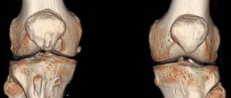
- knee;
- ankle joint;
- inner and outer areas of the ankle;
- dorsum of the foot and sole.
The leg skeleton consists of bones connected by joints. A complex system of muscles includes muscles, fascia, ligaments, tendons, etc. The trophism of soft tissues is provided by veins and arteries, the functions of the legs are provided by nerve fibers.
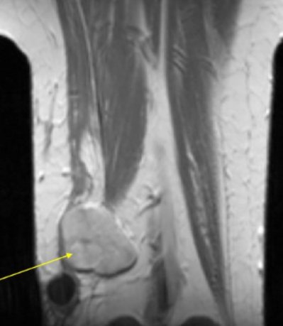
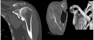
Liposarcoma (indicated by an arrow) of the lower thigh
The characteristics of the area under consideration predispose to:
- injuries of a traumatic nature (fractures, sprains and ruptures of ligaments, penetrating wounds, etc.);
- degenerative-dystrophic lesions of joints (arthrosis);
- bone diseases (osteoporosis, etc.);
- vascular pathologies (atherosclerosis, venous insufficiency, varicose veins, thrombosis);
- muscle diseases (myopathies, myositis, dystrophies);
- damage to neuromuscular junctions;
- infectious and inflammatory processes of any localization (tuberculosis, phlegmon, abscesses, etc.);
- benign and malignant tumors.
Pathological changes can affect any structural element of the limbs. Causes: genetic disorders, developmental abnormalities, infectious, autoimmune diseases, excessive stress, injuries, etc.
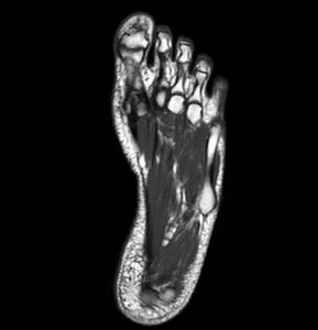
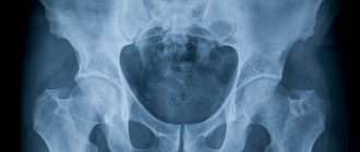
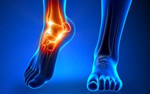
MRI image of the foot
How is an MRI performed?
There is no need to prepare for an ankle examination without contrast. When undergoing a procedure with contrast, it is advisable to come to the clinic on an empty stomach (at least 4-6 hours after your last meal).
Before scanning, you need to remove all jewelry with metal objects from your clothes. It is worth wearing comfortable clothes that will not fit your legs and that can be easily rolled up.
The patient lies down on a special tomograph couch, the lower leg is fixed with special belts. The couch moves into the tunnel and the machine begins to operate. A series of photographs are taken - in one or different projections at the discretion of the attending physician.
An MRI is completely painless for a person; the only discomfort may be from prolonged immobility and the noise of the operating device. To prevent its crackling and clicking noises from disturbing you, you can use earplugs or headphones. During scanning, communication is constantly maintained between the patient and medical staff using an intercom.
When all the pictures are taken, the procedure is completed (its duration is about 30-50 minutes), the doctors only have to analyze the results obtained - describe the examined area, the anatomical features of the joint and the detected pathologies. Study data is recorded on a disk or flash memory card. In this format, it is convenient to analyze them, compare them with previous results, and send them to doctors in other clinics (for example, to request a second opinion).
What can be seen in the photographs
After MRI, images of the lower leg can be used to examine in detail the bone and cartilage tissues, ligaments, nerve endings, soft tissues and blood vessels, and MRI of the ankle joint clearly shows the cavities of the corresponding tissues. The images reflect the presence of the following pathologies:
- heel spur, plantar fasciitis, inflammation;
- the presence of a tumor - the technique shows disorders in the early stages, helps to establish the type of tumor (malignant/benign), and diagnose the involvement of certain structural formations in the process;
- infectious lesions of soft tissues connected by tendons, the method shows the stage of spread of the purulent process, the degree of damage to articular and bone tissues;
- arthritis, arthrosis, bursitis;
- flat feet;
- Sever's disease - occurs as a result of microtraumas of the heel bone, the disease is common among athletes;
- congenital anomalies of the heel bone and finger structure;
- diabetic foot is a complication of diabetes mellitus;
- foreign bodies - the study shows the location of the objects, the doctor can determine the surgical approach from the images.
Approximate cost of diagnostics
The cost of MRI of the ankle depends on the region, the pricing policy of the medical institution, and the quality of the equipment. The average price of an examination varies between 2500-4000 rubles; it must also be taken into account that the price may increase due to the inclusion of additional services (injection of a contrast agent, printing of an image, recording on an electronic medium). Some clinics provide a connection to a personal account, in which images are added after each re-examination.