Formation of the fetal heart by week
The fetus needs autonomous blood circulation, so first of all its cardiovascular system is formed. Already at the 3rd week, an ultrasound will show the rudiments of the organ, and after 5 weeks, that is, by the 8th week of the embryo’s life, the heart will be formed.
In the 3rd week, the heart looks like a hollow tube, which will soon begin to beat.
By the 4th week, the tube begins to bend, and on the 5th week the first septum appears - and now the mother can hear the baby’s heartbeat for the first time.
By the 6th week, the interventricular septum is formed, followed by the interatrial septum.
By the 8th week, the fetal heart is formed, like that of an adult.
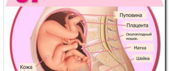
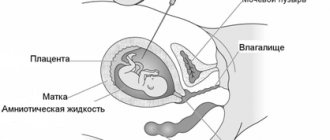
The difference is that the fetus does not breathe on its own - blood comes to it from the mother. And this will happen until the baby is born and takes his first breath - only then his blood circulation will be divided into 2 circles - large and small. Then the oval window will also close - the hole through which blood flows from one atrium to another while the baby is in the mother’s stomach.

From the moment of conception, a unique person who has never existed before appears. Mothers are looking forward to hearing the cherished heartbeat of the baby on an ultrasound scan! It turns out that he is ready to give this joy to his mother very soon after he was born. Let's reveal the secret of intrauterine development!
Previously, it was believed that the embryo’s heartbeat appears in the third week after conception, but according to recent studies, it turned out that the heart beats already on the 16th day. The study was conducted at the University of Oxford. The scientific work was published in the journal “Life” (eLife).
This discovery makes a significant contribution to the treatment of congenital heart disease. Based on the data obtained, researchers will be able to study the initial functions of the main organ of a developing child.
“Every day, many children are born with heart defects,” scientists say. “Knowledge based on new scientific findings will help health care providers treat these children and also prevent heart attacks later in life.”
How the study was conducted
The scientists concluded that, due to a number of similar parameters, it is possible to compare mouse pregnancy with a human embryo. The heart in rodents in the womb begins to beat at the end of the first week after the start of its development, and this is comparable to the 16th day after conception in humans. This calls into question the early conclusions of specialists, indicating the onset of fetal heartbeats only at 21 days.
The study was conducted on laboratory mice, which make it possible to compare pregnancy with a human embryo due to a number of similar parameters. In particular, the heart of rodents in the womb begins to beat at the end of the first week after the start of its development. Thus, in humans, this activity begins on the 16th day, which casts doubt on the early conclusions of specialists indicating that the fetal heartbeat begins only on the 21st day.
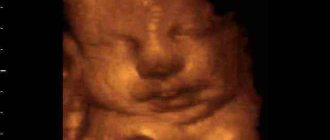
2D ultrasound at 3 weeks of pregnancy: fetal heartbeat
The embryo's heart is already pulsating on the 16th day after conception!
Great scientists from all over the world claim that it is at the moment of fusion of an egg with a sperm that a unique personality is formed, with unique genetic material, and that this little embryo is already a person. This small cell already contains all the genetic material: from the shape of the nose to temperament.
After ovulation, the egg enters the fallopian tube, where fertilization can occur. For this to happen, the sperm needs to penetrate the egg. A one-cell embryo is formed - a zygote with a normal human karyotype of 46 chromosomes. He is already unique from the point of view of genetics - already at this stage it is laid down what traits and characteristics this person will have.
Under the influence of the corpus luteum hormone progesterone, its mucous membrane thickens 3–4 times, swells, and becomes loose. Many additional blood vessels develop in it, and glands grow.
By the 4th day, when the embryo reaches the uterus, it is a lump of 8–12 cells. By the middle of the 6th day, the embryo consists of more than a hundred cells. By the time the baby enters the uterus, it is prepared to receive him.
By the 7th day after fertilization, the embryo changes its structure again. Now it is no longer a cluster of cells, but a vesicle - a blastocyst.
10–14 days. The embryo announces its development and existence through the chemical composition of the placenta and placental hormones, and the woman stops menstruating.
From the 16th day, the second, or actually embryonic, period of the child’s intrauterine development begins, which ends by the 13th week. Changes in the embryo grow like an avalanche, but following a clear plan. Here is a brief chronology of events.
Day 20 The formation of the brain, spinal cord and nervous system occurs.
Your baby is growing and developing quickly! And in less than nine months you will be holding him in your arms!
Salekh Yulia Vladimirovna, Obstetrician-gynecologist, Krasnodar:
You often hear the following expression: “Oh, my time is still very short, there is no heartbeat yet, which means that all this can be interrupted without any special consequences for yourself.” This is fundamentally wrong. A person is given a soul at the moment of conception, and not at the moment when a heartbeat appears on an ultrasound. Abortion is murder, and there can be no other expressions or apologies for it. At any stage, starting from the smallest stage, abortion is harmful to a woman’s health.
There is no need to chase men to fly into space and find incredible happiness there, because being a mother is the greatest happiness and purpose in a woman’s life.
Source
When do you start tracking your heart rate?
The baby’s heartbeat during pregnancy is monitored as part of routine screenings:
- In the 1st trimester - from the 11th to the 13th week;
- In the 2nd trimester - from the 18th to the 20th week. At this stage, developmental disorders are identified;
- In the 3rd trimester - from the 28th to the 35th week, when the heart and the arteries that are connected to it are assessed.
On an individual basis, an extensive diagnosis of the baby’s heart is necessary. For example, if there is a suspicion of a heart defect, with an enlarged cervical fold in the early stages, with disturbances in the child’s heartbeat during pregnancy, or with abnormal development of other organs.
Particular attention is paid to expectant mothers with heart disease, diabetes and those who have suffered infectious diseases during pregnancy or are taking anticonvulsant therapy.

Baby's heart rate by week
Depending on the trimester and phase of activity of the fetus, its heart pulsates at different frequencies. Gynecologists focus on the following indicators of the baby’s heartbeat in the womb:
- 4th - 6th weeks - in the early stages, heart rate is 80 - 85 beats per minute;
- 6th week - from 100 to 135 beats;
- 7th week - 115 - 130;
- 8th week - no higher than 170;
- 9th - 10th weeks - from 170 to 190;
- from 11th to the birth of the baby - an average of 150 beats.
The baby's heartbeat during pregnancy, starting from the 2nd trimester, becomes stable. A pulse of 140 - 160 beats per minute is considered normal. But any fluctuations in one direction or another indicate a serious condition of the fetus, for example hypoxia. In the 3rd trimester, the baby's heartbeat in the abdomen may fluctuate, but not by more than 10 units (+/-).
What pathologies does heartbeat help diagnose?
Heartbeat is the most important indicator; it is used to determine whether pregnancy is developing. If, with a heart size greater than 8 mm, the heartbeat cannot be heard, additional studies and careful monitoring are indicated, perhaps the pregnancy is frozen. Other pathologies can be determined by the nature of the heartbeat:
- When high fetal heart rate appears over a long period of time, fetoplacental insufficiency is diagnosed. This is a problem of blood supply to the fetus, often chronic.
- If during the third trimester the heartbeat becomes too slow, the child is said to be exhausted, and a decision is made to perform an emergency delivery.
- The pulse can be used to determine whether fetal hypoxia is present and whether it is life-threatening.
- A muffled heartbeat indicates low or polyhydramnios and breech presentation of the fetus.
- Echocardiography determines pathologies in the structure of the heart muscle, for example, defects in the chambers of the organ.
When symptoms of pathology appear, a diagnosis is not made. The doctor must perform a repeat ultrasound, assess the fetal heartbeat, and do a CTG. If the diagnosis is confirmed, then medications are prescribed to facilitate the functioning of the fetal heart. The main task for the mother and the doctor is to monitor the child’s condition; if necessary, it is important to take timely measures. A caesarean section or emergency delivery may be indicated.
Methods for monitoring fetal heart rate
Monitoring a child’s heartbeat during pregnancy allows you to assess his well-being and development, identify risks in time and, if possible, correct them.
In each trimester, gynecologists use different methods to assess the baby's heartbeat. Also, modern technologies allow expectant mothers to listen to the hearts of their babies at home.
Listening to the baby's heartbeat in the clinic
Gynecologists use the following methods:
- Ultrasound examination, or ultrasound. The most popular method that allows you to calculate your heart rate in the early stages. Using the device, the structure of the heart is assessed, abnormalities in development are identified, and sounds are listened to. There are 2 types of examination: vaginal (the sensor is placed in the woman’s vagina) and abdominal (the sensor is moved along the patient’s abdomen). The woman is lying on her back at this time. The procedure takes up to 20 minutes;
- Auscultation with a stethoscope. The device allows you to determine the approximate frequency and clarity of the baby’s heartbeat and his presentation. Diagnostics is informative only in the 3rd trimester. If the pregnant woman has polyhydramnios, oligohydramnios, or is overweight, this assessment method is not suitable. The procedure goes like this: the woman empties her bowels and bladder in advance, lies down on the couch and bends her knees. The doctor determines the position of the fetus and the listening point, then applies the wide side of the stethoscope to the patient’s abdomen and the narrow side to his ear;
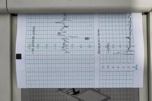
- Cardiotocography, or CTG. Used from the 32nd week. In addition to the baby’s heartbeat during pregnancy, the device evaluates the movement of the fetus and determines oxygen starvation. This allows you to immediately take measures to improve the baby’s condition. The first CTG is performed at the 32nd week, the second - before birth. In this case, the frequency of uterine contractions, fetal activity and sleep phases are monitored. If necessary, diagnostics are carried out more often. The procedure is carried out as follows: the woman lies on her side or half-sitting, and a sensor is attached to her stomach. During the procedure, the heartbeat is audible to the naked ear. It lasts 40 minutes.
If heart defects are suspected, echocardiography, or echocardiography, is performed. Ultrasound examination allows us to study the structure of the heart and blood flow. Informative in the 2nd - 3rd trimester. The procedure takes 25 - 40 minutes. The woman is asked to lie on her back, her stomach is lubricated with gel and a sensor is attached. If the procedure is prescribed in the early stages, the sensor can be inserted vaginally.
Devices for assessing heart rate at home
If you are overly worried about your baby's condition and constantly think about his well-being, purchase a portable device to measure the baby's heartbeat during pregnancy. Manufacturers offer several options:
- Portable fetal doppler. You can listen to your baby’s heart at any time and make sure everything is okay with him. Or react instantly if something goes wrong. The device can be used from 12 weeks; models with particularly sensitive sensors can be used from the 10th week.
- Stethoscope. At 18 - 20 weeks, you can listen to the baby's heartbeat using this simple but informative device. The best time to listen is before bed or 1 - 2 hours after eating. Don’t be alarmed if you don’t hear your baby right away—move the stethoscope over your belly in different directions. Give preference to devices from well-known manufacturers.
In later stages of pregnancy, you can download a mobile application. Some versions even allow you to make an audio recording of your baby's heartbeat during pregnancy.
At what stage can you hear the heartbeat of an embryo?
How many times during pregnancy is the fetal heart rate determined? If a woman has no indications for additional studies, then she will have 3 scheduled ultrasounds within 40 weeks. They are held on the following dates:
- 11–14 weeks;
- 18–21 weeks;
- 30–34 weeks.
Usually the first heartbeat of the fetus is determined by ultrasound. If there are grounds for early diagnosis, the procedure is performed earlier. After the first trimester, the gynecologist must listen to the fetal heartbeat at every appointment.
Deviations from the norm: when to sound the alarm¹
If your baby's heart beats 1 - 2 units slower or faster than normal, there is no reason to worry. You should sound the alarm if the fluctuations are too significant, the child’s heartbeat has sharply increased/decreased, or the condition is persistent.
Decreased heart rate, or bradycardia
There are many reasons for a decrease in heart rate. Only doctors will identify the true cause of bradycardia in a particular fetus. Here are possible reasons for a baby's heart rate to decrease during pregnancy:
- Mom's unhealthy lifestyle. The fetus suffers greatly if the mother smokes and drinks alcohol during pregnancy, does not walk in the fresh air, or does not move;
- Mother's health status. Vitamin deficiency, anemia, pulmonary and infectious diseases, heart defects, cardiovascular diseases can cause pathologies in the fetus;
- Psychological state of a woman. The expectant mother should not be nervous, because stress has a detrimental effect on the development and health of the child.
Heart rate may decrease due to taking medications, Rh-conflict, multiple pregnancy, severe toxicosis, premature placental abruption, oligohydramnios, polyhydramnios, or when the umbilical cord is entangled. All these conditions need to be either excluded or confirmed and urgent action taken.
If the pulse is non-monotonic and rhythmic, bradycardia does not threaten the baby’s life - it is enough to correct it with drugs.
If persistent bradycardia is diagnosed, it almost always indicates a pathological condition of the fetus: hypoxia, metabolic acidosis, or congenital pacemaker pathology.
Increased heart rate, or tachycardia
If the pulse reaches 170 - 200 beats per minute, the child’s heart rate is said to be increasing. This condition is caused by the same reasons as bradycardia. The child is simply trying to cope with the situation differently.
In addition to the reasons listed above, tachycardia is caused by severe bleeding, cardiovascular pathologies, endocrine diseases and dehydration against the background of toxicosis in the mother.
An increased heart rate of a child does not directly threaten his life, but the reasons that caused it can lead to serious consequences. Timely drug therapy can correct the condition. If it worsens and cannot be corrected, the woman is offered to induce labor early.
Persistent tachycardia with a heart rate above 180 beats per minute is dangerous for the fetus. It can lead to heart failure, dropsy, or fetal death in the womb.
Monotonous heartbeat of a child
Depending on the phase of activity, the baby’s heart beats at different frequencies. If he is sleeping, the pulse slows down and becomes more monotonous; if he wakes up and moves, the pulse quickens and becomes spasmodic. These “slides” are clearly visible on CTG: every movement of the fetus leads to an increase in heart rate. The indicators do not change if the fetus is deeply asleep. In this case, they try to wake him up: they ask his mother to take a walk, stroke his belly, and eat something sweet. But if these attempts do not lead to anything, and the baby continues to sleep, this is a very alarming sign, since it may indicate severe hypoxia.
Irregular heart rhythm, or arrhythmia
This condition is characterized by irregular and irregular contractions or changes in heart rate in the form of tachycardia or bradycardia.
If the heart rate is above 180 or below 100 beats per minute for a long time, this is a dangerous condition for the baby.
Irrhythmic contractions are a sign of pathology of pacemakers and pathways.
Arrhythmia appears against the background of structural defects, ischemia, electrolyte imbalance, inflammatory processes, and acute viral infections.
Determining gender by heartbeat
Previously, when there were no hardware diagnostic methods, gynecologists determined the gender by the child’s heartbeat. Modern doctors and mothers trust ultrasound examination more, which can almost accurately determine the sex of the unborn baby already in the 15th - 16th week. However, there are still gynecologists who use the predecessor method in practice.
According to the approach, girls and boys' hearts beat at different frequencies. For boys it is slower and more clear - from 130 to 150 beats, for girls it is faster and more subdued - from 150 to 170 beats.
However, this technique does not take into account the dependence of the pulse on the phase of activity and the position of the baby, the formation of his cardiovascular system, the condition of the mother, as well as differences in the baby’s heartbeat from week to week. All these factors affect heart rate, so they must be taken into account. Therefore, the method of determining sex by heartbeat cannot be considered reliable.
If you want to find out who will be born in your family, wait until the second scheduled screening - in most cases, at this time the ultrasound doctor can already clarify.
Sources:¹https://journals.lww.com/clinicalobgyn/Fulltext/2020/09000/Physiology_of_Fetal_Heart_Rate_Monitoring.18.aspx