What is barium used for?
X-ray examination of the esophagus with a contrast agent is an indicative and highly informative method of visualizing the condition of the walls of the organ. This examination method makes it possible with a high probability to identify pathologies in various parts of the esophagus, assess the stage and intensity of the disease and identify its localization.
On an empty stomach, the esophagus is a tube with collapsed walls and is poorly visualized on an x-ray. Data obtained with natural contrast of the esophagus do not allow us to reliably assess the condition of its walls and identify diseases of various localizations.
If there is air in the esophagus, only its contours appear unclear on the image. This is also not enough to make a correct diagnosis in many cases.
The most revealing x-ray method is an x-ray of the esophagus with contrast - a barium suspension. This substance delays x-rays and appears as a shadow on an x-ray. What does a barium x-ray of the esophagus show? This image is more revealing and provides more information compared to a conventional x-ray examination.
Barium follows the walls of the esophagus and creates the most indicative picture of the condition of the esophagus, therefore, when many diseases are suspected, experts prefer this diagnostic method.
It should be noted that the patient's intake of a thick suspension of barium sulfate makes it possible to leisurely examine various parts of the esophagus in various projections and positions. In this case, it is possible not only to perform fluoroscopy, but also to perform video magnetic recording.
About the procedure
Before performing an X-ray of the stomach, the patient requires careful preparation, which is described in detail below. The study itself consists of several stages, during which the patient first stands in front of the device screen, then lies on a special table.
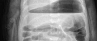
- First, a survey X-ray of the abdominal cavity is taken to identify the most noticeable and dangerous pathologies - intestinal obstruction, perforation of the stomach or intestinal wall.
- The following steps are carried out using a barium preparation. First, the patient drinks one or two sips of the solution, after which the equipment records the passage of the contrast agent through the esophagus to the stomach.
- The patient drinks a contrast solution to tightly fill the esophagus to detect the slightest disturbance in the structure of the mucous wall. In order to record them, several photographs are taken in different projections.
- The patient is placed in the Trendelenburg position with the head lowered below the level of the legs, and several more photographs are taken to study the lower esophageal sphincter
- The next step is an X-ray of the stomach with barium, which is performed in standing and lying positions on the back, and then on the side.
The study lasts for a total of about one and a half hours. After its completion, it is recommended to drink at least one and a half liters of water to speed up the process of removing the contrast agent from the body.
Indications for X-ray examination with barium
There are a number of indications that require an X-ray examination of the esophagus with barium. If there are suspicions regarding the disease and diagnosis, this type of diagnosis can also be performed. We list some conditions in which it is recommended to take an X-ray of the esophagus with barium.
Swallowing disorders: feeling of retention of a bolus of food, pain when swallowing food
Various disturbances in the act of swallowing, the appearance of a lump in the throat, pain when swallowing food may indicate an obstacle to the natural flow of food during the act of swallowing. The cause of this pathological condition may be the presence of a foreign body in the esophagus, thickening of its walls, or the growth of neoplasms of various origins.
An X-ray of the esophagus with barium will clearly show the location of the obstruction in a certain part of the esophagus, and will also make it possible to assess its size and shape, which will help specialists in making a reliable diagnosis and selecting rational treatment.
Chest pain not associated with pathology of the respiratory system and cardiovascular system
If there is a history of pain in the chest and, with the help of differential diagnosis, it was found that they are in no way related to the cardiovascular and respiratory systems, then it is possible to assume a pathology of the esophagus. Since the esophagus is located directly behind the trachea and descends all the way to the diaphragm and stomach, the cause of pain can very likely be localized there.
Diseases such as hiatal hernia, esophagitis and peptic ulcers can be accompanied by pain and are detected using contrast-enhanced radiography of the esophagus.
Repeated vomiting, heartburn, belching
If the clinical picture contains symptoms such as repeated heartburn, vomiting and belching, then this may indicate a violation of the motor function of the esophagus, as well as its anatomical sphincters or the presence of various pathologies of the walls. Thus, this symptomatology can occur with diverticula of the esophagus, dyskinesia and achalasia. All these diseases can be detected using a barium X-ray of the esophagus.
X-ray of the stomach
X-ray examination of the esophagus, stomach and duodenum is a contrast study aimed at determining the presence or absence of pathology in the upper digestive tract, and in the presence of pathology, at clarifying the nature of the identified changes. X-ray examination of the esophagus, stomach and duodenum is a completely safe, non-invasive procedure. Allergic reactions to contrast media are extremely rare due to the fact that barium sulfate is not absorbed from the digestive tract into the blood. The radiation dose received during the research using a modern digital device is extremely small and does not lead to the development of negative consequences.
The study is carried out in both vertical and horizontal positions of the patient (on the stomach, on the back, on the left and right side). In some cases, the study is carried out in the Trendelenburg position - on the back with the head end of the table lowered (up to -30 degrees). During the examination, the patient, at the command of the radiologist, takes a contrast agent - an aqueous suspension of barium sulfate. The contrast agent taken can range from a thick consistency to the consistency of liquid sour cream. The volume of accepted contrast is from 150 to 300 ml. Swallowing a contrasting suspension, which tastes like chalk, usually does not cause much difficulty. Indications for fluoroscopy of the esophagus, stomach and duodenum are the following clinical symptoms:
- Suspected gastroesophageal reflux.
- Abdominal pain.
- Discomfort in the epigastric region.
- Digestive disorder.
- Nausea.
- Vomit.
- Symptoms of bleeding from the upper digestive tract.
- Anemia.
- Weight loss.
X-ray of the esophagus, stomach and duodenum is informative for the following diseases and pathological conditions:
- Suspected or known gastritis or duodenitis.
- Peptic ulcer of the stomach and duodenum.
- Hiatal hernia.
- Varicose veins of the esophagus.
- Suspected perforation (usually a plain radiography of the abdominal cavity is sufficient; a contrast study is carried out with a water-soluble contrast agent).
- Tumors of the esophagus, stomach, duodenum.
- Symptoms of obstruction of the upper digestive tract.
- Examination before gastric surgery.
- Examination after operations on the upper digestive tract.
ATTENTION!
Research preparation required! To prepare for the study, you must abstain from food and liquids from 12 o'clock on the night before the study. On the morning of the test, you should not brush your teeth, smoke, or chew gum. On the morning of the test, you are allowed to take medications prescribed by your doctor (if any). The study is planned for the morning.
REMEMBER!
The quality of preparation affects the amount of information obtained and, as a consequence, the diagnostic value of the study.
Preparing for an x-ray of the esophagus
Before a barium x-ray of the esophagus, certain preparations should be made.
Stop eating and drinking 8 hours before the procedure
It is advisable to avoid eating any food in preparation for a barium x-ray of the esophagus for at least 8 hours before the procedure. This will provide a more accurate picture of the condition of the esophageal walls and prevent distortions that could be caused by food remaining in the esophagus. This will also help to avoid difficulties when barium suspension flows around the walls of the esophagus.
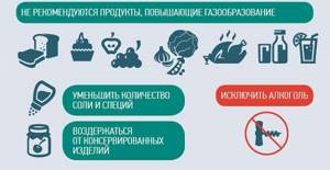
Exclusion from the diet of foods that stimulate gas formation in the gastrointestinal tract
Also, when preparing a patient for an X-ray of the esophagus with barium, it is worth excluding from the diet foods that contribute to gas formation in the organs of the gastrointestinal tract. It is necessary to understand that air masses interfere with obtaining informative images, which can cause difficulties in making a diagnosis.
Advantages of the clinic
Patients who need an X-ray of the stomach strive to undergo the examination in the safest possible conditions, so they contact the Medicina clinic, choosing:
- highly professional radiologists with many years of experience in gastrointestinal diagnostics;
- modern digital equipment for radiography;
- high-quality medical service, the high level of which is confirmed by multiple accreditation according to JCI standards;
- the opportunity to undergo examination after working hours or on a weekend;
- affordable prices for stomach x-rays and other diagnostic procedures.
Contraindications
There are a number of contraindications to X-rays of the esophagus with contrast.
Pregnancy (the procedure is prescribed only for health reasons)
Barium, like X-ray radiation, has a detrimental effect on the intrauterine development of the fetus and can lead to abnormalities in the child. Therefore, this research method is used extremely rarely and only for health reasons.
If there is a risk of contrast reflux into the respiratory system
If there is a high risk of barium suspension being thrown into the respiratory tract, which can lead to asphyxia and suffocation (for example, with fistula tracts, etc.).
Serious condition of the patient
The extremely serious condition of the patient is also a contraindication for this study. This can only make things worse.
Continued heavy bleeding from the gastrointestinal tract
Heavy bleeding can only become more intense with the introduction of barium and during fluoroscopy.
Indications
- suspicion of an ulcerative process;
- detection of a neoplasm;
- protrusions or other deformations of the gastric walls;
- inflammatory processes in the stomach;
- state of dysphagia (functional impairment of swallowing);
- abdominal pain;
- severe constant heartburn;
- sudden involuntary release of gas into the oral cavity from the stomach or esophagus with a sour odor;
- the appearance of scarlet blood in the stool;
- decrease in red blood cells;
- sudden weight loss without objective reasons.
- pain in the epigastric region;
- detection of mucous and purulent discharge, as well as blood impurities, in feces;
- chronic increase in intervals between acts of defecation, hardening of stools, feeling of incomplete bowel movement;
- frequent diarrhea with changes in stool color (black, tar-like);
- rapid weight loss not due to dieting.
X-rays of the large and small intestines are necessarily indicated for suspected congenital malformations, oncopathology, polyposis, saccular protrusions of the intestinal wall, granulomatous inflammation with segmental damage to different parts of the digestive tract, chronic colitis and enterocolitis.
results
- abnormalities in the structure of the digestive tract;
- acute expansion or narrowing of the lumens of the stomach/esophagus;
- malformations of certain organs of the gastrointestinal tract;
- hypertonicity/hypotonicity of the muscular wall of the stomach;
- tumors, papillomas, foreign bodies;
- reduction or radial arrangement of shell folding;
- cicatricial changes at the site of tumors, ulcers, chemical burns.
- pathological narrowing;
- saccular protrusions and elasticity of the walls;
- the introduction of one section of the intestine into another with the possible development of gastrointestinal obstruction;
- intestinal motor function;
- the presence of ulcerative and inflammatory processes;
- tumors, polyposis.
An examination can show how the bauhinium valve functions. This is the structure that separates the small and large intestines and is responsible for passing food between them. If it has pathological changes, then the food gets back access, and this poses a danger to the patient’s life.
In children
Some difficulties may arise when it is necessary to conduct radiography in young children (up to 5-6 years).

It is important to explain to them in advance that the procedure is absolutely painless. If the child remains anxious, you can show how an x-ray is done to another patient in order to calm him down.
also important for parents to remain calm themselves , since their emotional state is easily transmitted to their children. Also, medical personnel should be understanding of the child’s fear and remain friendly. Cases where a doctor scares or yells at a child are completely unacceptable.
An important aspect of the examination is the presence of parents nearby during the x-ray. This will significantly calm the child and follow all the necessary instructions from the doctor. To protect against X-ray radiation, parents or accompanying persons are given special gowns with lead plates , which protect the most important parts of the body.
Help You can also take your child’s favorite toys with you. This will create a more comfortable environment in the X-ray room.
The time for complete refusal of food in children is shorter than in adults and depends on their age.
| Child's age | How long should you refrain from eating? |
| Up to a year | 2-3 hours |
| 1-2 years | 3-4 hours |
| 3 years | 4 hours |
| 4-6 years | 4-5 hours |
| 7-8 years | 5 o'clock |
| 9-10 years | 6 hours |
| 11-12 years old | 7 o'clock |
| 13 years and older | 8 ocloc'k |
Important At the same time, if a one-year-old child is very hungry , then instead of milk or formula, he can be given a small amount of water to drink, which will reduce the feeling of hunger for a certain time.
How to prepare for the examination?
General rules
Preparing the patient for radiography of the esophagus is aimed at increasing the diagnostic information content. Why is it important? The presence of food or air in the digestive tract affects the quality of the images that the doctor receives.
24 hours before the X-ray examination, the consumption of alcoholic beverages is prohibited. They can irritate the mucous membrane, provoke the development of new ulcers, and also stimulate the production of gastric juice.
The patient is also advised to refrain from smoking on the day of the diagnosis. Substances contained in smoke are factors of aggression and also lead to vasospasm of the mucous membrane. also not recommended chewing gum , since chewing reflexively activates the production of gastric juice, and to brush your teeth immediately before the procedure.
Important The doctor must explain to the patient that numerous
folk tips that are aimed at reducing radiation exposure to the body are ineffective and can only reduce the quality of the image .
Preparation can be neglected only in emergency situations, when delaying time can lead to serious consequences for the health and life of the patient.
Features of the DIET

An important factor that often reduces the quality of the X-ray image is the accumulation of food and gases in the stomach cavity. Therefore, before a planned study, you need to refrain from eating for 8 hours . During this period of time, you can only drink water (but not strong tea or coffee) in small volumes.
Additionally, it is important to prescribe a diet 2-3 days in advance , to exclude foods from the diet that can contribute to increased gas formation. This is especially important for older patients who have chronic digestive problems. Therefore, they are prohibited from the following products:
- alcoholic and carbonated drinks;
- legumes;
- fresh bakery products;
- cuts, whole grain wheat;
- fermented milk products (kefir, yogurt, fermented baked milk);
- juices from apples, pears;
- grapes, raisins;
- fatty meats and fish;
- most types of cabbage (fresh, pickled and sour), Jerusalem artichoke;
- onions, turnips, radishes, Chinese salad;
- ice cream;
- products containing chocolate;
- barley;
- pasta.
Additionally, unload your diet by increasing the frequency of meals up to 5-6 times a day in smaller portions . This will allow the digestive tract to more quickly digest and break down food products, which will reduce the time they spend in the stomach cavity and the severity of gas formation.
At the same time, the diet should remain as balanced . The patient should consume sufficient amounts of nutrients and vitamins with food.
An example of a diet for 2 days before an x-ray of the stomach is given in the following table:
| 1 DAY | |
| First breakfast (8-9 am) | 2 soft-boiled eggs, buckwheat porridge with butter, a glass of warm still mineral water |
| Second breakfast (10-11 am) | Lightly warmed cottage cheese with sugar and weak green tea |
| Lunch (14-15 pm) | Vegetable soup (can be with thoroughly cooked meat), mashed potatoes with a steamed cutlet and a cup of weak tea |
| Afternoon snack (17-18 pm) | A few fresh bananas, a glass of compote |
| Dinner (20 pm) | Oatmeal (cooked in water), lean fish fillet and a glass of still water |
| Before bed (10 pm) | Dry cookies with a mug of weak herbal tea |
| DAY 2 | |
| First breakfast (8-9 am) | Boiled rice porridge with vegetable salad (cucumbers, tomatoes, carrots), a glass of still mineral water |
| Second breakfast (10-11 am) | Baked apple, a cup of weak green tea |
| Lunch (14-15 pm) | Vegetable soup with buckwheat and poultry (for example, chicken), buckwheat porridge with chicken Kiev, carrot and beet salad, a glass of compote |
| Afternoon snack (17-18 pm) | Scrambled eggs and a glass of still water |
| Dinner (20 pm) | 3-4 cheesecakes with sour cream and a cup of weak herbal tea |
Taking medications
Often, before an X-ray examination, the doctor additionally prescribes certain groups of medications:

- Adsorbents (activated carbon, Smecta). This group of drugs is capable of absorbing toxic substances, gases and food particles in the lumen of the digestive tract. They are usually given 2-3 hours before the expected start of the diagnosis.
- Antispasmodics (platifilin, drotaverine) reduce the tone of the smooth muscle muscles of the digestive tract. This is especially important if the patient has pain, or if a study using contrast agents is planned. They are given to the patient or administered 60-90 minutes before an x-ray of the stomach.
- Drugs that reduce the production of gases in the digestive tract (simethicone, espumizan). They prevent their accumulation in the lumen of the stomach. The medication is taken 6-8 hours before the test.
Please note: When scheduling an x-ray, be sure to tell your doctor what other medications you are taking. This is important, since the use of certain drugs (antacids, non-steroidal anti-inflammatory drugs) can reduce the diagnostic information.