To make correct diagnoses, doctors use various examination methods. It is not possible for a specialist to draw a conclusion about the state of health of your body based on one clinical analysis. Diagnosis of diseases of the urinary system especially requires a comprehensive approach and includes laboratory and instrumental studies.
Kidney ultrasound is prescribed in almost all cases when the patient complains of irregularities in the functioning of this organ. This method helps to identify inflammatory and sclerotic processes, developmental abnormalities, the presence of tumors and stones. Modern ultrasound machines are equipped with special color Doppler scanners, which allow us to examine the vascular structure of the kidneys in detail and study blood flow. However, it is not advisable to use ultrasound in all cases. When is this diagnosis useless?
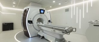
Computed tomography and magnetic resonance imaging: which is more effective?

Many people mistakenly believe that MRI is an analogue of CT. This is not entirely true; these methods are not completely interchangeable due to the principle underlying them. Thus, MRI diagnostics is based on the properties of the magnetic field, and CT is based on the high penetrating power of X-rays. The sensitivity of these techniques to certain pathologies depends on the density and composition of the structure being studied.
Magnetic resonance imaging is more effective in studying soft tissue and cartilaginous formations, while the capabilities of computer imaging are more extensive in diagnosing pathologies of skeletal bones, hollow and fluid-filled structures (for example, the largest arteries, bladder, intracranial sinuses, lungs).
When it comes to diagnosing diseases of the cervical spine, abdominal organs and retroperitoneal space, the information content of CT and MRI is approximately the same. In the absence of contraindications, preference is given to magnetic resonance examination, as it is safer in terms of radiation exposure to the body. The exception is in emergency cases when specialists choose CT, since the procedure takes several times less time compared to MRI.
CT, MRI, ultrasound – what, where and when?
“It’s easier to find a needle in a haystack,” each of us has heard this famous saying more than once. All Sashalber, an Italian painter, was always interested in checking this expression for veracity. And in order to confirm or refute it, he went on an experiment.
In one of the galleries in Paris, Jean de Losy, they placed a stack of real hay, inside which the director of the museum personally hid a needle. The search was successful, but it took... 48 hours.
But what to do if we are talking about health and you need to get a “needle” out of your body quickly and for sure? Just a century and a half ago, doctors never dreamed of looking inside a living person without making any incisions. Modern diagnostic methods make it possible to do this.
Computed tomography, magnetic resonance imaging and ultrasound – which to choose? We addressed this question to the executive director of MRT Expert Lipetsk, Oksana Egorovna Volkova.
Ultrasound diagnostics, unlike CT and MRI, requires special training. The better prepared the patient, the better the research will be conducted. Thus, an ultrasound of the abdominal organs is performed on an empty stomach or, if the diagnosis is scheduled for lunch or evening, the last meal should be six hours before the examination.
How to properly prepare for an abdominal ultrasound? The ultrasound diagnostics doctor at the Expert Orenburg Clinic, Anna Viktorovna Poskrebysheva, tells
Ultrasound of the pelvic organs in women and men requires a full bladder.
You can read more about pelvic ultrasound in women (gynecological ultrasound) here
There are diseases in which ultrasound is superior to other diagnostic methods, in particular CT, in most informative parameters. This is, for example, diseases of the thyroid gland. In case of diseases of the thyroid gland, ultrasound diagnostics makes it possible to determine the “good” or “evil” node and the pathology of the lymph nodes.
When is a thyroid ultrasound indicated? Read here
But the same cannot be said, for example, about the diagnosis of adrenal diseases. Due to the fact that these organs are located deeply, it is difficult to see a reliable picture of adrenal tissue using ultrasound. This is where CT and MRI of the adrenal glands come to the rescue.
So what should you choose? CT or MRI? With or without contrast? In fact, both the first and second studies, to enhance the differentiation of organs from each other, as well as normal and pathological structures, can be carried out both with and without contrast. However, the difference is in the contrast agent itself. The contrast used in MRI is hypoallergenic and practically does not cause complications. When performing a CT scan, a contrast agent containing iodine is used, which can cause complications.
Why is contrast prescribed during MRI? Find out here
What are the differences and advantages of CT and MRI?
Outwardly, these studies are similar, but the principle of their work is completely different.
The CT machine is based on an X-ray tube that rotates around the patient and makes 64 slices per rotation. The speed of computed tomography makes this diagnosis painless for people with claustrophobia and irreplaceable in case of emergency pathology and in intensive care patients.
How to get an MRI if you are claustrophobic? Find out here
When performing an MRI, a magnetic field is used, after which the device emits a radiofrequency pulse and the molecules of human tissue come into resonance. MRI diagnostics last longer, depending on the study - from 8-10 minutes to an hour, and you cannot move during the diagnosis. But the undoubted advantage of MRI is the absence of radiation during the study, which allows the method to be used as often as necessary without fear of consequences, including in pregnant women. In addition, MRI allows you to obtain sections of organs and tissues in a variety of planes, which makes the method highly informative.
There are organs for the diagnosis of which only one of the methods under consideration is most informative or applicable. For example, only CT is used to study the lungs. MRI clearly shows bones, soft tissues, muscles, ligaments, brain and internal organs. It is also possible to undergo a full body MRI. MRI diagnostics is a safe and painless procedure that allows you to scan the body as much as possible.
When is a full body MRI necessary? The chief doctor of MRI Expert Yelets, Vladimir Vladimirovich Tulinov, tells
“The diagnosis is unclear,” is a phrase none of us would like to hear from our attending physician, because living under the yoke of such ignorance is scary. Ultrasound, CT and MRI - all these methods allow you to detect the disease at an early stage, and the diagnosis is painless. And specialists and “Clinic Expert” will always help you decide on the choice of research method in each specific case.
Other related articles:
Contraindications for MRI
What to do if none of the research methods reveals the cause of the pain?
Why undergo MRI diagnostics of the brain for prevention?
Ultrasound or CT: which is better?
Ultrasound methods are used to study internal organs, superficial vessels and soft tissues. A CT scan, more precisely than an ultrasound, shows the condition of the bones, brain, deep-lying vessels and lymph nodes. Tomography often acts as a clarifying diagnostic method after receiving ambiguous ultrasound results. At the same time, ultrasound has absolutely no contraindications, while X-ray diagnostics have enough of them (pregnancy, early childhood). The attending physician decides which procedure to prescribe in this particular case.
When is a kidney ultrasound not informative?
Even taking into account all of the above, ultrasound of the kidneys in some situations cannot give accurate answers to the questions that arise from the treating doctor. For example, it is of little information if a tumor is detected in an organ, since it cannot reliably tell about its benignity or vice versa.
Prescribing a kidney ultrasound is advisable to confirm diagnoses, but not to identify the causes of the patient’s illness. Under such circumstances, it is better to conduct a more accurate study, that is, an MRI.
Difference between X-ray and computed tomography
X-ray and CT are based on x-ray radiation, in this these techniques are similar. The difference is that radiography creates images in one plane, not allowing one to look inside the area under study and obtain information about the composition of the tissue.
The result of tomography is cross-sectional images taken in different planes. Its main advantage over x-rays is that organs do not overlap each other in the images, and it is possible to differentiate structures even if their density differs by tenths of a percent. X-ray does not provide such information, giving only a general picture.
At the same time, radiography is much cheaper and more accessible. For example, fluorography is carried out in any clinic, while only large medical centers have computed tomographs. That is why X-rays are used for primary diagnosis, and CT scans are used to clarify the results of radiography.
What will a kidney CT scan show?
CT scan of the kidneys shows:
- appearance of the kidneys and adrenal glands;
- renal cysts - their presence, number, how many there are, where they are located;
- benign tumors;
- malignant neoplasms - CT scan of the kidneys will show their size, character, presence of necrosis, metastases, hemorrhages;
- salts, stones, crystals located in the kidneys and urinary tract;
- abscesses and other pathologies of an infectious nature;
- consequences of injuries - vascular damage, ruptures of the fibrous capsule, parenchyma;
- enlarged retroperitoneal lymph nodes;
- congenital anomalies - a CT scan of the kidneys shows incorrect location, fusion of the kidneys into a single whole, the absence of one of the two organs and other abnormalities;
- hydronephrosis.
Multislice tomography (MSCT) of the kidneys helps to detect foci of vascular tumors (angiomas), blockage of the urinary canals, and excessive accumulation of fluid in the tissues.
MSCT and CT: what is the difference?
Many patients who are scheduled for examination are interested in what kind of computed tomography is available and which one is better to choose. As for the second point, it makes sense to rely on the opinion of the attending physician. Let's try to figure out the first question here.
Depending on the type of equipment, there are several types of CT:
- Linear computed tomography is an outdated method that is practically not used in modern clinics due to the high radiation exposure and long duration of the procedure. Only one slice is captured in one step.
- Spiral and multilayer computed tomography are improved types of x-ray diagnostics. The main difference between CT and SCT is that the emitter moves in a spiral around the area under study, scanning it in different planes. This allows you to significantly reduce the procedure time and the amount of radiation exposure. Today, patients are most often prescribed MSCT. What kind of examination is this? Multislice tomographs differ from spiral tomographs in that they have several rows of detectors, due to which scanning and data collection are carried out continuously. The examination takes a minimum of time, while it is possible to study the smallest structures. MSCT allows you to perform a three-dimensional reconstruction of the studied area and examine the desired area from different angles.
- Computed positron tomography is the most modern diagnostic method based on the use of radioisotopes. Unlike standard CT, PET studies not the physical features of organs and tissues, but the chemical processes occurring in them. This makes it possible to identify cancer before a tumor forms, while other methods detect the problem after the tumor has appeared and reached a certain size. The examination is also effective in searching for distant metastases.
Benefits of Kidney Ultrasound
There is no doubt that ultrasound has become widely available due to its low cost and lack of age restrictions. The most significant advantages of this method are:
- high information content, allowing the doctor to see not only the structure, but also the functional abilities of the organ;
- recognition of pathology in the early stages due to the good sensitivity of ultrasound;
- safety, which makes it possible to conduct research repeatedly within a short time;
- non-invasiveness, which implies maintaining the integrity of the skin;
- painlessness;
- lack of special preparation for the procedure;
- speed of obtaining results.
Kidney CT scan with contrast
Enhanced CT scan of the kidneys has higher image quality. The examination will show:
- malignant processes;
- proliferation of connective tissue;
- blood clots in the veins;
- salt deposits in the collecting system;
- narrowing of the ureters.
In medical CT in St. Petersburg, kidney CT with contrast is fully automated. The contrast agent for MSCT is injected into a vein by bolus injection according to a schedule programmed by the doctor.
For CT scans of the kidneys with contrast, a drug with iodine is used, so side effects such as rash, metallic taste in the mouth and a rush of heat at the site of the bolus injection of the contrast agent cannot be excluded. In rare cases, blood pressure drops or breathing becomes difficult. Any discomfort during a CT scan of the kidney area should be reported to your doctor.
It is also worth warning a specialist about the presence of diabetes. The medications you take may not work well with the contrast agent.
Check with the administrator for the price of a kidney CT scan with contrast.
Preparing for a CT scan of the kidneys
No preparation is needed for a standard kidney CT scan. You just need to make sure that the woman is not pregnant, since X-ray studies (including conventional and multispiral tomography of the kidneys) are contraindicated.
If a CT scan of the kidneys with bolus contrast is planned, then preparation is necessary:
1. The patient must donate blood to determine the creatinine level. The analysis can be done directly in St. Petersburg at 6th Krasnoarmeyskaya, building 7. Find out how much it costs by phone. If creatinine levels are elevated, the procedure is prohibited.
2. A few days before a CT scan of the kidneys with contrast, it is advisable to start following a diet, excluding gas-forming foods - legumes, carbonated drinks, apples, grapes. This will reduce flatulence and make the photos more informative.
3. An important point in preparation is to refuse food and drinks 4 hours before MSCT or CT scan of the kidneys with contrast. To avoid nausea, you can eat something light just before leaving the house.
You must come to a CT scan of the kidneys in comfortable clothing without metal inserts or other metal objects in the scanning area.
Kidney CT scan procedure
A CT scan of the kidneys is done in the supine position. During diagnosis, the patient’s main task is to remain still and follow the specialist’s instructions. If contrast is involved, the procedure is performed twice. First, CT scans the kidneys in their native state, then the study is repeated with an intensifier.
A kidney CT scan is performed as follows:
- a mobile table with a patient is brought into the chamber of the device for multislice computed tomography of the kidneys;
- the scanning system rotates around the table and takes a series of pictures from different angles;
- a drug for CT scanning with contrast of the kidneys is injected into a vein;
- After the substance is distributed throughout the tissues, scanning continues.
Photos, descriptions and conclusions are issued two hours after the CT scan of the kidneys. You can come to St. Petersburg on any other day at any time convenient for you. We also send information to email addresses.