Detailed description of the study
Alpha-fetoprotein is a special protein that is synthesized by the tissues of the embryo and then by the fetus.
From the amniotic fluid, alpha-fetoprotein enters the woman's bloodstream through the placenta. During pregnancy, the level of AFP in the blood increases, reaching values of 150–250 IU/ml, while after the birth of a child its level sharply decreases. The AFP concentration corresponds to gestational age, and its abnormal change may indicate a violation of the normal development of the fetus.
The physiological increase in AFP in a pregnant woman begins around the 10th week of pregnancy and reaches its highest level by the 34th week, then a gradual decrease in its concentration in the blood is noted.
An increase in AFP levels can be observed in multiple pregnancies, large fetuses or malformations. A low level is characteristic of Down syndrome, as well as delayed fetal development. Abnormal changes in AFP levels are not sufficiently specific and conclusive, so additional laboratory and instrumental studies are required.
Thus, a study of the levels of AFP, human chorionic gonadotropin and estriol at 15-20 weeks of pregnancy allows us to identify defects in fetal development and chromosomal abnormalities.
A decrease in AFP levels may be associated with obstetric complications such as:
- Threatened spontaneous abortion;
- Hydatidiform drift;
- False pregnancy.
In the body of an adult, AFP is normally not detected or is detected in small quantities. It can be produced by liver cells - hepatocytes - during regenerative processes.
In liver diseases such as cirrhosis and chronic hepatitis, the level of AFP may be slightly increased. A significant increase in concentration may indicate the presence of cancer: liver cancer, testicular cancer, etc.
Liver cancer usually develops in patients with already diagnosed chronic liver disease. An AFP level test is prescribed for suspected cancer, as well as for dynamic monitoring of such patients, since they are at risk of developing liver cancer.
AFP, as a tumor marker, helps to detect malignant neoplasms, allows one to assess the effectiveness of therapy, and also assess the risk of developing relapse of the pathology.
In addition, the study is used to evaluate the effect of hepatotoxic drugs on the liver.
4.Risks and what can affect the result?
Risks of AFP analysis
If you are donating blood for an alpha-fetoprotein test, then possible risks may only be associated with taking blood from a vein. In particular, the appearance of bruises at the site of blood sampling and inflammation of the vein (phlebitis). Warm compresses several times a day will relieve phlebitis. If you are taking blood thinning medications, you may bleed at the puncture site.
What can affect the results of an AFP analysis?
Alpha-fetoprotein levels may become abnormal if:
- A woman carries more than one child;
- The woman has gestational diabetes;
- The patient smokes. Smoking increases AFP levels.
References
- Liver cancer (hepatic cell). Clinical recommendations. Association of Oncologists of Russia, 2021. - 36 p.
- Orlov, A.E., Kozlov, S.V., Kaganov, O.I. etc. Dependence of tumor markers on the level of metastatic liver damage. Coloproctology, 2021. - No. 28(64). — P.42.
- Lersritwimanmaen, P., Nimanong, S. Hepatocellular carcinoma surveillance: benefit of serum alfa-fetoprotein in real-world practice. Euroasian journal of hepato-gastroenterology, 2021. - Vol. 8(1). — P. 83-87.
Interpretation of results
Important! Standards vary depending on the reagents and equipment used in each particular laboratory. Therefore, when interpreting the results, it is necessary to use the standards adopted in the laboratory where the analysis was carried out. You also need to pay attention to the units of measurement.
| Floor | Age | Normal values, IU/ml |
| Man | Up to 1 month | 0,5 — 13600 |
| From 1 month to 1 year | 0,5 — 23,5 | |
| Over 1 year old | 0,9 — 6,67 | |
| Woman | Up to 1 month | 0,5 — 15740 |
| From 1 month to 1 year | 0,5 — 64.3 | |
| Over 1 year old | 0,9 — 6,67 |
Important! The interpretation of the results is always carried out comprehensively. It is impossible to make an accurate diagnosis based on only one analysis.
The standard units for measurement are IU/ml, however, some laboratories use ng/ml. In order to convert units of measurement, you must use the formula: 1 ng/ml * 0.83 = IU/ml.
How to convert ng/ml to IU/ml and vice versa for AFP:
- IU/ml=0.83 * ng/ml
- ng/ml=IU/ml / 0.83
Factors influencing the result
- In patients of the Negroid race, there is an increased content of alpha-fetoprotein, and in representatives of the Mongoloid race, on the contrary, it is decreased.
- Against the background of some endocrine pathologies, false-positive results of an AFP test can be determined.
- Taking monoclonal antibodies
- Insulin-dependent diabetes reduces AFP in the blood of pregnant women
Basic Concepts
This fetal glycoprotein in men, children and non-pregnant women has high sensitivity against primary liver cancer and germ cell tumors. In adult patients, the primary form of liver cancer in 90% of cases is hepatocellular oncology, and in children - hepatoblastoma.
You may be interested in: Pancreatic duct of Wirsung: normal, causes of expansion
In the presence of a DI of 20 units per milligram, hepatoblastoma is always, and hepatocellular cancer in 80% of cases, associated with an increased level of alpha-fetoprotein. But in most situations, hepatocellular cancer is diagnosed at a late stage, and the results of therapy, unfortunately, are unsatisfactory. In order to improve early diagnosis, screening programs based on AFP are used among patients with an increased risk of developing a primary form of liver cancer (we are talking about people with chronic active hepatitis, cirrhosis of any etiology, etc.).

Where can I get tested for alpha-fetoprotein?
An alpha fetoprotein test can be taken free of charge at the clinic to which you are geographically assigned. For pregnant women, alpha-fetoprotein can be determined in the place where they are observed during pregnancy.
If you have the financial means, you can get tested for alpha-fetoprotein for a fee. On average, the price ranges from 500 to 1000 rubles.
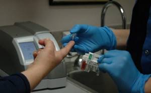
Photo: https://pixabay.com/photos/nurse-diabetes-diabetic-test-a1c-527615/
Low AFP during pregnancy
An indicator that is too low indicates the following deviations:
- malnutrition (malnutrition);
- hypoxia (oxygen starvation);
- Down syndrome;
- Edwards syndrome (trisomy 18 chromosome),
- Patau syndrome (trisomy 13 chromosome),
- frozen pregnancy;
- intrauterine death;
- diabetes mellitus or gestational diabetes in a pregnant woman;
- obesity of various types;
- low placenta previa;
- endocrine disorders.
When is it necessary to test AFP?
Indications for determining the presence of alpha protein in serum biological fluid are the following manifestations:
- Symptoms of suspected perinatal pathology,
- Pathologies of the chromosomal type in the embryo,
- The development of the fetal brain, as well as all brain cells of the body, is disrupted,
- Intrauterine defects of the internal organs of the unborn baby,
- Genetic hereditary pathologies in the family,
- When cancer cells metastasize to liver cells,
- To recognize and exclude neoplasms in the cells of the genital area, cancers such as teratomas, oncological pathology germinoma,
- Exclusion of malignant oncology in liver cells,
- Constant monitoring against oncological therapy testing is carried out before the start of therapy, during this treatment and at the end of the drug therapeutic course.
Alpha-fetoprotein testing is also carried out for liver cell pathology:
- With cirrhosis of liver cells,
- For hepatitis C,
- With hepatitis A pathology,
- With the development of hepatitis B.
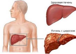
These types of liver cell diseases can provoke cancerous tumors in the affected cells.
In this situation, constant clinical monitoring of blood composition will allow not to miss the development of cancer cells at the initial stage, which will help to prescribe drug treatment in a timely manner.
With existing oncology, this type of clinical analysis is ineffective because it does not determine the level of development of malignant tumors, but only produces a marker value for the presence of such cells in the body.
This testing is carried out only at the stage of identifying cancer and establishing an oncology diagnosis.
Tumor markers in children, men and women
Germ cell tumors of newborns and infants are represented mainly by sacral and coccygeal formations. In this case, AFP-negative teratomas and AFP-positive teratoblastomas are detected. The marker of choice in the differential diagnosis of these formations is AFP, since its sensitivity in relation to teratoblastoma is close to 100%.
Determination of AFP can contribute to the choice of therapeutic tactics: AFP-negative teratomas require surgical treatment, while AFP-positive teratoblastomas require combination therapy.

Germ cell tumors in adolescents and adults can be distinguished by a rich morphological form, and, in addition to AFP, it often produces hCG, so the simultaneous establishment of both of these tumor markers is a mandatory requirement. In diagnosing germ cell tumors with an AFP DI of 10 units per milliliter, for hCG at 10 mIU/ml, the overall sensitivity of alpha-fetoprotein in women, men and children will be 60%.
The combined determination of both of these indicators allows achieving 86% sensitivity for primary germ cell tumors. The simultaneous determination of tumor markers AFP and hCG among adults and adolescents helps to confirm the diagnosis in the case of the development of gonadal (ovarian and testicular), extragonadal (retropitoneum, nervous system) and germ cell oncological formations.
High AFP in pregnant women
An increased level of AFP by more than 2-3 times is observed in the following diseases:
- anencephaly (severe pathology of the formation of cranial bones and cerebral hemispheres);
- hydrocephalus;
- spinal malformation (spina bifida);
- kidney and liver defects in the embryo;
- atresia of the esophagus or intestines;
- umbilical hernia, gastroschisis (defect of the anterior abdominal wall),
- teratocarcinoma (cancer) of the yolk sac;
- pathology of the placenta;
- encephalocele (cranial hernia);
- threat of miscarriage or premature delivery;
- large fruit;
- multiple pregnancy, etc.
Important: to diagnose pathology of fetal development, it is necessary to know the exact duration of pregnancy. The AFP level alone cannot serve as a diagnostic criterion.
How to decipher the REA analysis
In order to independently understand the test results, the patient must know the normal values of the CEA tumor marker and have an idea of what a deviation from the norm may mean.
The normal levels of this substance are the same for both sexes; the patient’s age also does not play a role. Normally, the concentration of CEA in the body of a man or woman should not exceed 6.5 ng/ml. Normal CEA values in blood serum indicate a low risk of developing cancer. The result may also be due to the fact that the testing is not sensitive to a specific tumor type.
What does exceeding the REA norm indicate?
If the levels of cancer embryonic antigen in the patient’s test results exceed the norm, this may be a signal of the following diseases:
- cancer of the rectum, colon;
- cancer of the stomach, pancreas;
- lungs' cancer;
- mammary cancer;
- cancer of the uterus, cervix, ovaries;
- spread of tumor metastases to bone tissue, liver;
- recurrence of cancer.
An increase in CEA values is not always evidence of the presence of cancer in the patient’s body. The level may also increase due to the development of benign diseases:
- liver cirrhosis, chronic hepatitis;
- chronic renal failure;
- chronic pancreatitis;
- colorectal polyps;
- tuberculosis;
- ulcerative colitis;
- Crohn's disease;
- emphysema, pneumonia, chronic bronchitis;
- autoimmune diseases;
- cystic fibrosis;
- rheumatoid arthritis;
- ovarian and breast cysts.
In addition, a slight increase in the values of the CEA tumor marker in test results is possible if the patient smokes or suffers from alcoholism. A slight increase in the concentration of antigen in the patient’s blood may indicate benign diseases in the acute stage, but the initial stage of the development of cancer cannot be completely excluded.
A serious increase in CEA concentration is observed in the presence of malignant tumors. If metastases are present, the rate can increase tenfold. If the indicators of this tumor marker increase in dynamics, which is detected during repeated tests, this indicates the low effectiveness of treatment directed against the malignant disease, the development of relapse, the onset of metastasis (often detected 3-6 months before the onset of clinical symptoms of metastases).
What can we say about the decrease in REA indicators?
The concentration level of this substance in the patient’s body may also decrease, which usually indicates the following:
- the patient underwent surgical removal of a malignant tumor;
- effective therapy for cancer was carried out;
- the benign tumor is in remission.
It is important to know
A positive result of examining a patient for a tumor marker of cancer embryonic antigen cannot yet be considered as a sufficient basis for making a medical verdict. This is just an indicator for further examination. Also, a negative CEA test result does not at all prove that the patient does not have cancer.
It is also worth considering that research methods in different laboratories may differ, which leads to different results. Therefore, doctors recommend repeat testing in the same medical center where the first analysis for the CEA tumor marker was done. The price of research for the CEA tumor marker depends on the specific laboratory.
<
p style=”text-align: justify;”>

Tips for preparing for the test
The material in which alpha fetoprotein can be determined is blood serum, cerebrospinal fluid, amniotic fluid. Blood is most often used.
There are no special events to prepare for the analysis. It is important that the blood serum is suitable for determining alpha-fetoprotein. Therefore, it is worth donating blood on an empty stomach or donating after a four-hour fast is allowed.
Some also advise avoiding smoking half an hour before blood collection.
There are a number of factors that can distort the results of the study. For example, the presence of type 1 diabetes mellitus in an expectant mother may contribute to an underestimation of the level of this fetal protein. It is also noted that for people of the Mongoloid race this indicator will be lower than average, and for the Negroid race, on the contrary, it will be higher.
Drug treatment of AFP
The drug alpha-fetoprotein is used when its trace level in the adult body is reduced. This drug has a wide range of regulated effects in the body.
Since this alpha protein is involved in the transport of prostaglandin molecules in the human body, its normal level should be in the blood.
Use to correct this protein in the body is recommended for the following pathologies:
- For diseases of the endocrine system, diabetes mellitus,
- In autoimmune-type pathologies such as thyroiditis, myasthenia gravis, rheumatic carditis,
- For bronchial type asthma,
- With fibroids in the cervix,
- With the development of urological infections and infectious pathologies of the genital area in the body,
- To ensure good blood flow and for prevention of arterial thrombosis,
- In the treatment of pathology multiple sclerosis,
- When there are ulcers in the intestines.
Drug treatment with this drug for oncological lesions of organs is also darkened. This drug is often used to treat pathologies on the skin.

The drug treatment regimen and dosage are prescribed by the doctor individually.