PET/CT (positron emission tomography - computed tomography) is a method for diagnosing diseases that combines the study of the structure and functional characteristics of tissues. This technology is most in demand in oncology for diagnosing and determining the extent of spread of malignant tumors.
- Description of PET/CT technology
- Advantages and disadvantages of PET
- Types of drugs for PET
- When is PET/CT indicated?
- Contraindications to PET/CT
- Preparing for PET
- Passing PET/CT diagnostics
- Complications and side effects of PET/CT
- Radiation exposure to the patient during PET scanning
Description of PET/CT technology
PET technology is based on the study of both structural and functional characteristics of tissues. Tissue function is assessed through metabolism. For example, a universal substance is selected that is necessary for all cells of the body. It is labeled with a radioactive label, injected into the body, and the places of its maximum accumulation are observed.
One of the most versatile substances in the human body is glucose. It is necessary to nourish almost all cells and tissues. But its greatest consumption is in malignant neoplasms, since a lot of energy is spent on the growth and reproduction of the tumor.
In PET/CT studies, glucose is labeled with radioactive atoms with a short half-life, such as fluorine-18. And after introduction into the body, it accumulates in large quantities in tissues with the most intense metabolism, i.e. in malignant neoplasms.
The radioactive tag decays, emitting energy in the form of gamma rays. The radiation is recorded by the device, and based on the data obtained, a visual model is built that shows the location of the tumor, its size and metastases.
Radioactive tracers accumulate only in pathological cells, while healthy tissues are not visualized. When the doctor wants to see both healthy and changed structures, a computed tomography scan is performed. It allows you to obtain a detailed image with millimeter accuracy.
After receiving data from both scanning systems, the software overlays them to create an image that gives the doctor a clear picture of where the pathological lesions are located.
Book a consultation 24 hours a day
+7+7+78
The essence of the PET method
As is known, in 90% of cases PET is used in oncology to determine the extent of cancer tumor spread and metastases. Tumor cells have a high metabolism and consume a lot of glucose during their life processes. This feature allows PE tomographs to detect tissue affected by cancer. The metabolite molecule (usually glucose, but ideally a substance related to a certain type of cancer cell) is modified in a special way - atoms are replaced with their radioactive analogues - isotopes with an ultra-short life period. After entering the body, the resulting drug is integrated into the metabolism, at the same time, re-emitted photons are scanned using gamma radiation and receiving sensors. Thus, areas with increased metabolism become visible on tomography as foci. Depending on the level of metabolism, lesions are divided into 4 levels:
- 1-intensity similar to soft tissue and non-working muscles
- 2- intensity as in the liver
- 3- at the level between the liver and brain
- 4- intensity, as in the brain.
The 4th level indicates the primary focus of the tumor or metastasis, the 3rd is more likely a cancerous lesion, but inflammation is also possible, the 2nd is more likely inflamed tissue.
Advantages and disadvantages of PET
Advantages:
- The ability to combine several types of diagnostics in one study, which allows you to obtain highly accurate data.
- The examination is painless and does not cause discomfort.
- Possibility of diagnosing cancer at an early, pre-symptomatic stage.
- Possibility of studying all organs at once.
- The procedure is performed on an outpatient basis and does not require hospitalization.
The disadvantages of the method include the need to use radioactive drugs. To reduce risks, the study is carried out in strict accordance with radiation safety standards. In general, the side effects of PET scans are not commensurate with the valuable information this research method provides. In this case, three rules apply:
- The research must be justified. The benefits of diagnostics must outweigh the harm from exposure.
- Optimization. Medical staff are obliged to take all measures that will minimize radiation exposure to the body.
- Rationing. All radiation safety standards and regulations must be strictly observed.
What does a second opinion give when interpreting CT and MRI?
Interpretation of computed and magnetic resonance imaging data at the Medica24 International Clinic will allow you to:
- avoid medical errors,
- establish an accurate diagnosis in a complex case of cancer or other disease,
- receive the most effective treatment,
- avoid complications and unnecessary side effects,
- improve the prognosis of the disease through optimal treatment.
A second opinion when deciphering CT, MRI, PET-CT data is especially valuable when prescribing expensive treatment for oncological diseases, on which the preservation of a person’s life depends, including surgical operations.
Expert doctors at the Medica24 International Clinic have extensive experience and exceptional qualifications in the interpretation of diagnostic data in oncology.
We will call you back, leave your phone number
Message sent!
expect a call, we will contact you shortly
This can not only significantly speed up treatment and improve its results (due to correcting the diagnosis), but also save money.
As the experience of our clinic shows, obtaining an expert opinion when deciphering MRI, CT, PET-CT data for oncological diseases in most cases helps to prescribe more effective treatment.
Types of drugs for PET
The potential of PET studies is determined by the arsenal of radiopharmaceuticals used—pharmaceuticals that are labeled with unstable isotopes that make the substances radioactive.
Currently, isotopes of the following elements are widely used in PET studies:
- carbon-11;
- nitrogen-13;
- oxygen-15;
- fluorine-18.
Fluorine is most widely used in oncological practice because it has the longest half-life and the lowest radiation energy. This, firstly, allows you to obtain images of high quality and spatial resolution. And secondly, the relatively long half-life (109.8 min) makes it possible to transport the drug from the production site to scanning centers.
The most common radiopharmaceutical in oncology is fluorodeoxyglucose FDG. This is an analogue of glucose, which is a universal substance absorbed by all cells of the body. Cancer cells absorb it faster, so they accumulate the drug in a higher dosage, which is clearly visible during scanning. The disadvantage of this substance is that it accumulates in slightly larger quantities in brain tissue and nephrons, which can cause illumination of these organs even in normal conditions. Therefore, the question arises of searching for other, more specific radiopharmaceuticals.
For the brain, such a drug is 18F-FET. It contains the amino acid tyrosine, labeled with the fluorine-18 isotope. Tyrosine has a very high selective accumulation in brain tissue, which is used for imaging neurotumors. In addition, 18F-FET is used for the diagnosis of oropharyngeal tumors, in the search for metastases and for the diagnosis of lesions of the lymph nodes of the neck.
In addition, there are a number of specific radiopharmaceuticals for the diagnosis of individual tumors:
- 68GaPSMA - used in the diagnosis of prostate cancer.
- 68Ga-DOTA-TATE, -NOC, TOC, -LAN, etc. are used for the diagnosis of neuroendocrine tumors.
- 18 FCH - used to diagnose hepatocellular carcinoma.
The choice of radiopharmaceutical is made by a specialist doctor at the PET center based on the histological type of cancer, sensitivity and specificity of the tracer.
Scope of application of radiopharmaceuticals
The isotopes most often used in PET are (18)F, (11)C, (13)N, (15)O, whose half-lives are 109, 20, 10 and 2 minutes, respectively. A variety of radiopharmaceuticals allows you to choose the best method depending on the purpose of the examination:
- glucose labeled with (18)F and (11)C- isotopes is used to calculate the intensity of glucose metabolism;
- (15)O-labeled water allows you to assess cerebral blood flow, 15(O2) - is used to determine oxygen metabolism;
- (11)C-labeled methionine and leucine, (18)F- labeled tyrosine and fluorocholine to study amino acid metabolism;
- thymidine with (18)F- to assess the rate of cancer cell proliferation;
- (18) F-fluoromizonidazole - detects tissue hypoxia;
- (11) C-DOPA, (18) F-DOPA, (11) C-raclopride - for studying post- and presynaptic processes in the dopaminergic system;
- (18)F-flumazenil- in the benzodiazepine system.
PET computed tomography with choline or acetate has been used successfully in the study of prostate cancer and brain tumors; (18) F-DOPA- for detection of medullary tumor of the thyroid gland, neuroendocrine oncological processes; methionine - for tumors of the lungs, cervical region, head, breast. In the vast majority of cases, fluorine-labeled (18F) glucose is used.
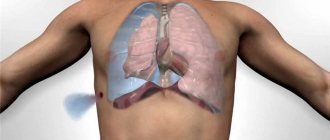
When is PET/CT indicated?
- Search for the primary tumor site when metastases are detected.
- Staging of an already identified malignant process.
- Differential diagnosis of relapse and post-therapeutic or post-surgical changes.
- Monitoring the course of the disease, detecting relapse (for example, with an increase in the level of tumor markers).
- Planning for radiotherapy and surgery.
- Planning a biopsy of the tumor and determining its most “aggressive” area.
When is a CT scan prescribed?
Computed tomography is more accessible and universal. It is suitable for diagnosing a wide range of diseases: bones, joints, internal organs. By contrast, the possibilities of this type of examination are almost limitless, as they are used to detect internal bleeding, traumatic injuries, skeletal abnormalities, brain studies, abdominal and pelvic scans. The main convenience is that this diagnostic is relatively inexpensive and is available even in small towns. However, when identifying malignant neoplasms and studying their dynamics, this examination is not very effective - it is not possible to differentiate the type of tumor, evaluate its dynamics and identify other features of the pathological process.
Contraindications to PET/CT
There are absolute and relative contraindications to PET/CT. The following states are considered absolute:
- Pregnancy. The study is not carried out when a diagnosis is established, and in the case when a woman suspects its presence.
- Lactation. It is recommended to stop breastfeeding 6 hours before the intended examination. The timing of resumption of feeding is determined by the tracer used.
A relative contraindication to the study is decompensated diabetes mellitus when planning to use fluorodeoxyglucose (FDG) as a radiopharmaceutical. In this case, consultation with an endocrinologist is required and stabilization of blood glucose levels below 7.7 mmol/l.
The study is carried out with caution in cases of impaired renal function, since the data obtained may be distorted due to retention of the radiopharmaceutical in the tissues.
Book a consultation 24 hours a day
+7+7+78
Indications for the study
| Indications | Objectives of the study |
| Oncopathology | Detection, staging of tumors, assessment of treatment effectiveness, differential diagnosis of radiation necrosis and relapse |
| Neurological diseases | Early diagnosis of Alzheimer's disease, identification of the epileptogenic focus during the interictal period, visualization of functional areas (speech, motor functions) |
| Heart diseases | Detection of myocardial hibernation |
| Inflammatory diseases | Search for an unknown inflammatory focus; vasculitis |
Contraindications
- childhood;
- pregnancy, lactation;
- decompensated diseases;
- general serious condition;
- high blood glucose levels;
- less than 3 months have passed since the operation;
- less than 6 months have passed since the completion of radiation therapy.
Preparing for PET
The day before the scheduled date of the study, it is necessary to avoid physical activity. If you plan to use FDG, you should also avoid foods rich in carbohydrates. This includes not only sweets and sugary drinks, but also fruits, vegetables, baked goods, legumes, etc. On the day of the procedure, eating is strictly prohibited. You can only drink clean water.
The patient should arrive at the clinic wearing comfortable clothing. It is advisable that it does not contain metal fittings and decorations (zippers, buttons, buttons, fasteners, etc.). But if necessary, staff can provide a disposable gown.
If the patient has diabetes, glucose levels should be measured before the test. If it is above 7 mmol/l, you need to take half the dose of insulin. The minimum time between insulin injection and radiopharmaceutical administration is 5 hours. If the patient is taking tablets to lower glucose levels, it is necessary to discuss with the endocrinologist the possibility of discontinuing them on the day of the study.
False PET results
Both false positive and false negative results are possible. Sometimes PET scans reveal foci of radiopharmaceutical accumulation in organs that are considered tumorous, although they are not. This is primarily due to improper preparation for the study.
Before PET, it is important to adhere to a diet low in simple carbohydrates and avoid physical activity. If these conditions are not met, unreliable results may occur. In addition, intense accumulation of glucose is typical of inflamed tissues. Such an inflammatory focus can mimic a malignant tumor.
Attention! It is important to correctly follow the preparation method for the procedure so as not to get false results.
False positive results are observed when:
- granulomatous diseases;
- postoperative changes;
- inflammatory diseases (abscess);
- metformin therapy (in the intestines);
- reactions to foreign bodies;
- fat necrosis.
A false negative result is the absence of radiopharmaceutical accumulation in the tumor. This situation can be observed with low-grade tumors of a non-aggressive nature. Some tumors may be hidden due to high physiological accumulation of radiopharmaceuticals in certain organs.
It is difficult to interpret PET results against the background of normal accumulation of radiopharmaceuticals in the brain, tonsils, muscles (especially after physical exercise), bone marrow, and thymus. To visualize the tumor, another radiopharmaceutical, for example, based on amino acids, may be required.
Passing PET/CT diagnostics
The patient must arrive at the clinic exactly at the appointed time. In this case, it is necessary to take all medical documents that are of interest within the framework of the disease - data from previous CT scans, histology data, laboratory tests, a statement of treatment, as well as data on concomitant pathology that may affect the diagnostic results (diabetes mellitus, kidney disease) .
After completing the paperwork, the patient is taken to the treatment room. There they take a blood test for glucose and measure anthropometric data. If everything is in order, the patient is taken to a relaxation room. There he is comfortably positioned, and a peripheral intravenous catheter is installed, through which the radiopharmaceutical is administered. To ensure it is evenly distributed, you need to drink a glass of water (or a contrast agent that the nurse will suggest) every 15 minutes. After administration of the radiopharmaceutical, you must be in a relaxed state and not talk. Any physical activity can lead to uneven distribution of the tracer and distort the results.
After 60–90 minutes, the patient is invited for a scan. First, a CT scan is performed, and then a PET scan. A CT scan takes a matter of minutes, while a PET scan takes up to half an hour. In this case, it is important to lie still so as not to provoke the formation of artifacts in the photographs that can distort the image. Both studies are carried out in the same tomograph and do not cause pain. After the procedure is completed, the patient must remain in the clinic for another hour, as a repeat scan may be necessary to clarify the data.
After a PET examination, the patient should not be in crowded places or have contact with children and pregnant women for 24 hours. In a family circle, you should keep a distance of at least 1 m from each other. Such restrictions apply for 24 hours after administration of the radiopharmaceutical.
In addition, during this period it is necessary to take a sufficient amount of liquid, at least 2.5 liters. This will help remove the contrast and radiopharmaceuticals faster and safer. You can drink more than just water. You can consume any caffeine-free products - juices, sparkling water, fruit drinks, mineral water, etc.
Carrying out the procedure
- Before the start of the study, in the absence of contraindications, the patient is injected with a contrast agent. The dosage is selected individually. The drug can be administered intravenously through a special catheter, orally, or inhaled through a mask.
- Next, the patient is asked to rest for about an hour. This is necessary for even distribution of contrast and muscle relaxation.
- After this, a series of photographs are taken on a special table. The patient should remain motionless for 20-40 minutes.
- The device itself is similar to a CT scanner; it can also be of an open type (such a device is approved for patients with claustrophobia).
- After the procedure is completed, the patient remains free for some time within the walls of the diagnostic center. This is necessary to remove the entire dose of the drug.
Complications and side effects of PET/CT
Complications during PET/CT are mainly related to the administration of CT contrast. As a rule, these are allergic reactions and complications associated with vein puncture - vascular damage, hematoma, pain, neuritis, etc. Renal dysfunction occurs extremely rarely. If contrast is taken orally, nausea, vomiting and diarrhea may occur.
Dizziness and weakness may occur after a PET scan due to the need for the test on an empty stomach. Therefore, it is recommended to bring a snack with you that can be eaten after all procedures related to the study are completed.
What is the difference between PET-CT and CT
PET, as a separate technique, has poor anatomical information, since it is not always able to correctly determine the primary tumor process, namely its location and exact size. To overcome this barrier, combined PET-CT devices were created, allowing two examinations to be carried out simultaneously. The difference between the PET CT method is that this device combines images obtained on PET and CT, as a result you can see an anatomical section with a map of metabolic activity superimposed on it.
Radiation exposure to the patient during PET scanning
Radiation exposure during PET/CT depends on the radiopharmaceutical used and the scope of the study. For example, scanning the head will expose you to less radiation than scanning the whole body. To minimize harm from PET/CT, the following standards are used:
- The research is carried out using ultra-short-lived isotopes, the half-life of which is several minutes or hours.
- An individual dose calculation is used for each patient, taking into account weight, height and age.
- The scanners use special filters and programs that reduce X-ray doses.
- Applying the required amount of liquid not only improves the quality of the resulting image, but also helps to remove the radiopharmaceutical faster.
In general, PET/CT is a fairly safe procedure, but it is performed only for strict indications when other studies do not provide the necessary information.
Book a consultation 24 hours a day
+7+7+78
PET/CT and CT with or without contrast?
What exactly does contrast do for PET/CT diagnostics?
— A contrast agent is introduced to obtain a more accurate image; changes at the cellular level and minor pathological processes become visible. The injected substances accumulate in the foci of pathology and, thanks to the contrast, it is possible to more clearly identify problem areas. The standards for conducting studies with contrasting necessarily include the recommendations of the NPCC DIT DZM. As a rule, it is not advisable to conduct a contrast-enhanced study if it was carried out earlier, within the last 2-3 weeks before, and its results are in the patient’s hands on a disk or any other medium.
Most studies are carried out with contrast only if the radiologist makes such a prescription, based on the clinical situation and the significance of the outcome of the study.
Rules for preparing for PET/CT for patients living in the Kemerovo region-Kuzbass:
- Order of the Ministry of Health No. 1823 of June 17, 2021
- Referral for PET/CT (according to the order of the Ministry of Health of Russia dated December 2, 2014 No. 796n)
- Memo for patients
- Statement of consent to the processing of personal data
Rules for preparing for PET/CT for patients living in other regions of the Russian Federation:
- Referral for PET/CT (form 057у/04)
- Memo for doctors of the referring medical organization
- Memo for patients
- Statement of consent to the processing of personal data
The direction in form No. 057у/04 must indicate:
- “for examination” and not for hospitalization;
- name of the medical institution where the patient was sent, i.e.: LLC “Medical Center for Oncologic Diagnostics”;
- Patient's name;
- Date of Birth;
- compulsory medical insurance policy number;
- permanent residence address;
- ICD diagnosis code;
- justification for the direction indicating the type of research and area of research.
This referral must be signed by the doctor (indicating the full name of the doctor) who referred the patient and certified by the doctor’s seal, and must also be signed by the manager. clinic (indicating your full name) and certified by the seal of a medical organization.