Rhythms of the brain EEG rhythm (English rhythm) is a regular (having a constant frequency) type of electrical activity that corresponds to a certain state of the brain and is associated with certain cerebral mechanisms. When describing a rhythm, its frequency is indicated, typical for a certain state and region of the brain, amplitude and some characteristic features of its changes over time with changes in the functional activity of the brain. The main EEG rhythms are associated with various human conditions.
Short description
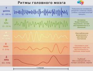
Alpha rhythm is regular, sinusoidal in shape, with a frequency of 8-13 Hz (oscillations per 1 s) and an amplitude of 20-80 μV (microvolts). The alpha rhythm is recorded when biopotentials are withdrawn from all areas of the cerebral cortex, but more constantly from the occipital and parietal areas. The alpha rhythm is recorded in a person under conditions of physical and mental rest, always with eyes closed and in the absence of external irritations.
The beta rhythm has an oscillation frequency of 14-35 Hz. Amplitude 10-30 µV. It can be recorded in any areas of the brain, but is more pronounced in the frontal lobes. When opening the eyes, mental work and other stimuli, the alpha rhythm is quickly replaced by the beta rhythm. This phenomenon of changing a rare rhythm to a more frequent one is called the activation reaction (or desynchronization).
The theta rhythm has a frequency of 4-7 Hz and an amplitude of 100-150 μV. Observed in a state of shallow sleep, with oxygen deprivation of the brain, under anesthesia.
The delta rhythm is characterized by slow oscillations with a frequency of 0.5-3 Hz, high amplitude 250-300 μV, up to 1000 μV. It is found in all areas of the brain, during deep sleep, as well as during anesthesia. In children under 7 years of age, the delta rhythm can also be recorded while awake.
An alpha equivalent of similar frequency, recorded over the central and central-parietal regions, is called the sensorimotor or Rolandic rhythm, since it is most pronounced in the projection of the Rolandic sulcus. It is believed that the Rolandic rhythm was first described in detail by Gastaut in 1958. This rhythm has a characteristic wave shape: arched waves with rounded tops and “sharp” bases, reminiscent of the Greek letter μ (mu). Therefore, there is another name for the sensorimotor rhythm - mu rhythm . The mu rhythm, like the alpha rhythm, is subject to depression during the activation reaction. But unlike the alpha rhythm, the mu rhythm is suppressed during motor activity: voluntary (clenching fingers into a fist), reflex, and even with the intention of movement.
kappa waves with a frequency of 8-12 Hz and a tau rhythm can be recorded over the midtemporal regions . It is believed that the recording of these rhythms on the EEG is hampered by the bones of the skull, and more often alpha equivalents can be recorded in the presence of defects in the underlying bone structures. The tau rhythm is selectively sensitive to auditory stimuli, mental arithmetic, and other types of mental activity.
There is another phenomenon: lambda waves are sharp single-phase oscillations, usually in the alpha or, less commonly, theta frequency ranges with an amplitude of up to 30-50 μV, recorded in the occipital leads during operation (!) of the visual analyzer (for example, when demonstrating visual images). Lambda waves are thought to be associated with saccadic eye movements when viewing complex images.
Comparison of EEG rhythms
| Rhythm | Frequency Hz) | Typical location | Common manifestations | For what pathologies |
| Delta δ | 0 — 4 | Frontally in adults, posteriorly in children; high amplitude waves | Deep sleep in adults In children Deep anesthesia and comatose states Present during attention tasks | Subcortical damage Diffuse damage Metabolic encephalopathy deep midline disorders Hydrocephalus |
| Theta θ | 4 — 7 | Hippocampus, cortex | In infants Drowsiness or awakening in adolescents or adults Simple (inaction) Associated with suppressed response to stimuli (has been found in situations where the subject is actively trying to inhibit some action or reaction) In hypoxia and shallow anesthesia | Focal subcortical lesions Metabolic encephalopathy deep midline disorders Some cases of hydrocephalus |
| Alpha α | 8 — 12 | Posterial areas of the head, on both sides, but with greater amplitude on the non-dominant side. Central locations (c3-c4) during rest | In a relaxed state With eyes closed Also involved in the control of inhibition, probably for the purpose of planning inhibitory activity in various parts of the brain Borderline state between sleep and awakening Meditation Immersion in dreams and fantasies | Coma |
| Beta β | 13 — 30 | Both sides, most frontally; small amplitude waves | Increased attention Active concentration, busyness or anxious thinking Mental activity Solving complex problems | Benzodiazepines Dup15q syndrome |
| Gamma γ | 30 — 100+ | Somatosensory cortex | Active learning Creative activity Cross-modal sensory processing (perception combines two different sensations, such as sound and a visual image) During short-term memory (recognition of objects, sounds, tactile sensations) During a conversation | Decreased cognitive abilities |
| Mu μ | 8 — 13 | Somatosensory and motor cortex | Motor neurons during inactivity | Autism |
EEG normal alpha rhythm
➥ Main article: Alpha rhythms
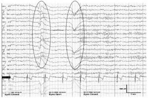
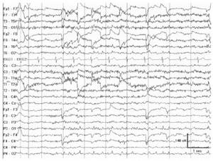
Rice. 1. Normal 10-Hz α-ri when the eyes open and is restored when the eyes are closed. Noteworthy is the increase in rhythm frequency immediately after closing the eyes (squeak phenomenon)
Alpha rhythm remains the first step in EEG analysis. Normally, the EEG reveals a rhythm predominant in the posterior leads, with a bilateral distribution, with a frequency in the range of 8-13 Hz (α-frequency). If this rhythm weakens when the eyes open, it is designated as the α rhythm. During the normal development of a child, the α-rhythm with a frequency of 8 Hz is formed by the age of 3 years. The frequency of the α rhythm remains stable between 8 and 12 Hz even during the normal process of growing up and aging, even into old age. In approximately 10% of healthy adults, the alpha rhythm is poorly visualized, and in less than 10% its amplitude may be <15 μV. The alpha rhythm is maximally expressed in the occipital regions and shifts in the anterior direction in a state of drowsiness. Amplitude asymmetry >50% should be considered pathological, especially if the amplitude on the left is higher than on the right. The alpha rhythm is most pronounced in a state of relaxed wakefulness, and the differences in frequency on both sides do not exceed 1 Hz. Unilateral absence of a-rhythm indicates pathology on the ipsilateral side (Banquo phenomenon). Normally, the frequency of the α rhythm can transiently increase immediately after closing the eyes (squeak phenomenon). There are two variants of the α-rhythm: with a decrease in frequency by half (slow alpha variant) or with its doubling (fast α-variant), while maintaining the characteristic distribution and reactivity. According to the characteristics of the waves, the α-rhythm can have a sawtooth shape. The paradoxical α rhythm appears in a state of activation, and not in a state of relaxed wakefulness.
EEG norm of mu rhythm
➥ Main article: Mu rhythms
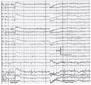
Rice. 2. Pronounced mu rhythm
in the left central leads when opening the eyes
The mu rhythm is localized in the central leads, which corresponds to the sensorimotor cortex, at rest, has an arched shape and a frequency characteristic of the alpha rhythm (usually 8-10 Hz) (Fig. 2). Although it resembles the α rhythm, it is not blocked when the eyes are opened; on the contrary, this rhythm is blocked during movements in the contralateral limb. It can be detected only on one side, and can be asymmetrical and asynchronous, despite the absence of an underlying structural lesion. The mu rhythm can slow down with age and usually has a lower amplitude than the α rhythm. A persistent, nonreactive mu-like rhythm associated with focal slowing is considered pathological.
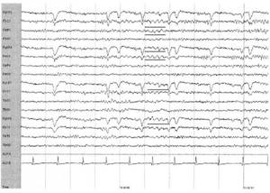
Normal EEG beta rhythm
➥ Main article: Beta rhythms

Rice. 3. Breach rhythm
in the right temporal region (with a maximum in T4) after craniotomy and temporal lobectomy
The beta rhythm exceeds 13 Hz in frequency. This is a common rhythm, normally its frequency varies between 18-25 Hz, and the amplitude does not exceed 20 μV. An amplitude above 25 µV is considered pathological. Benzodiazepine drugs, barbiturates and chloral hydrate provoke the appearance of pronounced generalized β-activity (fast activity) with an amplitude of >50 μV, which occupies >50% of the wakefulness recording and is in the frequency range of 14-16 Hz. Beta activity normally increases during drowsiness, light sleep, and mental activity. A persistent decrease in amplitude >50% suggests pathological changes in the gray matter of the cerebral cortex of the hemisphere in which the amplitude of the β rhythm is lower; however, less severe asymmetry may simply reflect normal cranial asymmetry. Defects of the skull can lead to the appearance of a breach rhythm (breach rhythm) of focal localization, asymmetric, high-amplitude (the amplitude can increase more than three times); β-activity in the area of skull defects can reach higher frequencies. This is considered normal in the absence of association with spikes (epileptiform activity) or focal slowing.
Theta rhythm on EEG
➥ Main article: Theta rhythms
Rice. 4. Normal θ rhythm
in the fronto-central leads in an 18-year-old patient in a state of wakefulness
The theta rhythm is characterized by a frequency of 4 to 7 Hz and varies in amplitude and morphology. In approximately one third of cases in healthy young adults, an intermittent theta rhythm is detected in a state of wakefulness, with a frequency of 6-7 Hz, an amplitude of <15 μV, maximally expressed in the frontal or fronto-central regions. Theta activity in the frontal areas can increase during emotions, concentration, and when solving intellectual problems. Theta activity normally increases during hyperventilation, in a state of drowsiness, and during sleep. Intermittent activity of 4–5 Hz, bitemporal, or even unilaterally predominant (usually left > right), can occur in approximately one third of cases in the elderly in the absence of clinical manifestations and is not considered pathological.
Lambda rhythm
Rice.
5. Lambda waves with biocipital location in a 28-year-old patient with complaints of dizziness. Noteworthy is the frequent appearance of an artifact associated with pursuit eye movements in leads F7 and T8
Lambda waves were originally described as superficially positively directed peaked θ waves appearing in the bilateral occipital region. These potentials have a duration of 160 to 250 ms, and at times may be peaked, asymmetric, and have a higher amplitude than the predominant resting rhythm in the posterior leads. When λ waves appear asymmetrically, they can be confused with interictal epileptiform discharges, which can lead to erroneous interpretation of the EEG. λ waves are best expressed in young adults (in cases where they are detected), although they are more often found in children. Lambda waves are better expressed when the patient is viewing a textured or complex image with the appearance of rapid saccadic eye movements. If, in this case, a white sheet of paper is placed in front of the subject’s eyes, this leads to the cessation of the flow of visual information necessary for the formation of λ waves.
Variant of EEG delta rhythm norm
➥ Main article: Delta rhythms
Rice. 6. Intermittent δ waves
left medial temporal leads during transition to drowsiness in a healthy 84-year-old patient undergoing evaluation for syncope.
The delta rhythm is activity of less than 4 Hz that accounts for <10% of the normal waking EEG recording in individuals aged 10 years or older. In the waking state, δ activity can be considered normal in very early and older ages. Normally, in older people, rare irregular δ-complexes can be detected in the temporal leads. This activity is similar in distribution to the temporal θ rhythm, often more pronounced in the left than in the right temporal areas, but in the Normal does not exceed 1% of the recording. In some cases, δ activity is considered normal - in people over 60 years of age, when a state of drowsiness occurs, in response to hyperventilation, and also during slow-wave sleep. Excessively expressed generalized δ activity is pathological and indicates encephalopathy of nonspecific etiology. Focal arrhythmic δ activity usually indicates a structural lesion involving the white matter of the ipsilateral hemisphere, especially if the activity is continuous and unreactive.
The nature of rhythms and connections with brain functions
New discoveries in the field of electroencephalography (EEG) made over the past several decades have significantly changed the classical concepts on which the training of psychiatrists and neurologists in medical schools was based. According to classical concepts, information processing in the human brain is realized through the impulse (spike) activity of individual neurons. Oscillatory electrical activity (eg, EEG rhythms) was, at worst, rejected and ignored, and at best, considered background activity. An example is the book Principles of neuroscience, edited by E. Kandel, J. Schwartz and T. Jessel, which is considered the “bible” of scientists involved in the study of brain activity. The chapter on EEG electrical oscillations was missing from the fourth edition, and John Martin's chapter, “Collective Electrical Activity of Neurons: The Electroencephalogram and the Mechanisms of Epilepsy,” presented in the third edition, was discarded.
Over the past few years the situation has been slowly changing. We are now experiencing an EEG renaissance, driven by new methods for assessing human EEG and new experimental findings in animal studies that have allowed electrophysiologists to discover that changes in EEG oscillatory patterns play a critical role in maintaining brain function and can therefore be used to a powerful tool for diagnosing brain dysfunctions.
From a general point of view, oscillations are present in all physical and biological systems that strive to achieve equilibrium. In almost all cases, oscillations occur when a system is driven by two opposing processes: one that takes the system out of equilibrium and one that brings it back to equilibrium. In this respect, EEG fluctuations are no different from those in other biological systems. In the case of any observed EEG rhythm (such as alpha, beta or theta), we can always detect the factor that throws the neuron or neural network out of balance and the cause that brings it back.
However, oscillations can not only be a reflection of the action of two opposing forces in neural networks, but hypothetically can also serve as a source of a unifying principle in the organization of neural networks. For example, changes in the total local electrical potential created by neurons that generate a given rhythm can involve other neurons in this process that are not directly involved in rhythm generation. This entrainment synchronizes the activity of all neurons in the neural network with the rhythm generators. Despite attempts to prove this assumption, we still do not know whether it is correct or not.
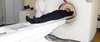
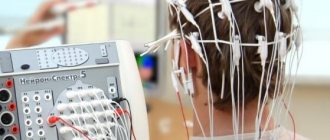
HOW IS A BRAIN EEG DONE?
A special cap with electrodes-antennas connected to the device itself is put on a person’s head. Signals coming from the cerebral cortex are transmitted to an electroencephalograph, which converts them into a graphic image (waves). This image resembles the rhythm of the heart on an electrocardiogram (ECG).
During the registration of brain biocurrents, the patient is in a chair in a comfortable position (reclining). However, he should not:
- be under the influence of sedatives;
- being hungry (in a state of hypoglycemia);
- be in a state of psycho-emotional arousal.
Types of bioelectrical activity of the brain
➥ Main article: Bioelectrical activity of the brain
EEG rhythms in the frequency range from 0 to 70 Hz cover several categories of electrical phenomena recorded from the scalp. These bioelectrical phenomena are traditionally divided into the following types: shifts of DC potentials, decasecond oscillations and slow waves, delta, theta, alpha and beta EEG rhythms. It must be emphasized here that the concept of rhythm assumes that rhythm represents regular changes in electrical potential measured by electrodes from the scalp. If a Fourier transform or wavelet transform is applied to an EEG recording containing rhythms, then these rhythms are detected in the corresponding spectra in the form of peaks.
To record decasecond oscillations, special amplifiers are required. Decasecond fluctuations are generally not considered in conventional EEG. Delta rhythms cover the frequency range from 1 to 4 Hz, theta rhythms - 4-8 Hz, alpha rhythms - 8-13 Hz and beta rhythms have a frequency above 13 Hz. Theta, alpha and beta rhythms are present in normal EEG, recorded at rest (with eyes closed or open) and while solving various problems. Delta rhythms in the normal brain are expressed in spectrograms in the form of peaks only during the state of deep sleep. Although the basic EEG rhythms have been known since Berger (late 1920s), their neurophysiological basis has only recently begun to be elucidated, starting around the 1980s.
It must be emphasized that the EEG is a sensitive marker of a person’s state: EEG rhythms change significantly when a person falls asleep and moves from one stage of sleep to another. For example, in stage II sleep, certain oscillations appear, called sleep spindles. Sleep spindles disappear while theta and delta rhythms develop in later stages of sleep. During wakefulness, rhythms can determine the extent to which the brain responds to various psychological tasks. For example, occipital alpha rhythms are suppressed (desynchronization response) while frontal beta rhythms are increased (synchronization response) in response to behaviorally relevant visual stimuli.
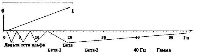
Rice. 7. Frequency ranges in the EEG spectrum
Frequency limits are not strictly defined. However, the usefulness of frequency classification is proven by the entire history of EEG.
In the damaged brain, the normal mechanisms for generating EEG rhythms may be disrupted, and the rhythms may: 1) become slower in frequency (EEG slowing); 2) have an unusual localization (for example, alpha rhythms in the temporal regions); 3) become higher in amplitude (hypersynchronization) and in greater synchrony with other areas (hypercoherence). In some severe cases (characterized, for example, by disconnection of cortical areas and subcortical structures, as a result of trauma or tumor), a slow rhythm with a frequency of the delta range (1-3 Hz) may appear. Also, normal synchronization mechanisms may be enhanced, causing spikes or spike/slow wave patterns to appear on the EEG, indicating a focus of epileptiform activity in the human brain, which can sometimes lead to an epileptic seizure. Normative databases can help the electroencephalographer identify the listed abnormal patterns and assess the level of statistical significance of the abnormality for each individual studied. The spatial localization of EEG rhythm generators can be assessed by various methods, such as dipole approximation or low-resolution electromagnetic tomography (LORETA).
Paroxysmal activity
➥ Main article: Paroxysm
In a normal brain, the processes of cortical excitation and inhibition are well balanced. The cortex, with balanced processes of inhibition and excitation, in a state of quiet wakefulness generates normal regular electrical oscillations: alpha rhythms, beta rhythms and mid-frontal theta rhythm. If this balance is disturbed so that excitation exceeds inhibition, the cortex begins to produce abnormal patterns of electrical activity called paroxysms. In most cases, they can be recorded using regular scalp EEG in the form of certain electrographic patterns. There are several types of epileptiform activity, the most common of which are spikes, sharp waves, and spike-slow wave complexes. Spikes are typically generated by a local cortical region called the focus. Using modern techniques such as electromagnetic tomography, LORETA or dipole analysis, epileptogenic foci can be localized in the cortex with good accuracy. Since the 1950s, for many years, spike detection by visual inspection of the EEG remained the only method. Over the past 10 to 20 years, highly reliable methods for automatic spike detection have been developed. Currently, most modern QEEG systems have additional tools for automatic spike detection, which help practitioners detect and analyze paroxysmal activity in the brain.
Reflection of the functioning of brain systems
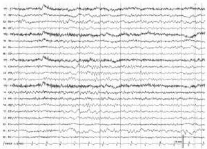
Rice.
8. An EEG recording duration of three minutes provides reliable and reproducible EEG spectra Power spectra and EEG topograms recorded from one subject for 3 minutes, with an interval between recordings of 7 days.
EEG is a complex combination of rhythms. For example, the patient's EEG spectra shown in Fig. 8 include frontal theta rhythm, occipital alpha rhythm, posterior temporal low-frequency beta activity, and central high-frequency beta activity. When compared with a normative database, frontal theta activity and posterior temporal low-frequency beta activity were found to be outside the normal range.
Different EEG rhythms reflect different mechanisms. Alpha rhythms reflect the state of the thalamocortical pathways. The midfrontal theta rhythm reflects the functioning of the limbic system. Beta rhythms are more local and reflect the state of local cortical areas. Determination of abnormal rhythmic EEG activity and the connection of these abnormalities with various brain systems corresponds to the second criterion of endophenotypes as biological markers of disease.
EEG of the brain (Electroencephalography)
Electroencephalography (EEG) is a method for studying the functional state of the brain, based on recording its bioelectrical activity through intact integumentary tissue of the head.
The EEG records the electrical activity of the brain, generated in the cortex, synchronized and modulated by the thalamus and reticular activating structures. Registration of bioelectric potentials of the brain and their graphic representation by photographic method or by ink recording are carried out by a special device - an electroencephalograph.
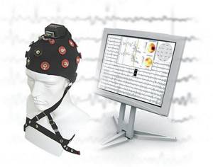
Its main components are highly sensitive electronic amplifiers, which make it possible to obtain on paper tape in real time a picture of changes in biopotential oscillations in different areas of the cerebral cortex, and oscillographic recording systems. Modern electroencephalographs are multi-channel devices (usually having 8 or 16, sometimes 20 or more amplification-recording units - channels) that allow simultaneous recording of biocurrents discharged from several symmetrical parts of the head. The study should be carried out in a light- and sound-proof room. Since EEG is one of the methods that allows one to highly accurately distinguish epilepsy from other paroxysmal conditions, therefore, undergoing an EEG (electroencephalogram) has become mandatory to obtain a psychiatric examination to obtain/replace a driver’s license (according to the Letter of the Ministry of Health and Social Development of the Russian Federation dated April 5 2012 N 14-5/10/2-3374).