Radiography appeared 5 years before the end of the 19th century and became the first study in the history of mankind of the internal structure of the body, without introducing it into it. For a whole century, X-rays had a reputation as the most informative study, and even today CT and MRI are not able to displace radiography from clinical practice.
- What is radiography?
- Indications and contraindications for
- Advantages and disadvantages of x-ray diagnostics
- Types of radiography
- Preparing for X-rays
- Research technique
- How are radiographic results interpreted?
Among diagnostic procedures, the combined share of CT with MRI does not exceed 3%, while X-rays are performed 15 times more often. Every year, more than 125 million X-ray examinations are performed in public medical institutions alone, and every fifth one is performed on a digital device. The frequency of x-rays is comparable only to the number of ultrasounds.
What is radiography?
Radiography is an image of a specific part of the body made using ionizing radiation directed from a machine.
Fluoroscopy is an X-ray examination lasting several minutes, when the doctor, through a special screen, visually monitors what is happening inside the patient’s body, occasionally photographing what is happening.
Recording the internal state of an organ on film in real time - an x-ray or radiograph.
You can take an x-ray of any part of the body and get a true image; in fact, it is a black and white “photograph” of all layers of the anatomical area of interest.
The reliability of the image can be increased by adjusting the shutter speed and power of the X-ray tube. Tissues and organs filled with air will be dark in the image, bones will be light, that is, the greater the density of the tissue, the lighter it will be on the x-ray.
Radiography provides an image fixed at a point in time; several images in different positions of the patient will help to get an idea of the deviations of processes from the norm in dynamics, but the idea will be somewhat incomplete without the introduction of contrasting solutions into the vascular bed, hollow organ or ducts of the studied area.
Modern X-ray machines have not become completely harmless to the subject, but the dose of radiation received during the study is minimal and is offset by the importance of the diagnostic information obtained. Analogue devices are gradually becoming a thing of the past, giving way to digital ones, where everything is calculated by a computer program, the image is displayed on the monitor without the need to develop the film, and the pictures taken during the process are stored on electronic media.
Indications and contraindications for
The main indication for radiography in oncology is the need to obtain reliable information about the state of the anatomical area of interest in real time, that is, the diagnosis of deviations from the anatomical norm that cause certain clinical symptoms.
All cancer patients undergo radiography of several anatomical areas already at the examination stage:
- zones of development of the primary malignant process;
- zones of maximum probable metastasis, which must include chest x-ray, usually called “lung x-ray” by patients;
- areas where, according to the clinical signs or complaints of the patient, metastases are suspected.
During treatment, it is also impossible to do without radiography:
- choosing the optimal volume of surgical intervention is impossible without knowledge of the relationship of the cancer tumor with nearby anatomical structures;
- after surgery with intubation and anesthesia, the condition of the lungs is monitored; if inflammation is suspected, X-ray examinations are carried out at intervals of several days;
- after surgery, the installation of internal catheters or stents is monitored;
- during cyclic chemotherapy, assessment of the dynamics of metastases in the lungs and bones is impossible without X-ray examination;
- in the organs of the gastrointestinal tract and pulmonary system, complications of special treatment or the natural course of the malignant process are diagnosed;
- Dynamic monitoring after completion of treatment requires images of areas of possible cancer relapse and metastasis.
The very presence of a malignant tumor, the process of its treatment and subsequent follow-up is an absolute indication for regular and repeated X-ray diagnostics.
Contraindications to examination in oncology are relative and are almost never encountered in clinical practice:
- studies are not carried out on pregnant women, who extremely rarely have malignant neoplasms;
- the patient’s severe and extremely serious condition is also not a reason to refuse examination, since it may be due to an underlying disease and quick diagnosis using x-rays helps in choosing optimal therapy.
Oncological disease is an absolute indication for X-ray examination in the absence of absolute contraindications.

In which bronchi do malignant tumors occur?
The air that a person inhales passes through the nose, pharynx, larynx, and trachea. At the level of the upper edge of the fifth thoracic vertebra, the trachea ends and divides into two main bronchi . This location is called the tracheal bifurcation . The main bronchi are the first order bronchi , they are divided into lobar (second order) , then into segmental (third order), subsegmental (fourth order), lobular, and finally into terminal (end) bronchioles.
All these branches together make up the bronchial tree . The walls of large and small bronchi are structured in the same way: on the inside they are lined with mucous membrane , under it there is a framework - fibrocartilaginous membrane , on the outside - the adventitial membrane.
Malignant tumors that originate from the mucous membrane are called cancer. They can occur in any part of the lung, but are most often found in the hilum - where the main bronchus enters the lung. In two thirds of cases, cancer develops in the bronchi of the first, second and third order.
Advantages and disadvantages of x-ray diagnostics
Radiography, in addition to reliable diagnostic information about the real state of the anatomical region under study, allows inexpensive and fairly rapid monitoring of the effectiveness of antitumor therapy. A few minutes are enough for the examination.
It is correct to take more than one direct photo - a “photo” from front to back, but also a side one, in order to have a complete understanding of the localization of the pathology inside the organ.
During the tumor process, layer-by-layer images are taken - tomograms, when the X-ray beam seems to cut the organ and surrounding tissues, forming a slice of any anatomical part at a certain distance from the skin. Several tomograms are taken in “steps” of several centimeters. Additional options increase the cost of the examination, which is still incomparable with the price of standard CT and MRI.
Not all organs are accessible to X-rays, for example, the pancreas is not visible at all, the intestines are noticeable only by their contents, additional contrast allows you to see many hidden internal organs.
Patients treat X-ray diagnostic procedures with ease because they do not hurt and there is no risk of infection. Each X-ray examination complements the total radiation exposure of the patient, but a cancer patient has to take many pictures and quite often, because without high-quality diagnostics it is impossible to either select the optimal treatment or evaluate its result. Nevertheless, from the point of view of the harm-to-benefit ratio, a cancer patient benefits from radiography more than he loses health due to radiation.
Radiography is inferior in image quality to CT and MRI, but there is not always a need to clarify “100%”; often the speed and accessibility of the examination is sufficient. Each method has been found to have an optimal place in the diagnostic and treatment process for an oncological patient.
CT scan for lung oncology
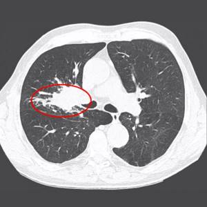
The best diagnostic data for cancer tumors is provided by lung tomography. This study also helps to detect accompanying signs that may, to one degree or another, illustrate the pathological process. According to the results of tomography, you can find:
- narrowing of the bronchi;
- complete obstruction of the bronchial lumen;
- problems filling the lungs with air;
- blurred contour of the bronchi due to damage by the tumor process;
- shadow of a tumor in the area of bifurcation of the trachea;
- increase in the angle between the bronchi;
- abnormal cavities;
- compression by bronchial metastases.
Lung cancer is not always visualized on x-rays, and if darkening is visible, the doctor still needs to differentiate it. That is why tomograms receive such important attention in diagnosing respiratory oncology.
Types of radiography
The types of research are determined by the characteristics of the anatomical zone being studied and the process of taking the image.
Standard radiography is a survey, that is, a picture that gives an idea of the condition of the entire organ, or rather pictures in two mutually perpendicular projections - anterior and lateral.
In dentistry and for oral tumors, a version of the overview image is known as orthopantomography, but in radiography technology it is a panoramic image, during which the beam emanating from the apparatus passes along a wide arc, appearing on film in the form of the upper and lower jaw, and not an individual tooth.
The opposite of the survey view, targeted radiography allows you to “photograph” a specific area, for example, the upper mediastinum or the root of the lung altered by a tumor. This type is performed only after a survey x-ray.
Essentially the same targeted contact radiography is used in dentistry, when a film is placed in the mouth and only the diseased tooth is removed. In oncology, intraoral technique is used for cancer of the oral mucosa and oropharynx to assess the involvement of bone in the tumor conglomerate.
Similar to sighting is close-focus radiography, usually of small structures and at a close distance from the beam tube. In a small focus, the pathology is seen more clearly.
The introduction or intake of a contrast agent visualizes the cavitary organs of the gastrointestinal tract, urinary system and vascular network - contrast study of the intestine - irrigoscopy, bile ducts - cholecystography, urinary tract - urography, fistulas - fistulography.
X-ray according to Vogt is not used in oncology; a photograph of the eye without skull bones is necessary for injuries. For malignant eye processes, CT is used.
There is also rarely a need for x-rays with functional tests when the patient is filmed in a certain position, for example, with spinal tumors.
Soft tissue radiography is also not a widely used method, but may be useful in sarcomas.
Main features of nodular forms of peripheral cancer associated with the anatomy of the respiratory system
There are differences in the anatomical structure of the left and right lungs:
- the right main bronchus is wider, straighter and located higher;
- Three lobes stand out on the right, two on the left, while the upper lobe of the left lung bordering the heart has the so-called “cardiac notch.”
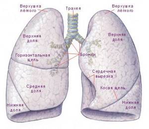
The structure of the human lungs
Cancer of any type most often affects the right paired organ, since it is easier and easier for carcinogenic substances to penetrate here.
The development of carcinoma in the upper lobes does not cause significant enlargement of the lymph nodes, but leads to expansion of the root.
Tumors of the lower lobes, as the disease progresses, affect nearby lymph nodes, which is manifested by a significant increase in the latter.
Preparing for X-rays
Modern digital devices require virtually no patient preparation. Diagnosis of the condition of the chest organs takes place “on the fly,” that is, it is possible at any time and only requires the removal of clothing and jewelry.
To study the state of the gastrointestinal tract, there is a need to empty the organs; when examining the esophagus, stomach, and gallbladder, you should not eat for several hours.
In case of oncological pathology of the colon, contrast is required, so the intestines are cleansed 2 hours before diagnosis. It is advisable to avoid foods that promote gas formation for a couple of days.
Signs of the disease
Many patients ignore the first symptoms that arise from tumors, mistaking them for a common cold.
The first signs of lung cancer in women and men that should not be ignored:
- Prolonged cough.
- Shortness of breath, especially with minor physical exertion.
- Breathing is wheezing.
- General weakness, depressed state.
- Lack of appetite.
- Temperature surges.
Bloody streaks are found in the coughed up sputum.
Over time, these symptoms become more severe. These include severe pain in the chest, difficulty swallowing food, and thinness.
Research technique
Before diagnosis, the patient must undress, since all tissues repel some of the X-rays; this rebound creates secondary radiation, which reduces the clarity of the image. Attenuation of ricochet rays from the skin, soft tissues, and organs is provided, and each X-ray machine has its own correction table.
Metal decorations also get in the way, creating artifacts; they are removed.
I place the patient on the table in a certain position that allows optimal visualization of the pathological area. The X-ray room staff goes into a room with special protection, from where they communicate with the patient via speaker, offering to freeze at a certain moment to take the image. This is followed by developing the film and describing the x-ray picture.
Causes of bronchial cancer
A normal cell turns into a tumor cell when certain changes occur in its genes. It is impossible to say exactly why, in one case or another, genetic defects arose that led to malignant transformation. But there are known factors that significantly increase the risk of the disease. They are called risk factors .
The main risk factor for bronchopulmonary cancer is smoking. The likelihood that a person will be diagnosed with a malignant tumor directly depends on the length of smoking, the daily number of cigarettes smoked, the age at which the person started smoking, and the brand of cigarettes (tobacco quality, carcinogen content). Not only active but also passive smoking is dangerous. If someone constantly smokes in an apartment, the risk of lung cancer is increased for all its residents.
Other risk factors:
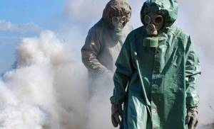
- Occupational hazards: workers in mines, factories producing cement, glass and fiberglass.
- Contact with chemicals, some volatile substances, asbestos.
- Air pollution with radon , a radioactive gas that is naturally released from the soil when uranium decays.
- It can accumulate indoors.
How are radiographic results interpreted?
Depending on the diagnostic task, radiography can be performed with an increase or decrease in the image, increased rigidity, several series and in several projections. Specialists know what to do and how to do it, they know all the capabilities of the equipment.
A blurry image is not only a waste of film and wasted time; blurriness requires re-examination and is associated with additional radiation exposure. For a better image, special filters are used, the capabilities and characteristics of which must be known to the X-ray room personnel and correlate them with the individual characteristics of the patient - his anthropometric data, weight and, ultimately, with the distance to the organ being studied.
Since complete immobility of the subject is impossible, the internal organs also move at their own rhythm, the image will be of high quality only with high power of the device and a short shutter speed.
A high-quality radiograph can be spoiled by incorrect interpretation. A standard for describing images has been developed, but the final result is related to the professionalism of the doctor, his experience and knowledge, observation and intuition, and of course, knowledge of clinical oncology. Our clinic has excellent equipment and staff, so X-ray reports are always of an expert level.
Book a consultation 24 hours a day
+7+7+78