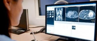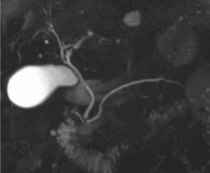
Three-dimensional MR cholangiopancreatogram shows a normal gallbladder. Its duct appears as a bright line and connects the organ with the common bile duct. Ultrasound, which has sensitivity and specificity for both gallstone disease and inflammation, has been the traditional method for assessing gallbladder diseases, but if sonography results are ambiguous, MRI is justified. Diagnostics based on the interaction of a magnetic field and hydrogen protons in water molecules with subsequent computer processing is absolutely safe for most patients who do not have metal components in their bodies. Magnetic resonance scanning allows you to simultaneously conduct an anatomical and physiological study of the condition of the organs of the biliary system. To improve visualization, diagnostics can be performed with contrast enhancement.
An MRI of the gallbladder should be done in patients with fever, unexplained pain in the right hypochondrium, accompanied by or without jaundice, or a palpable formation in this anatomical area.
Gallbladder diseases
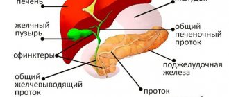
The gallbladder serves as a storage facility for bile produced in the liver. Violation of its function is diagnosed after operations and due to diseases of the hepatopancreatobiliary system, duodenum or the organ itself.
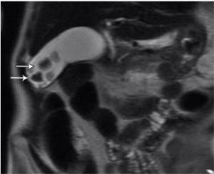
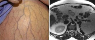
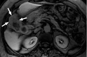
Gallbladder MRI shows stones (arrows)
Common gallbladder pathologies include:
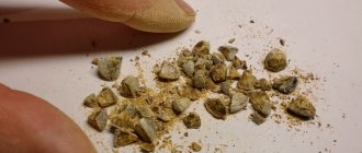
- cholelithiasis. The process of stone formation triggers the oversaturation of bile and the precipitation of crystals. Obesity, rapid weight loss, pregnancy, and impaired estrogen production are known risk factors. Gallstones may consist primarily of cholesterol or the pigments calcium bilirubinate and glycoproteins. Clinical manifestations are absent or manifested by paroxysmal pain in the right hypochondrium, associated with errors in nutrition. MRI is an important tool for assessing the density of stones; based on the data obtained, the doctor decides on the possibility of conservative treatment, endoscopic crushing or surgery;
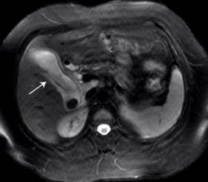
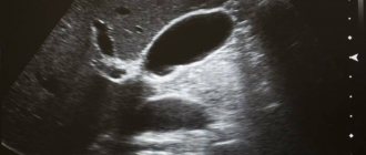
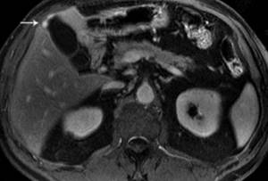
MRI of the gallbladder: acute cholecystitis. The image shows wall thickening, uneven contours, and swelling (arrow)
- cholecystitis: acute inflammation of the gallbladder is often a complication of cholecystolithiasis (cholelithiasis). The clinic presents with severe pain in the area of the right hypochondrium, increased body temperature, and the level of blood leukocytes. In the absence of stones, they speak of non-calculous cholecystitis. In the first hours, the gallbladder increases in size and fills with exudate or pus. The contents contain a large number of bacteria. In the absence of timely diagnosis and treatment, necrosis and perforation of the organ develops;
- in chronic cholecystitis, MRI shows a decrease in the size of the gallbladder and thickening of its walls. Symptoms are vague and include nausea, bloating, epigastric pain;
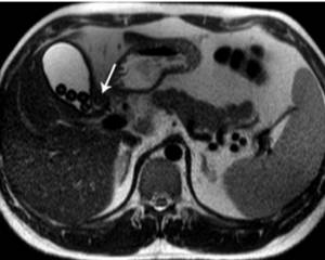
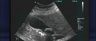
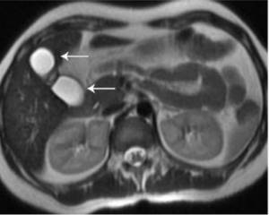
MRI: multiple stones in the lumen of the gallbladder and a stone contributing to the formation of a fistula tract (arrow)
- Mirizzi syndrome. The destructive-inflammatory process is associated with compression of the common hepatic duct by stones located in the cystic duct or Hartmann's pouch. As a result of compression, a stricture or fistula is formed - vesicocholedocheal fistula;
MRI scan with contrast: gallbladder cancer. Wall thickening and swelling in the fundus (arrows)
- Gallbladder tumor. Gallbladder cancer is a malignant neoplasm, most often located in the fundus or cervix. Tumor invasion occurs in the common bile duct, cystic duct, liver and anatomically closely located organs - the stomach and intestines. Distant metastases in advanced cases are found in the omentum, ovaries (in women), pleura, and peritoneum. Histologically, gallbladder cancer is most often represented by adenocarcinoma. MRI allows you to detect a tumor before the development of clinical manifestations, at an early stage, which improves the chances of recovery. In 90% of cases, examination reveals background cholecystolithiasis. Symptoms as they progress include pain in the abdomen, right hypochondrium, weight loss, weakness, low-grade fever;
MRI: adenomyomatosis of the gallbladder (arrow)
- Hyperplastic adenomyomatosis. Adenomyomatosis of the gallbladder is a benign, non-inflammatory process characterized by the simultaneous proliferation of the muscular and mucous layers, which leads to dysfunction and digestive problems. There are no clinical manifestations for a long time. As cystic cavities, growths, and nodes form, interfering with the contraction of the organ and the release of bile, complaints develop that correlate with the severity of the pathological process. The danger is the appearance of areas of atypia, which increases the likelihood of malignancy. Hereditary predisposition, cholelithiasis, chronic pathologies of the hepatopancreatobiliary region, and poor nutrition are considered the main factors of adenomyomatosis;
MRI of the gallbladder demonstrates a congenital malformation - duplication (arrows)
- Developmental anomalies. Anomalies in the development of the gallbladder include: agenesis (absence), aplasia (underdevelopment), organ duplication. Unfavorable factors influence the formation of malformations of the bile ducts. Sometimes pathology is a diagnostic finding, but more often an extended assessment is required when symptoms manifest that are similar to the clinical manifestations of pancreatitis, cholecystitis, or hepatitis. MRI can reveal atresia (underdevelopment), stenosis (narrowing), the appearance of additional channels for bile drainage, a common bile duct cyst, etc. Ultrasound in these cases is not informative.
Classification
According to the modern classification, cholelithiasis is divided into three stages:
- the initial physicochemical stage (pre-stone stage, characterized by changes in the composition of bile) is not clinically manifested; it can be identified by biochemical analysis of the composition of bile;
- the stage of stone formation (latent stone carriage) is also asymptomatic, but with instrumental diagnostic methods it is possible to detect stones in the gallbladder;
- the stage of clinical manifestations is characterized by the development of acute or chronic calculous cholecystitis.
Sometimes a fourth stage is identified - the development of complications.
Classification of cholelithiasis by localization:
- Concretions of the bottom and body of the gallbladder (usually asymptomatic).
- Gallbladder neck stones (usually symptomatic).
Classification of gallstones:
- Based on their chemical composition, they are distinguished: Cholesterol (pure and mixed): Non-calcified.
- Calcified.
- Non-calcified.
- Non-calcified.
- Single stones (1-2).
- Small (less than 3 cm).
Do they do MRI of the gallbladder?
An MRI of the gallbladder can be done free of charge upon referral from the attending physician at a recommended clinic with which an agreement has been concluded, or by self-referring to a private diagnostic center. The examination is justified if it is necessary to clarify ultrasound data, clinical and laboratory parameters, in case of a recurrent course of the disease, as part of preoperative diagnostics, etc. In St. Petersburg, there are more than a hundred places where MRI of the gallbladder is performed. When choosing a clinic, you should clarify the characteristics of the magnetic resonance imaging scanner: the higher the field strength, the more accurate the results. It is better to choose expert-class equipment.
Alternative ways to diagnose liver conditions
MRI of the liver and biliary tract is the most accurate, safe and effective diagnosis, but it is quite expensive and not all patients can afford. In this case, the doctor may suggest alternative research methods, such as CT.
CT, like MRI, can show the presence or absence of liver disease, but its cost is an order of magnitude lower. The pricing policy is determined by the fact that computer tomography equipment costs less than magnetic resonance imaging equipment. But it is important to understand that during a CT scan the patient is irradiated with X-rays, therefore, it cannot be called safe for the body.
Best materials of the month
- Coronaviruses: SARS-CoV-2 (COVID-19)
- Antibiotics for the prevention and treatment of COVID-19: how effective are they?
- The most common "office" diseases
- Does vodka kill coronavirus?
- How to stay alive on our roads?
Often, studies need to be repeated after a short period of time, and in this case, diagnosis using computed tomography is no longer suitable. All doctors recommend, despite the cost, to undergo safer and more reliable diagnostics through the use of magnetic resonance equipment.
Preparing for an MRI of the gallbladder

For MRI of the gallbladder and liver, you should appear on an empty stomach, but you can take a small snack with you - it will be useful while waiting for the result.
Preparatory measures include following a diet 4-5 days before diagnosis, excluding from the diet foods that contribute to stagnation of bile or its excessive production :
- fatty meats and fish;
- legumes;
- raw vegetables, herbs and fruits;
- mayonnaise, margarine;
- spices and marinades;
- canned food;
- milk and its derivatives;
- yeast-containing baked goods;
- confectionery products, etc.
Alcohol, strong coffee and tea are prohibited. You must appear for an MRI of the gallbladder on an empty stomach; your last meal should be no later than 6-12 hours before. Smoking contributes to organ contraction and unreliable results. For patients suffering from flatulence and constipation, the possibility of cleansing the intestines with an enema or taking medications (antispasmodics, sorbents) should be checked with the doctor who referred for diagnosis.
Why, in fact, about the old?
It is known that behind large volumes, as a rule, “traps” are hidden. There are situations when a seemingly simple intervention turns into a complex process with a possible serious outcome. This is due to pronounced scar-infiltrative changes that occur as a result of ongoing or previously suffered exacerbations of the disease.
A new galaxy of young surgeons is growing, for whom the topic of cholecystitis seems to have been resolved long ago. It is for them that it is necessary to return to the topic again and again. There is nothing more dangerous than a frivolous attitude before surgery. Everyone knows the wisdom: “you need to learn from other people’s mistakes,” but for some reason people still prefer to learn from their own...
We should not talk about “blue” blisters, but about complicated and insidious forms of this pathology. Today, experts are increasingly talking about the so-called “difficult” gallbladder.
MRI of the gallbladder: how is it done?
MRI diagnostics is absolutely painless.
At the appointed time, the patient arrives for the MRI procedure. At the reception you must fill out a questionnaire that allows you to identify contraindications and voluntary consent to the diagnosis. All objects made of or containing metal (coins, jewelry, watches, cell phones, keys) that can cause artifacts on films are deposited. It is preferable to wear loose-fitting clothing that does not put pressure on the stomach - you will have to lie still for 15-30 minutes.
An MRI of the gallbladder is performed in an isolated room; the medical staff monitors the progress of the procedure through the glass of an adjacent room. There is a button for speakerphone at hand.
When a contrast is planned, the amplifier is injected into a vein by a nurse or the drug is given as a bolus using special equipment. You may be asked to hold your breath for a few seconds. The noise from a working tomograph can be leveled out using headphones. Before the procedure, the x-ray technologist will place intensifying coils in the abdomen and fasten straps to ensure immobility. On command, the tomograph table will move the subject into the drum tunnel. There are no painful sensations during the MRI. The procedure does not involve the use of x-rays, therefore it is absolutely safe.
Contraindications
Contraindications for MRI can be divided into two categories:
- Absolute – the first trimester of pregnancy, the presence of cardio and neurostimulators, vascular clips, metal pins, fragments and other metal and other electronic devices in the body. They can be damaged by exposure to a magnetic field, so MRI is prohibited.
- Relative – there is no strict prohibition on the examination, but the feasibility of it is decided by the clinic doctor. The main relative contraindications are the second and third trimester of pregnancy, claustrophobia, panic attacks and other conditions in which the patient may experience severe discomfort during the procedure or involuntary body movements that can reduce the clarity of the images.
MRI after gallbladder removal

Each pain has its own cause, the sooner it is found out, the greater the chance of recovery.
After removal of the gallbladder, if complaints of pain and heaviness in the epigastric region, digestive disorders, persistent abnormalities in liver tests, the doctor may prescribe an MRI of the abdominal organs to clarify the cause. unpleasant symptoms. During cholecystectomy, special metal staples (clips) are often applied to cut off the cystic duct from the biliary system. In modern surgery, titanium devices are used, which does not in any way interfere with MRI after removal of the gallbladder and does not pose any risk to patients.
Clinical manifestations after surgery can be caused by:
- postcholecystectomy syndrome;
- progression of pathology in gallbladder cancer;
- activation of chronic diseases;
- non-compliance with the recommended diet;
- complications and medical errors.
An MRI will help determine the cause of pain in the right hypochondrium after gallbladder removal.
Why do you need contrast?
The contrast agent used for MRI examinations is practically safe for the human body. By injecting contrast into the blood before performing an MRI of the liver, it is possible to detect: the presence of narrowing of the bile ducts; the presence of neoplasms, polyps and stones; causes of obstructive jaundice; condition of the biliary tract and bladder.
The contrast agent, entering the vessels, carries out the process of staining them. The stronger the blood flow, the stronger the staining, therefore, the diagnosis will be more informative.
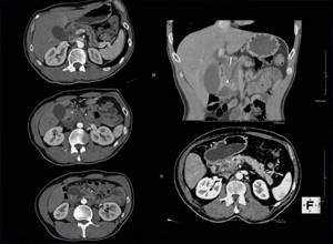
Differential diagnosis
Differential diagnosis: small stones with polyps.
T2-VI ax. scan: in the projection of the gallbladder body, the presence of multiple filling defects (calculi) is noted. Arrows indicate stones.
T2-VI cor. scan: the presence of multiple filling defects (calculi) of the gallbladder and deformation of the body of the gallbladder are noted. Arrows indicate stones.
MRCG: a few filling defects (calculi) are visualized in the CBD, the bile ducts are dilated. Arrows indicate stones.