Published: 06/20/2012 Updated: 05/20/2021
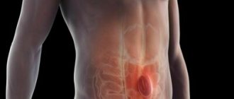
Diagnosis of infectious diseases is one of the most difficult problems in clinical medicine. Laboratory research methods play a leading role in a number of nosological forms, and in a number of clinical situations they play a decisive role not only in diagnosis, but also in determining the final outcome of the disease.
Diagnosis of infectious diseases almost always involves the use of a complex of laboratory methods.
The network of independent laboratories "CityLab" uses 3 groups of special laboratory research methods in its work to diagnose infectious diseases:
- bacteriological;
- serological;
- polymerase chain reaction (PCR) method for detecting DNA or RNA of the causative agent of an infectious disease in the material being studied.
In some patients, to diagnose the etiology of the infectious-inflammatory process, it is enough to conduct a bacteriological study; in other clinical situations, data from serological studies are decisive; thirdly, only the PCR method can provide useful information. However, most often in clinical practice, a clinician needs to use data from various laboratory research methods.
Bacterioscopic method (smear for infection)
Smear (bacterioscopic method) is the most common method of laboratory diagnosis.
With the bacterioscopic method, analysis is performed using a light microscope. The material to be tested is added to a drop of saline solution, thoroughly mixed and distributed over the glass. There should be good staining of parts of cells and bacteria in different colors to assess the composition of discharge from the urethra, vagina and cervix.
Analysis of scrapings from the urethra or vagina in gynecology allows you to determine: - the number of leukocytes (their increase indicates an infection, but in healthy women a small number of leukocytes are present in the flora); - the number of red blood cells (the main indicator of the inflammatory process with an increased number); - the composition of the flora (in women it depends on the menstrual cycle, but cell activity indicates a violation of the microflora); - the presence of Trichomonas, gonococci, fungus - the main goal for identifying the causative agent of infections; — the presence of lactobacilli (their absence indicates a disturbed microflora). The smear also determines the degree of cleanliness of a woman’s vagina: degrees 1 and 2 are characteristic of healthy flora, and degrees 3 and 4 reveal colpitis and inflammation.
The advantages of this method are its simplicity, accessibility and speed of obtaining results (30 - 60 minutes or less). However, the sensitivity of this method is limited (about 105 bacteria per 1 ml). Viruses, chlamydia, mycoplasma and ureaplasma are practically not detected in a urogenital smear, and the smear results alone are often not enough to make a diagnosis. To diagnose these infections, it is recommended to use PCR diagnostics and ELISA tests. The smear results can usually be used as a guide.
It is better to refrain from urinating before taking a smear.
MEDICAL CENTER
Interpretation of study results contains information for the attending physician and is not a diagnosis.
The information in this section should not be used for self-diagnosis or self-treatment. The doctor makes an accurate diagnosis using both the results of this examination and the necessary information from other sources: medical history, results of other examinations, etc. Limitation of the method
When studying biological fluids (seminal, pleural, peritoneal, ejaculate, etc.), the main purpose of the study is to identify bacteria. Due to the fact that in most cases these types of material are poor in cellular composition, smears on glass are poorly adhered and fixed; during the staining process, part of the smear may be lost, which leads to a low information content of the description of the cellular composition.
ATTENTION: this type of microscopic examination does not replace general clinical analysis, the main purpose of which is a microscopic examination of the cellular composition, carried out strictly in liquid (native) material.
When viewing a smear, pay attention to:
- the presence and type of epithelial cells present in the smear;
- the presence of cellular detritus and mucus;
- the number of leukocytes determined in one field of view;
- the presence of certain morphological types (morphotypes) of bacteria and fungal elements, their relationship to dyes (with Gram staining), as well as their relative quantity.
Types of bacteria that may be found in swabs:
- gram-positive and gram-negative cocci located extra- and intracellularly, in pairs, groups, chains;
- gram-negative coliform bacteria;
- lactomorphotypes (type of lactobacilli);
- morphotypes of diphtheroids or corynebacteria;
- morphotypes of strict anaerobic bacteria (mobiluncus, peptostreptococci, fusobacteria, bacteroides)
Interpretation of the microscopic picture of smears:
- The total amount of material on the glass is estimated.
- The condition of the epithelium and the presence of key cells are assessed. The number of epithelial cells is not taken into account, since it has no diagnostic significance (true for gynecological smears).
Assessment of leukocyte reaction:
- no leukocytes, units in p/z.;
- up to 10 in p/z. - a small amount of;
- 10 - 15 in p/z. - moderate amount;
- 30 - 50 in p/z. and more - a large number;
- moderate and large amounts are assessed as etiologically significant for the inflammatory response.
Quantitative assessment of microflora:
- + or up to 10 in p/z. insignificant/scanty amount;
- ++ or 11 - 100 in p/z. moderate amount;
- +++ or 100 - 1000 in p/z. large amount/significant;
- ++++ or more than 1000 in p/z. massive amount.
Seeding recommended:
- in the case of a moderate and large leukocyte reaction in the absence of microbes in the smear (since the microscopic method has a lower limit of detection > 105 CFU/ml, and pathogenic potencies of opportunistic microorganisms can appear in small quantities (at the level of 104 - 105 CFU/ml );
- if morphotypes of UPM are detected by microscopy, even as single ones in the field of view, to determine the genus and type of microorganism, determine sensitivity to antibiotics and estimate the quantity to identify etiological significance;
- for differentiated diagnosis of lactobacilli, clostridia, eubacteria, propionobacteria, etc., since they are represented by a single morphotype;
- to determine the type of fungi and determine their sensitivity to antimycotic drugs;
- in the case of bacterial vaginosis, if it is combined with a nonspecific inflammatory process, accompanied by a moderately pronounced leukocyte reaction.
Seeding is not recommended (or required):
- in case of bacterial vaginosis, if it is not combined with UPM;
- in case of cytolytic vaginosis;
- in the case of an intermediate form of microcenosis;
- in case of vaginal atrophy.
Bacteriological method (bacteriological culture)
Bakposev is a method of diagnosing STDs by growing microbes in an environment favorable for this. It is the gold standard in identifying infections because it detects not only the virus, but also its activity. The advantage of this method is the ability not only to identify the bacterium, but also the ability to obtain an antibiogram, on the basis of which the doctor prescribes precise treatment. Most often, culture is used to diagnose ureaplasmosis, mycoplasmosis, trichomoniasis, chlamydia, and various forms of candidiasis.
Rules for taking a culture test:
- do not douche during the day, do not use vaginal suppositories;
- a day before the test, abstain from sexual intercourse;
- stop taking antibiotics for a week;
- do not empty your bladder for 1.5-2 hours.
PCR
PCR diagnostics, or polymerase chain reaction, is aimed at determining the specific nucleotide sequence of the DNA section of the infectious agent inside the cell, which cannot be achieved with a smear or bacterial culture. PCR analysis is often used to detect: chlamydia, ureaplasmosis, mycoplasmosis, herpes genius, gonorrhea, trichomoniasis. DNA diagnostics is indispensable for detecting viral hepatitis and HIV infection. This method is capable of identifying the pathogen even at the stage of the incubation period, but it does not give an idea of the activity of the virus, as ELISA does. In addition, PCR may not detect genital herpes and cytomegalovirus in vaginal scrapings if the virus is in “hibernation”; it is recommended to use an enzyme-linked immunosorbent assay to detect these infections. The rules for submitting material for PCR diagnostics are the same as for bacterial culture.
PCR is the most accurate and modern diagnostic method. A summary assessment of the sensitivity of various diagnostic methods carried out in several foreign research centers showed that “rapid” or express tests have a sensitivity of 40-60%, ELISA - 50-70%, direct immunofluorescence (DIF) - 55-75%, culture - 60-80%, and PCR from 90 to 100%.
Serological research methods
All serological reactions are based on the interaction of antigen and antibody. Serological reactions are used in two directions.
1. Detection of antibodies in the blood serum of the subject for diagnostic purposes. In this case, of the two components of the reaction (antibody, antigen), the unknown is the blood serum, since the reaction is carried out with known antigens. A positive reaction result indicates the presence in the blood of antibodies homologous to the antigen used; a negative result indicates their absence. Reliable results are obtained by studying “paired” patient blood sera, taken at the beginning of the disease (3-7 days) and after 10-12 days. In this case, it is possible to observe the dynamics of the growth of antibodies. For viral infections, only a fourfold or greater increase in antibody titer in the second serum has diagnostic value.
With the introduction of the enzyme-linked immunosorbent assay (ELISA) method into laboratory practice, it became possible to determine antibodies belonging to various classes of immunoglobulins (IgM and IgG) in the blood of patients, which significantly increased the information content of serological diagnostic methods. During the primary immune response, when the human immune system interacts with an infectious agent for the first time, predominantly antibodies belonging to class M immunoglobulins are synthesized. Only later, 8-12 days after the antigen enters the body, class G immunoglobulin antibodies begin to accumulate in the blood. During the immune response to infectious agents, class A antibodies (IgA) are also produced, which play an important role in protecting the skin and mucous membranes from infectious agents.
2. Establishing the genus and species of a microbe or virus. In this case, the unknown component of the reaction is the antigen. Such a study requires a reaction with known immune sera.
Serological tests do not have 100% sensitivity and specificity for the diagnosis of infectious diseases, and may give cross-reactions with antibodies directed to antigens of other pathogens. In this regard, it is necessary to evaluate the results of serological studies with great caution and taking into account the clinical picture of the disease. This is what explains the use of multiple tests to diagnose one infection, as well as the use of the Western blot method to confirm the results of screening methods.
In recent years, progress in the field of serological research is associated with the development of test systems for determining the avidity of specific antibodies to pathogens of various infectious diseases.
Avidity is a characteristic of the strength of the connection of specific antibodies with the corresponding antigens. During the body's immune response to the penetration of an infectious agent, a stimulated clone of lymphocytes begins to produce first specific IgM antibodies, and somewhat later specific IgG antibodies. IgG antibodies initially have low avidity, that is, they bind the antigen quite weakly.
Then the development of the immune process gradually (this can be weeks or months) moves towards the synthesis by lymphocytes of highly specific (high-avidity) IgG antibodies that bind more firmly to the corresponding antigens. Based on these patterns of the body’s immune response, test systems have now been developed to determine the avidity of specific IgG antibodies in various infectious diseases.
The high avidity of specific IgG antibodies makes it possible to exclude recent primary infection and thereby, using serological methods, establish the period of infection of the patient. In clinical practice, the most widespread determination of the avidity of IgG class antibodies for toxoplasmosis and cytomegalovirus infection, which provides additional information useful in diagnostic and prognostic terms when these infections are suspected, especially during pregnancy or planning pregnancy.
mutual fund
PIF provides for the direct detection of chlamydia antigens.
The PIF method is the most important screening method for diagnosing urogenital chlamydia.
Indications for the purpose of analysis:
- Acute phase of the disease.
- Chronic phase of the disease.
- Establishing the etiology of a chronic infectious process of the urogenital tract.
- Pregnancy with a burdened obstetric history.
- Infertility of unknown origin.
Preparation for the study: In women, it is best to take biological material no earlier than 14 days after menstruation. Before taking the material, patients are advised to refrain from urinating for 1.5-2 hours.
Material for research: scraping from the urethra or cervical canal, urine sediment, prostate secretion.
The disadvantage of this method is the frequency of false positive results for chlamydia. When using the direct immunofluorescence method, a false-positive diagnosis of urogenital chlamydia is made in 36% of patients. In foreign practice, the diagnosis established by PIF is usually confirmed using a cultural study.
“Methods for examining sputum for tuberculosis”
Date: 07/19/2018
Sputum is a secretion released during inflammation of the trachea, bronchi and lungs. Its appearance is noted not only with damage to the respiratory organs, but also with disorders of the heart and blood vessels. Methods for examining sputum involve macroscopic, chemical and microscopic determination of its characteristics.
What does the analysis reveal?
Examination of sputum makes it possible to detect microorganisms that cause the pathological process, identify the presence of mycobacteria in tuberculosis, identify cancer cells, blood and purulent impurities, and also determine bacterial resistance to antibiotics.
For what conditions is analysis indicated?
Sputum examination for general analysis is carried out in the following conditions:
- cough;
- pneumonia;
- inflammation of the bronchi;
- suppuration of the lung;
- tuberculosis;
- bronchiectasis;
- pulmonary gangrene;
- tumor in the lungs;
- acute bronchitis;
- chronic bronchitis;
- chronic tonsillitis;
- tuberculosis;
- whooping cough;
- silicosis;
- acute form of obstructive bronchitis;
- pneumonia;
- anthrax.
Preparing for the study
Mucus will be better released if you take an expectorant the day before the test or drink a large amount of warm drink. Before collection, it is recommended to clean your teeth and mouth by rinsing with warm boiled water.
Basic collection rules
It is advisable to collect sputum for bacteriological examination in the morning (it accumulates the night before meals) in a sterile container provided by the laboratory. An amount of 5 ml is sufficient for analysis. The secretion is analyzed no later than 2 hours after its collection. Until sent for testing, the container with the contents should be stored closed in the refrigerator.
The amount of sputum in various diseases
The amount of secretion released varies depending on the nature of the pathological process. Usually it varies from several spits to 1 liter per day. A small amount is released during inflammation of the bronchi, congestive processes in the lungs and at the onset of an attack of bronchial asthma. At the end of the attack the volume increases. It can be up to 0.5 l, and can also be released in large quantities if there is pulmonary edema.
A lot of mucus is released during a purulent process in the lungs when communicating with the bronchi, during suppuration, bronchiectasis and gangrene.
Examination of sputum for tuberculosis shows the breakdown of lung tissue. In particular, this process is provoked by the cavity, which communicates with the bronchi.
What is the reason for the decrease or increase in secretion secretion?
An increase in the amount of secretion secreted may be associated with a deterioration in the patient’s condition and may be observed during an exacerbation. An increase may also refer to positive dynamics in the development of the disease.
A decrease in the amount of mucus secreted may indicate regression of inflammation or a violation in the area of drainage of a cavity filled with pus. At the same time, there is a deterioration in the patient’s well-being.
Nature of the discharge
Mucous secretion is released during acute or chronic bronchitis, bronchial asthma, pneumonia, lung cancer, bronchiectasis, pulmonary echinococcosis accompanied by suppuration, actinomycosis.
Sputum mixed with pus is observed with lung abscess, echinococcosis and bronchiectasis.
Mucus mixed with blood or consisting entirely of blood is characteristic of tuberculosis. The appearance of blood may indicate the presence of oncology, bronchiectasis, or suppuration of the lung. This phenomenon is also observed in middle lobe syndrome, pulmonary infarction, trauma, actinomycosis and syphilitic lesions. Blood can also be released during lobar and focal pneumonia, congestive processes, cardiac asthma and pulmonary edema.
Serous sputum is observed with pulmonary edema.
Sputum color
Examination of sputum reveals its different colors. Mucous and serous discharge has no color or has a whitish tint.
The addition of pus gives the secretion a greenish tint, which characterizes such pathological processes as lung abscess, gangrene, bronchiectasis, and actinomycosis of the lung.
Discharge with a rusty or brown color indicates that it does not contain fresh blood, but a product of its breakdown - hematin. Such a secretion can be released during lobar pneumonia, anthrax, or pulmonary infarction.
A greenish color mixed with dirt or a yellow secretion indicates a pathology of the respiratory system in combination with jaundice.
Sputum turns bright yellow in eosinophilic pneumonia.
Ocher-colored mucus occurs with siderosis of the lung.
A blackish or grayish secretion is noted when there is an admixture of coal dust. With pulmonary edema, serous sputum is observed in large quantities. As a rule, it is colored uniformly pinkish, which is explained by the presence of red blood cells. Such discharge is similar to liquid cranberry juice.
The secretion may also become colored due to some medications. For example, the antibiotic Rifampicin can give it a red color.
Smell
The nature of the pathological process in the respiratory organs can also be indicated by the smell of the secretion. Sputum gives off the smell of rot in cases of gangrene of the lung or putrefactive lesions of the bronchi, oncological neoplasms, complicated by necrosis of bronchiectasis.
Availability of layers
Often, examination of secretions reveals the presence of layers. With a stagnant nature, sputum mixed with pus is observed with suppuration of the lung and bronchiectasis.
The secretion, mixed with rot, contains three layers. The top layer is foam-like, the middle layer is serous, and the bottom layer is mixed with pus. This composition characterizes lung gangrene.
Impurities
An admixture of food can be observed in the presence of a malignant tumor in the esophagus when it communicates with the bronchi and trachea. When echinococcus enters the bronchi, hooks or scolex of the parasite can be found in the sputum. Very rarely, adult roundworms are found that penetrate the respiratory system of weakened people.
Pulmonary fluke eggs appear when a cyst ruptures, which forms in the lungs in the presence of parasites.
Gangrene and suppuration of the lungs cause the appearance of pieces of pulmonary necrosis. If there is a tumor, fragments may be present in the discharge.
Convolutions containing fibrin are found in patients with fibrinous bronchitis, tuberculosis and pneumonia.
Rice bodies, or Koch lenses, are characteristic of tuberculosis.
Dietrich's plugs, which include decay products of bacteria and fatty acid tissue cells of the lungs, are found in putrefactive bronchitis or gangrene of the lung.
The chronic form of tonsillitis involves the release of plugs from the tonsils, similar to Dietrich's plugs.
Chemical method
Chemical examination of sputum involves determining:
- A protein indicator that can help in the differential diagnosis of chronic bronchitis and tuberculosis. With chronic bronchitis, traces of protein are noted in the secretion, and with tuberculosis, the amount of protein in the sputum will be much higher, and it can be indicated by numbers (up to 100-120 g/l).
- Bile pigments. They are found in sputum when the respiratory system is affected in combination with hepatitis. In this case, the liver communicates with the lungs. Bile pigments are inherent in pneumonia, which is caused by the breakdown of red blood cells inside the lungs and the subsequent change in hemoglobin.
Cytological method for studying secretions
For the differential diagnosis of tuberculosis and many other lung lesions, the cytological method is widely used, which includes two stages: clinical and microscopic examination of sputum.
A clinical study helps determine which method should be used to collect material to obtain the correct test result.
There are two main types of material required by microscopic examination of sputum: spontaneous and reduced. The second type of secretion is obtained by exposure to various irritants (expectorants, inhalations, etc.).
Material obtained from needle biopsy
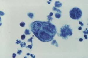
Cytological examination of sputum involves the study of macroscopic and microscopic analysis of its cells.
The most information for cytological analysis is provided by sputum taken in the morning on an empty stomach. Before testing, it should be stored for no more than 4 hours.
- Sputum contains squamous epithelial cells, which are examined microscopically. But they have no significance for making a diagnosis. Columnar epithelial cells - both individually and in groups - can be observed in diseases such as bronchial asthma, bronchitis and lung cancer. It should be noted that columnar epithelium can also appear due to the penetration of mucus from the nasopharynx.
- Alveolar macrophages are reticuloendothelial cells. Macrophages, which are contained in the protoplasm (phagocytic particles or dust cells), can be found in patients who have inhaled dust for a long time.
- Protoplasmic macrophages (formed during the breakdown of hemoglobin) are called cardiac defect cells. They can occur during congestive processes in the lungs, mitral valve stenosis, and pulmonary infarction.
- A small number of leukocytes are found in any sputum. Their increased content is observed in secretions mixed with pus.
- Eosinophils. The sputum of asthmatics is rich in such cells. Cells can be observed in the eosinophilic form of pneumonia, helminth damage to the body, tuberculosis and pulmonary infarction.
- Red blood cells. Single red blood cells do not reflect the picture of the disease. The appearance of an increased amount indicates the presence of bleeding in the lungs. In fresh blood, unchanged red blood cells are detected. If there is an admixture of blood that has stagnated in the lungs for a long time, then leached red blood cells are detected.
- Cancer cells. They can be found in secret groups. They indicate the presence of a tumor. When single cells are found, diagnostic difficulties often arise. In such cases, a repeat sputum analysis is performed.
- Elastic fibers, the appearance of which is caused by the breakdown of lung tissue, provoked by tuberculosis, abscess, gangrene, tumor. Such cells are not always characterized by gangrene, since due to the action of secreted enzymes, they can be dissolved.
- Kurshman spirals. These are special bodies that look like tubes. They are detected when examined under a microscope. Sometimes visible to the eye. Typically, spirals are associated with diseases such as bronchial asthma, pulmonary tuberculosis and pneumonia.
- Charcot-Leyden crystals are found in sputum with an increased content of eosinophils in diseases such as bronchial asthma, eosinophilic pneumonia. Opening a focus of tuberculosis in the lumen of the bronchi can be characterized by the presence in the secretion of elastic fiber-crystals of cholesterol, MBT and amorphous lime (the so-called Ehrlich tetralogy) - 100%.
Application of bacterioscopy
Collecting sputum for examination by the bacterioscopic method involves analyzing the secretion to detect mycobacteria characteristic of tuberculosis. They look like thin curved sticks of different lengths, thickened on the sides or in the middle, which are located both individually and in groups.
The detection of Mycobacterium tuberculosis is not a dominant sign for diagnosis and requires confirmation by bacteriological methods. Tuberculous mycobacteria are not detected in secretions under normal conditions.
The basis for the analysis is purulent particles, which are taken from forty-six different areas and thoroughly ground to a homogeneous mass using two glasses. Next, they are dried in air and fixed with a burner flame.
Bacteriological examination of sputum using the Ziehl-Neelsen method suggests that it is stained red. In this case, all secretion particles, with the exception of mycobacteria, acquire a blue tint, and mycobacteria acquire a red color.
If the body is suspected of being affected by tuberculosis, after testing three times for the presence of mycobacteria with a negative response, the flotation method (Pottenger analysis) is used.
The usual method of examining a stained smear for MTB gives a positive result only when the amount of MTB is at least 50,000 units in 1 ml of sputum. The presence of tuberculosis cannot be judged by the number of mycobacteria.
Bacterioscopy of patients with nonspecific lung diseases
Laboratory tests of sputum in the presence of nonspecific lung diseases during bacterioscopy can reveal the following bacteria:
- For pneumonia - pneumococci, Frenkel diplococci, Friedlander bacteria, streptococci, staphylococci (100%).
- With gangrene of the lungs, a spindle-shaped rod can be found in combination with Vincent's spirochete (80%).
- Yeast-like fungi (70%), to determine the type of which requires culture of the secretion.
Author: Seytimova Moldir
Regional anti-tuberculosis dispensary
Bachelor laboratory assistant
07/17/2018
ELISA
An enzyme-linked immunosorbent assay aims to detect antibodies in the blood. Accordingly, the higher the titer, the more antibodies to a particular infectious disease. IgG antibodies indicate a previous infectious disease, and IgM antibodies indicate the presence of an acute infectious process. ELISA allows you to diagnose:
- viral infections: hepatitis, HIV infections, herpes virus, rubella, cytomegalovirus;
- bacterial infections: tuberculosis, borreliosis, syphilis;
- other diseases: giardiasis and toxoplasmosis.
Such a blood test is necessary to determine the stage of the disease, evaluate the effectiveness of treatment, and obtain information about a previous disease.