Every person has undergone laboratory tests at least once in their life, although, without a doubt, these studies are carried out much more often. They are included in the group of mandatory preventive studies and help to identify pathology when there are no symptoms.
It is difficult to overestimate the importance of laboratory diagnostics, because it provides the doctor with up to 80% of important diagnostic information. It helps to confirm or refute the diagnosis, monitor the dynamics and effectiveness of treatment, identify the causative agent of the disease and determine the pathological processes occurring in the human body.
Laboratory diagnostics of biological materials in medicine is carried out using specialized equipment, one of which is an optical microscope device. Various studies in medicine under multiple magnification, which are carried out using a microscope, are called microscopy.
Detailed description of the study
The study of the leukocyte formula includes determination of the total number of leukocytes, the absolute and relative content of promyelocytes, metamyelocytes, myelocytes, band neutrophils, segmented neutrophils, eosinophils, basophils, monocytes, lymphocytes, plasma cells. If blast cells are present in the blood smear, they are also indicated in this study. The study of the leukocyte formula is of great importance in the diagnosis of hematological, infectious, inflammatory diseases, as well as in assessing the severity of the condition and the effectiveness of the therapy. At the same time, changes in the leukocyte formula are not specific - they may have a similar nature in different diseases or, on the contrary, dissimilar changes may occur with the same pathology in different patients. The leukocyte formula has age-related characteristics, so its changes should be assessed from the perspective of the age norm (this is especially important when examining children).
The leukocyte formula determines the following types of leukocytes:
- Neutrophils protect the body from infections. Most often, the number of neutrophils increases in acute inflammatory and infectious diseases, tissue necrosis, and intoxication.
- Eosinophils are involved in the development of allergic reactions. Their number increases most often in allergic conditions and helminthic infestations, less often in tumors and some collagenoses.
- Basophils are involved in allergic and inflammatory reactions. Their number increases with any allergic diseases, some blood diseases, hypothyroidism, etc.
- Lymphocytes are the main cells of the immune system, providing an adequate immune response to the body when exposed to foreign agents. The content of lymphocytes in the blood increases with infectious mononucleosis, many other viral infections, as well as with diseases of the lymphatic system and blood.
- Monocytes, cells that provide phagocytosis, i.e. “devouring” foreign microorganisms. Monocytosis - an increase in the number of monocytes - is characteristic of many infections: tuberculosis, infectious mononucleosis, sepsis. It occurs in blood diseases and collagenoses.
- Plasmocytes, cells of lymphoid tissue that produce immunoglobulins. Normally absent in peripheral blood, they appear there during viral infections, plasmacytoma, malignant tumors, and autoimmune diseases.
The study on calculating the leukocyte formula in the Hemotest Laboratory is carried out on automatic hematology analyzers, which use modern technology of flow cytometry and conductometry. The analyzer evaluates 10,000 cells in one sample, determining size, structural, cytochemical and other characteristics. After this, mandatory microscopy of a stained blood smear is performed to determine the morphological characteristics of leukocyte cells. When pathology is detected in the cytoplasm or nucleus of cells, this information must be indicated in the notes to the analysis. For microscopy of the leukocyte formula, Romanovsky staining is used. When calculating the leukocyte formula, laboratory keyboard counters are used. In a smear, 100 leukocyte cells are counted, followed by the percentage and absolute number of cells, based on the total number of leukocytes. Considering that the automatic and manual counting methods have different errors and reproducibility, automatic cell counting is more diagnostic. During microscopy, immature forms of granulocytes are differentiated into promyelocytes, myelocytes and metamyelocytes, band neutrophils, plasma cells are indicated and, if detected, lymphocytes of varying degrees of maturity are noted, a significant variation in the size of cells of the lymphoid series; polymorphism of lymphocyte nuclei; marginal basophilia and vacuolization of the cytoplasm of lymphocytes, uneven contour of the nucleus and cytoplasm. In visual differential counting, there are three main sources of error: uneven distribution of cells in the preparation, failure to recognize cells, and the presence of errors in repeated analyzes. This method is difficult to standardize and eliminate the “human” factor of analytical errors. The advantage of this analysis is the visual assessment of the morphology of leukocyte cells by the clinical physician.
Venous blood is considered the best material for laboratory research:
- To ensure the quality of the test result, it is necessary to donate venous blood (unless the child has special indications for taking capillary blood). When blood is taken from a finger, the blood cells are deformed, and some of the red blood cells are destroyed, forming microscopic clots in the test tubes. In this case, it is impossible to conduct research; in this case, repeated collection of biomaterial is required.
- In venous blood, blood cells are not destroyed, microscopic clots are formed much less frequently. That is why capillary blood is used only in children in isolated cases.
- Thanks to modern technologies, the procedure for collecting venous blood is painless and safe even for small children, since completely closed disposable BD vacuum systems are used, which eliminate infection and meet all international standards.
- Taking blood from a vein takes a few seconds.
Microscopy tests: what do they reveal?
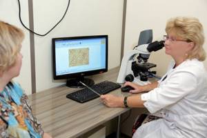
Microscopy studies are widely used in laboratory diagnostics. Why are they prescribed and what preparation do they require? Maria Vasilyevna Biserova, a doctor from the KDL laboratory network (partner of the Expert Clinic Perm), head of the medical department, told us about microscopy tests.
— Maria Vasilievna, what tests are carried out using microscopy?
— You can look at any biological material under a microscope: smears from mucous membranes, scrapings from the skin, urogenital smears. You can also examine sputum, pleural, synovial fluid, ejaculate, urine, feces, nails, and hair. Research allows us to identify signs of inflammation and determine the morphotype of the infectious agent (rod, coccal flora, fungal elements, protozoa). Cytological and histological studies can also be classified as microscopy. Most often they are used when examining surgical material or diagnostic biopsy material.
All microscopic tests are convenient, fast, but at the same time subjective, requiring the professionalism of a laboratory diagnostics specialist.
Read materials on the topic:
Urinalysis: frequently asked questions What does the Nechyporenko urine analysis show? Not only a colonoscopy: what will a stool test tell you? Histology: what kind of analysis is this?
— What does a general blood test (CBC) with microscopy show?
— CBC allows you to detect the presence of inflammation in the body, assess the number of red blood cells and platelets. Microscopy is traditionally used to evaluate the leukocyte count. By changing the composition of this formula, one can assume the presence of a viral or bacterial process in the body.
Read materials on the topic:
General and clinical blood test: frequently asked questions What does hemoglobin tell you? Leukocytes. What will a blood test tell you? Platelets: friends or enemies?
— Is microscopy performed as part of a general urinalysis (UU)?
— Of course, microscopy is performed in the OAM and is necessary to identify inflammatory diseases of the urinary tract. In a modern laboratory, microscopic examination of urine has been replaced by the automatic method of flow cytometry, which allows one to assess the cellular composition of urine with great accuracy, since the device identifies and counts up to 65,000 elements of urinary sediment. In some cases (when pathology is detected), the doctor still has to additionally examine the urinary sediment under a microscope.
— How to prepare for a general urine test?
— For research, an average portion of morning urine is collected in a sterile disposable plastic container. Preliminary hygiene procedures are mandatory.
— Who and why are swab tests from the throat and nasal mucosa prescribed?
— Based on the results of a rhinocytogram (microscopy of nasal smears), we can assume the infectious or allergic nature of the inflammatory process. For infectious diseases of the nasopharynx, methods of direct detection of the pathogen are more often used - these are flora cultures and PCR studies.
Read more about PCR in our article
— What other microscopic examinations are carried out for respiratory diseases?
— If the disease is accompanied by a cough, a general sputum analysis is important. Microscopy can reveal the presence of cells and microorganisms that should not normally be present in sputum (leukocytes, macrophages, atypical cells, Kurshman spirals, Charcot-Leyden crystals, etc.). This also allows you to assess the degree and suggest the cause of inflammation (viral, bacterial, allergic).
To confirm the infectious agent, bacteriological and molecular genetic (PCR) tests are accurate.
— How to prepare for such an analysis?
- Sputum is collected in a sterile container by coughing in the morning, before eating, after brushing your teeth and thoroughly rinsing the mouth with boiled water. In this case, saliva from the oral cavity should not get into the container.
— You mentioned microscopic analysis of synovial fluid. How and why is it carried out?
— A general analysis of synovial fluid allows us to identify the nature of the disease (inflammatory, non-inflammatory). In this case, both external properties (color, viscosity) and chemical composition are assessed. But synovial fluid can only be obtained by puncture of the joint; this procedure is performed by a doctor. In modern diagnostics for joint diseases, the determination of markers of inflammatory, in particular rheumatic diseases, in the blood is widely used. This is a quick and reliable way to correctly diagnose and prescribe treatment.
Want to know more about tests? Read articles in our section
You can sign up and take laboratory tests here. ATTENTION: the service is not available in all cities
Interviewed by Daria Ushkova
The editors recommend:
Viruses and bacteria - what is the fundamental difference?
For reference:
Biserova Maria Vasilievna
Clinical laboratory diagnostics doctor, head of the medical department of Medical LLC, KDL laboratory network.
Types of microscopic examinations
Using a microscope, you can examine various cells of the human body. Various biological materials are taken for research - blood, urine, semen, smears, mucous secretions, etc.
In medicine today, several types of microscopic examinations are carried out - stereoscopic, infrared, luminescent, ultraviolet, x-ray, polarization microscopy.

A blood test allows you to determine the quantitative and qualitative composition of blood, the ratio of its formed elements, and identify atypical or immature cells. Microscopic analysis of urine allows you to determine the presence of salts, cellular elements and cylinders; the study allows you to identify existing problems in the water-electrolyte balance of the body, as well as metabolic disorders.
Despite the fact that special electronic devices for laboratory research - analyzers - are widely used today, visual inspection of biological materials to identify atypical or immature cells is still performed by medical personnel using microscopes.
In medical departments you can undergo all types of laboratory tests. At any convenient time and at an affordable price, you can perform any type of laboratory diagnostics, and based on its results, receive qualified advice from a specialist in the profile of the disease. You can sign up for a consultation by calling the numbers listed on the website.
Prices
| Blood smear microscopy | 150₽ |
All prices
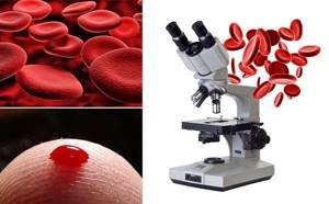
Blood smear microscopy is an examination under a microscope of a preparation prepared from a drop of blood.
The study allows you to morphologically evaluate blood cells (formed elements), as well as count them. Blood cells are formed and mature in the red bone marrow and are then released into the general bloodstream. Each type of cell has its own functions. Under physiological conditions, the number and morphological characteristics of blood cells are stable and do not exceed the reference values. With various diseases, the quantity and properties (shape, volume, color, presence of inclusions, their number, etc.) naturally change. For this reason, the assessment of cellular elements in a blood smear is a universal test for diagnosing many pathological conditions and is widely used in the practice of doctors of almost any specialization.
A peripheral blood smear is a “snapshot” of blood cells as they are when the sample is taken. To perform the study, venous or capillary blood is placed on a glass slide, which must be thoroughly degreased. Then another slide is placed on the slide at an angle of 45′ and drawn along the drop of blood so that it spreads in a thin layer across the width of the ground glass. Then the smear is fixed so that the blood cells are more stable. After this, the smear is stained with a special dye, which makes the cells and their elements brighter, and dried. After which the doctor examines the smear under a microscope in the laboratory.
When is the study scheduled?
There is a fairly wide range of diseases and disorders in which the properties of cells circulating in the bloodstream can change. Normally, only mature cells enter the blood from the bone marrow, but in a number of diseases, such as leukemia, immature cells—blasts—can enter the blood. In some conditions, for example during a massive infection, characteristic impurities may appear in leukocytes, and the cells themselves may become atypical, as in infectious mononucleosis. The detection of pathological cells in a smear in large numbers allows one to suspect the disease that caused them and prescribe additional examination.
A blood smear can be regularly prescribed to patients with cancer of the bone marrow and lymph nodes to monitor the dynamics of the condition and monitor the effectiveness of treatment.
The history of the microscope
It is not known for certain when the first microscope appeared, but attempts to examine small objects using a magnifying glass (a biconvex optical lens) were made back in Ancient Rome.
The first device vaguely similar to a microscope was created by the Dutch optician Hans Jansen and his son. They were the first to discover that by using two lenses they could obtain a multiply magnified image. Their device was more like a telescope, but unlike the latter, it did not bring objects closer, but magnified them.
A magnifying device with concave and convex lenses was developed by Galileo Galilei and called it “occolino” or “little eye”. The term "microscope" was coined by Galileo's friend Giovani Faber. The devices created were very primitive and made it possible to obtain an image magnified by 9-10 times.
The first microscope, similar to its modern counterparts, was a metal plate in the center of which a lens was located. It was created by the Dutch naturalist Antonie van Lieuwenhoek, with its help he was able to make important discoveries in the structure and functioning of the human body. He examined the composition of the blood, the structure of the tissues, and also saw bacteria. Using a Leeuwenhoek microscope, it was possible to obtain magnification up to 270 times.
Russian scientists Pyotr Kulibin and Mikhailo Lomonosov also contributed to the development of the microscope. The latter used a microscope for his research.
General (clinical) blood test: interpretation and norm

A complete blood count provides essential information about the types and numbers of blood cells, especially red blood cells, white blood cells and platelets. Using a general blood test, the treating doctor can detect the nature of symptoms such as weakness, fatigue, and bruising. A complete blood count is used to diagnose anemia, infection and many other disorders.
A general (clinical) blood test includes the following indicators:
- Absolute white blood cell count ( WBC ). White blood cells protect the body from infection. If an infection develops, these cells attack and destroy bacteria, viruses, or other pathogenic organisms. White blood cells are larger in size than red blood cells, but their number is less. When a person gets sick, the number of white blood cells increases very quickly. Sometimes white blood cell counts are used to detect infection or to evaluate the effectiveness of cancer treatment.
- Determination of the ratio of different types of white blood cells (leukogram). The main types of leukocytes are neutrophils, lymphocytes, monocytes, eosinophils and basophils. Counting immature neutrophils, also called band neutrophils, is part of this test. The number of each cell type can provide important information about the health of the immune system. Too high or too low the number of one type of cell may indicate an infection, an allergy or toxic reaction to a drug or chemical, or various diseases such as leukemia.
- Absolute red blood cell count ( RBC ). Red blood cells carry oxygen from the lungs to the rest of the body. They also carry carbon dioxide back to the lungs for exhalation. When the level of red blood cells is low (anemia), the body does not receive enough oxygen. When levels are high (polycythemia), there is a risk of red blood cell clots that can block tiny blood vessels (capillaries), also making it difficult to carry oxygen.
- Hematocrit ( HCT , precipitated red blood cell volume, PCV ). This test measures the volume of blood per red blood cells. For example, a hematocrit value of 38 means that 38% of the blood is red blood cells. Hematocrit and hemoglobin are the two main tests for detecting anemia or polycythemia.
- Hemoglobin ( Hgb ). Hemoglobin molecules are found in red blood cells; they carry oxygen and give the blood its red color. A hemoglobin test measures the level of hemoglobin in the blood and shows how the body copes with transporting oxygen.
- Erythrocyte index. There are three red blood cell indices: mean erythrocyte volume (MCV), mean erythrocyte hemoglobin content (MCH) and mean erythrocyte hemoglobin concentration (MCHC). All of them are calculated using special equipment, and the numbers are derived from other indicators of a general blood test. MCV reflects the size of red blood cells, MCH is the average hemoglobin content in an individual red blood cell, MCHC is the hemoglobin concentration in an individual red blood cell. These indices are used in diagnosing various types of anemia. The red blood cell distribution width (RDW) index is also calculated, which reflects the difference in size and shape between cells.
- Absolute platelet count. Platelets are the smallest type of blood cell. They play an important role in the blood clotting process. If bleeding occurs, the platelets are activated and bind together, forming a plug that prevents bleeding. If the number of red blood cells is insufficient, there is a risk of uncontrolled bleeding. If there is an excess amount, there is a risk of thrombus formation in a blood vessel. In addition, platelets can be involved in the formation of sclerotic plaques on artery walls (atherosclerosis).
- Mean platelet volume ( MPV ). This indicator reflects the average value of the measured platelet volume. MPV is used in conjunction with platelet count to diagnose certain diseases. If the platelet count is normal, the MPV may be low or high.
Your doctor may also order a blood smear microscopy at the same time as your complete blood count, but this is not part of your routine blood test. For this test, a drop of blood is placed on a plate and diluted with dye. The plate is examined under a microscope, noting the number, size and shape of red blood cells, white blood cells and platelets. Differences in the shape or size of blood cells can indicate various diseases, such as leukemia, malaria or sickle cell disease.
A clinical blood test is carried out for the following purposes:
- Finding the cause of symptoms such as fatigue, weakness, fever, bruising, or weight loss
- Diagnosis of anemia
- Establishing the amount of blood lost during bleeding
- Diagnosis of polycythemia
- Diagnosis of infections
- Diagnosis of blood diseases, such as leukemia
- Monitoring the body's response to drug treatment and radiation therapy.
- Monitoring the blood cell response to abnormal bleeding.
- Screening of indicators before surgery.
- Tracking high or low levels is used to detect various diseases, such as high eosinophil levels that may indicate allergies or asthma.
A complete blood count is performed as part of a routine medical examination. A complete blood count provides valuable information about the overall condition of the body and health.
Preparing for the examination
No special preparation required.
How is the examination carried out?
- The shoulder is tightened with an elastic tourniquet to stop the blood flow. The veins under the tourniquet become larger, making it easier to insert the needle.
- The puncture site is cleaned with alcohol.
- The needle is inserted into the vein. Sometimes more than one injection is required.
- A blood collection tube is attached to the needle.
- When enough blood has been collected, the tourniquet is removed.
- After the needle is removed, a cotton swab is applied to the injection site.
- The area should be pressed and a bandage applied.
If a baby's blood is being tested, a heel prick is done instead of drawing blood from a vein.
Feelings during the procedure
A blood sample is taken from a vein in the arm. The forearm is wrapped with an elastic band, so there may be a feeling of tightness. You may not feel any pain from the needle, but sometimes there will be a slight burning sensation or pain.
Complications
When taking blood from a vein, the chance of complications occurring is very small.
- There may be a slight bruise. By pressing the tampon to the puncture site for a few minutes, you can reduce the chance of its occurrence.
- In rare cases, the vein may become swollen, this is called phlebitis. The problem can be treated with several warm compresses.
- Continuous bleeding may occur in patients with blood disorders. Taking medications such as aspirin, warfarin (such as Coumadin), and other blood thinners increases the chance of bleeding. Tell your doctor before taking a sample if you have problems with blood clotting or are taking blood-thinning medications.
results
A complete blood count provides important information about the types of blood cells - red blood cells, white blood cells, platelets - and their numbers. A complete blood count helps determine the cause of various symptoms such as weakness, nausea, bruising and is used in the diagnosis of various diseases - anemia, infections and many others.
Normal results
IMPORTANT! The normal values, or range of normal values, given here are average values. These vary depending on the laboratory, so where you are being tested will have different readings. The laboratory report should contain information about the reference range used. In addition, overall health and other factors are taken into account when analyzing the results. Therefore, a value that does not fit into the range presented here may be normal for you personally.
The normal range depends on your age, gender, type of blood sample, and how high above sea level the area where you live is. The doctor can use all the indicators of the general blood test when checking the condition of the body. For example, the number of red blood cells, hemoglobin, hematocrit are the main indicators in diagnosing anemia, but erythrocyte indices and smear microscopy also help detect this disease and can show the possibility of its occurrence.
To assess the number and size of leukocytes, the doctor takes into account the results of counting the number of cells and the ratio of their types. To see if there are too many or few cells of a certain type, the doctor compares their number and percentage. There is a norm for each type of leukocyte.
Indicators may change during pregnancy. During each trimester, your doctor should report changes in your normal range.
| Absolute white blood cell count ( WBC ) | |
| Men and non-pregnant women: | 5,000–10,000 WBC per cubic mm (mm3) or 5.0–10.0 x 10⁹ WBC per 1 liter (L) |
| The ratio of different types of leukocytes | |
| Neutrophils: | 50%–62% |
| Band neutrophils: | 3%–6% |
| Lymphocytes: | 25%–40% |
| Monocytes: | 3%–7% |
| Eosinophils: | 0%–3% |
| Basophils: | 0%–1% |
| Absolute red blood cell count ( RBC ) | |
| Men: | 4.5–5.5 million RBC per µl or 4.5–5.5 x 10¹²/l |
| Women: | 4.0–5.0 million RBC per µL or 4.0–5.0 x 10¹²/L |
| Children: | 3.8–6.0 million RBC per µL or 3.8–6.0 x 10¹²/L |
| Newborns: | 4.1–6.1 million RBC per µL or 4.1–6.1 x 10¹²/L |
| Hematocrit (HCT) | |
| Men: | 42%–52% or 0.42–0.52 of the total volume |
| Women: | 36%–48% or 0.36–0.48 of total volume |
| Children: | 29%–59% or 0.29–0.59 of the total volume |
| Newborns: | 44%–64% or 0.44–0.64 of total volume |
| Hemoglobin (Hgb) | |
| Men: | 14–17.4 grams per deciliter (g/dL) or 140–174 grams per liter (g/L) |
| Women: | 12–16 g/dl or 120–160 g/l |
| Children: | 9.5–20.5 g/dl or 95–205 g/l |
| Newborns: | 14.5–24.5 g/dl or 145–245 g/l |
Typically, a normal hemoglobin level is one third of the hematocrit level.
| Red blood cell indices | |
| Mean erythrocyte volume ( MCV ) - Adults: | 84–96 femtoliters (fl) |
| Mean erythrocyte hemoglobin content ( MCH ) - Adults: | 28–34 picograms (pg) per cell |
| Mean erythrocyte hemoglobin concentration ( MCHC ) - Adults: | 32–36 grams per deciliter (g/dL) |
| Red blood cell distribution width by volume ( RDW ) | |
| Norm: | 11.5%–14.5% |
| Absolute platelet count | |
| Adults: | 140,000–400,000 platelets per mm3 or 140–400 x 109/L |
| Children: | 150,000–450,000 platelets per mm3 or 150–450 x 109/L |
| Mean platelet volume (MPV) | |
| Adults: | 7.4–10.4 µm3 or 7.4–10.4 fl |
| Children: | 7.4–10.4 µm3 or 7.4–10.4 fl |
| Blood smear microscopy | |
| Norm: | Normal indicators of the number, shape, color and size of blood cells. |
High performance
Absolute red blood cell count (RBC)
- Conditions and habits that cause high red blood cell counts include smoking, exposure to carbon monoxide, long-term lung disease, kidney disease, some cancers and heart disease, alcoholism, liver disease, a rare bone marrow disease (polycythemia vera), or rare problems related to with hemoglobin, which are associated with the transfer of oxygen.
- Diseases related to the water component of the body can also cause high red blood cell counts. These include dehydration, diarrhea, vomiting, increased sweating, and taking diuretics. Lack of fluid in the body causes red blood cell counts to appear high. This is sometimes called pseudopolycythemia.
Absolute white blood cell count (WBC)
- Causes of high white blood cell counts include infection, inflammation, tissue damage (for example, due to a heart attack), kidney failure, lupus, tuberculosis, rheumatoid arthritis, malnutrition, leukemia, and cancer.
- Taking costosteroids and certain medications, underactive adrenal glands, problems with the thyroid gland, and removal of the spleen can also cause an increase in white blood cell levels.
Absolute platelet count
- High platelet levels can occur due to bleeding, iron deficiency, cancer and bone marrow problems.
Low performance
Absolute red blood cell count (RBC)
- With anemia, the level of red blood cells decreases. Anemia can be caused by heavy menstrual bleeding, stomach ulcers, colorectal cancer, inflammatory bowel disease, tumors, Addison's disease, thalassemia, lead poisoning, sickle cell disease, or a reaction to certain chemicals and medications. One reason for low red blood cell counts may be removal of the spleen.
- Not getting enough folic acid or vitamin B12 can also cause anemia, such as pernicious anemia, which causes problems absorbing vitamin B12.
- Red blood cell indices and smear microscopy can help find the cause of anemia.
Absolute white blood cell count (WBC)
- Low white blood cell counts may be caused by chemotherapy and reaction to drug treatment, aplastic anemia, viral infections, malaria, alcoholism, AIDS, lupus, Cushing's syndrome.
- An enlarged spleen can reduce white blood cell counts.
Absolute platelet count
- Low platelet levels can be caused by pregnancy, immunopathological thrombocytopenic purpura and other diseases that affect the process of generating platelets or destroy them.
- An enlarged spleen can reduce platelet counts.
What can affect the results of the examination?
Reasons why the analysis is not possible or the results will be incorrect:
- When drawing blood, your arm was tied with an elastic tourniquet for a long time.
- Taking drugs that cause low platelet counts, such as certain antibiotics, steroids, thiazide diuretics, chemotherapy drugs, quinidine, meprobamate.
- Very high levels of white blood cells or fats (triglycerides) can cause falsely high hemoglobin levels.
- With an enlarged spleen, the level of platelets (thrombopenia) or white blood cells may be reduced; this condition can also cause some types of cancer.
- During pregnancy, the level of red blood cells is often reduced, and less often, the level of white blood cells is increased.
On a note:
- The absolute white blood cell count can reach up to 2000 WBC per microliter due to exercise, stress or smoking.
- Children tend to have higher levels of white blood cells than adults.
- Often, a red blood cell count test includes:
- Erythrocyte sedimentation rate (ESR). The ESR test measures how quickly red blood cells settle to the bottom of a tube. During inflammatory processes occurring in the body (for example, in the presence of infection or cancer), red blood cells settle more slowly than under normal conditions. Using ESR analysis, inflammatory diseases can be diagnosed if the blood test results are normal.
- Analysis of reticulocyte content. This is an analysis of the number of young red blood cells in a blood sample. As a rule, the content of reticulocytes is lower compared to the content of mature red blood cells. However, recent bleeding or destruction of mature red blood cells may cause increased reticulocyte production. This test is done to detect certain types of anemia and screen for the body's response to treatment.
- Hematocrit measurements may vary depending on the assay method and equipment used.
Microscopy of native blood
Blood is a whole world of cells that can be compared to space.
And each cell is a living being with its own life, with its own purpose, performing its own function. When using traditional analysis methods, many indicators remain normal for a long time, so early signs of the disease ( fatigue, sleepiness during the day, memory impairment - the most common and obvious signs of a blood disorder ) go unnoticed...

And, probably, it has happened to many that when they were feeling unwell, their blood tests for some reason turned out to be within the normal range. What is the reason for the discrepancy?
What can be revealed by microscopic examination of blood?
A routine blood test is done after a few hours, when all cell movement has stopped. The study of a “live” drop of blood is an examination of a sample immediately after collection for no more than 5 minutes and is carried out with a magnification of more than 1000 times.
- An important difference between this method and conventional tests is the examination of a blood sample without any preliminary treatment and in the presence of the patient.
- The patient has a unique opportunity to look into the cellular world of his body, see deviations in the functioning of cells, and take care of his health at the earliest stages of illness.
- We managed to connect the microscope with a video camera and a computer, which will give you the opportunity to see your blood on the screen.
The “live” blood drop method allows you to evaluate:
- Qualitative analysis of blood composition.
- With the state of metabolism and hormonal imbalance.
- And changes in blood cells, which are observed under stress, exposure to radiation, electromagnetic radiation, inflammatory processes in the body, deficiency of vitamins, microelements and protein.
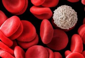
- of red blood cells: shape, size, their interaction, location in the blood plasma, aggregation (sticking together), leading to oxygen starvation.
- The condition of leukocytes, their size, activity (it is important to remember that a decrease in the number of leukocytes signals inflammatory processes in the body).
- The state of lymphocytes in the blood and the body’s ability to self-heal;
- With the state of platelets and the tendency to form blood clots, a predisposition to increased coagulation, vascular thrombosis, ischemia, hypertension;
- The degree of purity of blood plasma and various inclusions: cholesterol, sugar, urine salts;
- With the stage of accumulation and distribution of toxins in the human body;
- The causes of damage to red blood cells are lack of folic acid and vitamin B12, iron deficiency;
- With the state of fat metabolism and predisposition to atherosclerotic processes.
Preparation for the procedure:
Do not eat food 2 hours before ;
On the day of diagnosis, try not to eat meat, sweets and fatty foods.
Blood is the main living medium on which all processes occurring in organs and cells depend!
Using a microscopic diagnostic method using a drop of live blood, the doctor can select a more accurate and individual treatment plan.