Liver disease is one of the most common diseases of the digestive system, the steady increase of which is called “the second epidemic of our century.” A special place among them is occupied by various tumors and tumor-like lesions. In recent years, thanks to the introduction into clinical practice of more advanced diagnostic equipment and the fairly widespread use of various contrast agents in studies of the abdominal cavity using ultrasound, X-ray computed tomography and magnetic resonance imaging, the possibilities for correct diagnosis of various focal liver lesions have significantly increased. In addition, the sensitivity and specificity of various methods for diagnosing liver tumors reaches 88-97%.
According to many authors, benign tumors do not have clinical symptoms and manifest themselves mainly in the form of focal liver formations. In clinical practice, as focal liver formations are identified, the question of topical diagnosis and exclusion of malignancy of the process becomes acute.
Hemangioma
Hemangiomas are considered by many authors to be the most common benign vascular tumors of the liver of mesenchymal origin, occurring in various age groups. Capillary hemangioma consists of a plexus of a large number of branching capillary-type vessels with a narrow lumen and without directional or very slow blood flow; 90% of the area of the vascular bed is venous caverns. Cavernous hemangiomas are more common than capillary hemangiomas, and in some cases there is a mixed type. According to most authors, hemangiomas usually do not manifest clinically and are relatively more often detected in women.
On ultrasound, small (up to 3.0 cm) hemangiomas appear as homogeneous hyperechoic formations with clear contours. In some cases, the echogenicity of hemangiomas may be reduced, and the contours may appear blurred. In some hemangiomas, a pseudocapsule in the form of a hypoechoic rim can be clearly visualized, which makes their differential diagnosis with metastases difficult. Many researchers associate the prospect of improving diagnostics with the use of intravenous contrast with special echo contrast agents.
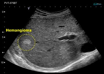
Capillary hemangioma of the liver is a homogeneous hyperechoic focus less than 3 cm with clear boundaries.
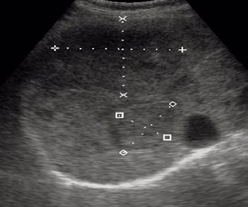
Two cavernous hemangiomas of the liver.
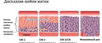
Degrees and causes of endometrioid formation of the ovaries
There are 4 degrees of development of endometrioid cysts:
- In the first degree, the ovaries are minimally affected; single foci of endometriosis are observed on the ovaries and in the peritoneum of the small pelvis.
- The size of cysts located on one ovary reaches 5-6 centimeters. In addition, endometriosis appears on the peritoneum and adhesions in the appendages.
- The third degree is characterized by the presence of cysts measuring 5-6 centimeters on both ovaries, as well as significant adhesions and damage to the uterus, fallopian tubes and peritoneum by endometriosis.
- Huge cysts are found on both ovaries, also affecting neighboring organs.
Regarding the causes of endometrioid cysts, they have not yet been precisely established. Some scientists are inclined to believe that this is a consequence of a mutation in endometrial cells, while others believe that it is formed due to menstrual blood entering the ovaries.
There is an increased risk of developing this disease in women who have undergone abortion, cesarean section, surgical removal of uterine polyps, etc. In addition, this type of cyst can occur as a result of endometritis, adnexitis, oophoritis, stress, weakened immunity, hormonal imbalance, genetic defects, etc. .d.
Focal nodular hyperplasia (FNH)
Focal nodular hyperplasia (FNH) is considered the second most common benign liver tumor (after hemangiomas). According to a number of authors, FH occurs in 0.9-5% of the population, and in 5-20% of cases it can be combined with cysts and hemangiomas. According to the literature, FUH, without having any signs of a tumor, is included in the group of benign hepatocellular tumors of epithelial nature. This is a single, round, non-encapsulated formation with impaired hepatic architecture, separated by septa reaching the central scar. The average size of the lesion is 5.7 cm (from 1.5 to 12.0 cm). It has been established that FUH is not prone to hemorrhage and has no malignant potential. Usually the tumor is an incidental finding during diagnostic studies. Small nodules are usually asymptomatic. Large tumors can cause abdominal pain.
With native ultrasound, in most cases, FUGs are difficult to visualize, appearing as a rounded area with clear contours, an isoechoic or slightly hypo- or hyperechoic structure. According to some authors, in diagnosing FEG, contrast ultrasound is more informative in contrast to other diagnostic methods. The emergence of second-generation echo contrast agents has significantly expanded the capabilities of the method. This makes it possible to differentiate different types of tumors, taking into account their nature of vascularization, which is important in the differential diagnosis of focal liver formations. Increased vascularization of the tumor in combination with the characteristic “wheel with spokes” pattern is a pathognomonic sign of FAG (71.4%). In this case, the detection rate of the “feeding” artery of the tumor increases to 98%. The sensitivity and specificity of ultrasound using intravenous contrast in diagnosing FUG reaches 83 and 98%, respectively.
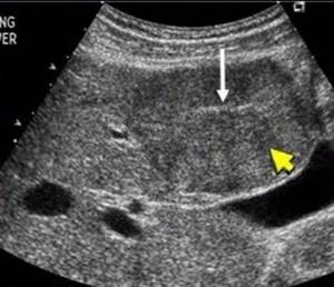
Hypoechoic formation in the liver is benign focal nodular hyperplasia. The literature describes that it is characterized by a central scar (white arrow).
B-mode. In the IV segment of the liver, reaching the contour of the liver, deforming it, a tissue density hypoechoic formation, somewhat heterogeneous in echostructure, measuring 50 x 40 mm, irregular in shape, with clear, even contours, is visualized.
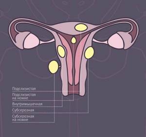
Submucous (submucosal) uterine fibroids
Such nodes are located under the mucous membrane and grow into the uterine cavity. Submucosal fibroids are the most unpleasant in terms of symptoms, since they lead to heavy periods, during which a woman loses a lot of blood, uterine bleeding, infertility, miscarriages, and premature birth.
A small nodule—only 2–3 cm in diameter—can lead to severe symptoms. Sometimes the bleeding is so severe that the woman has to be hospitalized and receive a blood transfusion.
Chronic bleeding from submucosal fibroids leads to anemia. Characteristic manifestations of this condition: pallor, constant feeling of fatigue and weakness, headaches and dizziness, shortness of breath, tinnitus, fainting, rapid heartbeat.
Hepatocellular adenoma (HCA)
Hepatocellular adenoma (HCA). HCA is considered a rare benign tumor originating from hepatocytes. There are suggestions about the influence of oral contraceptives, exogenous androgens, pregnancy and an imbalance of endogenous sex hormones on the development and growth of tumors. Conflicting data are provided on the size of adenomas - figures from 1 to 19 cm are indicated, with an average of 5.4 cm. In some cases, patients have multiple adenomas. The formations are well delineated. A number of authors note the characteristic distinctive features of adenomas - unlike hemangiomas, they are not located next to the hepatic vessels and do not occupy an entire lobe. The course of HCA is asymptomatic in most cases. When the tumor size is 5 cm or more or its subcapsular location, the risk of bleeding increases. The authors emphasize the practical importance of diagnosing adenoma due to the high risk of bleeding, rupture, malignancy and the need for surgical intervention.
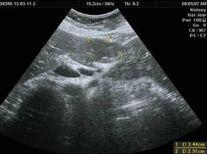
On ultrasound, GCA usually looks like a solitary heterogeneous formation with clear contours of varying degrees of echogenicity: 20-40% of adenomas appear hypoechoic, 30% are hyperechoic, which is due to the presence of fatty inclusions in the tumor tissues. According to a number of authors, when using CDK, peripheral peri- and intratumoral vessels are identified in some cases.
Liver adenoma
Thus, ultrasound of the abdominal organs is currently a screening method for detecting tumors and tumor-like processes in the liver. Due to its fairly high information content and accessibility, ultrasound is performed at the first stage of diagnosis if a liver tumor is suspected. Ultrasound allows not only to detect a tumor in the liver, but also to judge its size, topography, and operability of the process. Intraoperative ultrasound is informative for the diagnosis of intrahepatic tumors, since other research methods are of little use during surgery.
Ultrasound in the “gray scale” mode allows you to identify various changes in the parenchyma. However, in some cases, it is not possible to make a differential diagnosis between diffuse diseases due to the similarity of echographic signs.
B-mode echography with Dopplerography of the vascular system of the liver with assessment of the hemodynamic state makes it possible to clarify the nature of the pathological process. However, despite the high specificity (97%), the sensitivity of the method in detecting liver tumors remains quite low (60%), which allows preference to be given to dynamic CT or MRI techniques. In addition, according to some researchers, the informativeness of the diagnostic conclusion of ultrasound, as a subjective research method, depends on the experience and qualifications of the ultrasound diagnostic doctor.
How to recognize the symptoms of an endometrioid cyst?
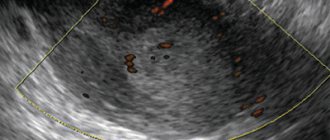
An endometrioid cyst is an echogenic formation of the ovary, which has symptoms characteristic of diseases of this kind. Among the most common are irregular menstruation, pain in the lower abdomen, and inability to conceive a child. In addition, abnormalities in the functioning of the gastrointestinal tract and bladder may be observed.
While the cyst is small, it may not cause the woman any discomfort and is discovered only during a gynecological examination. The formations can be on one of the ovaries or on both sides. The growth rate of an endometrioid cyst is impossible to predict. In some cases it increases rapidly, in others it can freeze for several years, and then grow very slowly or stop growing altogether. The transformation of cysts of this type into malignant ones is extremely rare, and is usually associated with hormonal changes due to menopause.
The danger of an endometrioid cyst is that it can burst, after which its contents end up in the abdominal cavity, causing inflammation. In addition, it is this type of cyst that can twist, causing sharp pain in the lower abdomen, nausea and vomiting. Such processes can only be detected using ultrasound.
Bibliography
- S. V. Shakhidzhanova, T. S. Pustovitova, “Some aspects of the diagnosis of focal liver pathology,” journal “Clinical Imaging” N19, December 2001.
- A.Z. Guseinov, T.A. Guseinov, “Modern diagnosis of liver tumors”, Bulletin of new medical technologies, electronic journal – 2016 – N 4, Tula.
- Zh.N. Kyzhyrov, B.B. Baimakhanov, M.M. Sakhipov, A.T. Chormanov, N.N. Birzhanbekov, E. Serikul, “Diagnostics of focal liver diseases”, Bulletin No. 1, 2021, Kazan.
- S.N. Berdnikov, V.N. Sholokhov, Yu.I. Patyutko, M.S. Makhotina, E.S. Chuchuev, K.E. Abirov, “Complex elastography and elastometry in the differential diagnosis of hyperechoic liver formations,” Journal: SonoAce Ultrasound No. 25, Moscow.
- O.M. Kurzantseva, “Some aspects of the diagnosis of focal nodular hyperplasia of the liver (fibronodular hyperplasia)”, Journal: SonoAce Ultrasound No. 30, Kemerovo.
Conservative therapy
The main goal of drug therapy is to stop the growth of education, eliminate the clinical manifestations of the disease, prevent possible complications and return the woman to a full life.
Conservative treatment of uterine fibroids is indicated in the following cases:
- the size of the node does not exceed 12 weeks of pregnancy;
- external manifestations of the disease are moderate or mild;
- there are contraindications to surgery.
The basis of drug therapy is hormonal agents. This is explained by the fact that often the growth of interstitial fibroids is associated with disorders in the endocrine system.
Most often, gynecologists use the following groups of drugs:
- androgens;
- gestagens;
- combined oral contraceptives;
- analogues of gonadotropin releasing hormone.
Androgens, or male sex hormones, can reduce the size of uterine fibroids. The minimum duration of treatment is six months. Gestagens are recommended to be taken with concomitant hyperplasia of the uterine mucosa. The therapy is long-term. Its main goal is to reduce the growth of the endometrium. Combined oral contraceptives can reduce the size of fibroids or stop their growth. The use of gonadotropin releasing hormone analogues is indicated only in the most severe cases, when premature menopause is possible. To increase effectiveness, conservative therapy is supplemented with herbal medicine and physiotherapeutic treatment of uterine fibroids.
Diagnostic methods
To confirm the diagnosis of subserous uterine fibroids, it is not enough to just undergo an examination by a gynecologist; more precise methods are needed. These methods include:
- ultrasonography;
- hysterosalpingography (introducing a contrast agent into the uterus and performing radiography);
- MRI;
- echography;
- CT scan;
- blood test (to detect anemia).
Only after passing all the necessary examinations can you begin to treat the subserous node of uterine fibroids.
What are the types of neoplasms in the reproductive organs?
- ectopic pregnancy, in which a fertilized egg implants outside the uterus;
- an abscess in the uterine tube or ovary, which occurs with inflammatory and infectious diseases of the pelvic organs;
- endometrioma - benign ovarian cyst;
- leiomyoma (fibroids) is a benign gynecological tumor;
- Ovarian torsion is an acute gynecological situation with impaired blood supply to the ovary;
- cancerous tumors.