Ultrasound of peripheral lymph nodes
The lymphatic system is one of the body systems, which, along with the circulatory system, ensures the maintenance of a healthy internal environment.
However, if the main function of the circulatory system is to provide nutrition and oxygen supply to tissues, then the lymphoid system acts as a kind of filter that prevents the spread of pathogenic pathogens and removes waste products from tissues. Palpation of superficially located nodes has long remained a priority research method, allowing one to obtain a general idea of the size of the nodes and its density. However, the technique is not able to provide detailed information about the shape and internal structure of the object under study; in addition, not all lymph nodes are accessible for external inspection. Ultrasound of lymph nodes today is a priority method that allows one to obtain detailed information about their location, size, shape, structure and relationship to nearby organs, and is of great diagnostic importance for determining the nature of metastasis, dynamic monitoring of a number of diseases and timely detection of acute infectious conditions.
What does ultrasound of lymph nodes show?
Normally, lymph nodes remain invisible on ultrasound. Healthy structures have high echogenicity and transmit ultrasound beams without distortion. Visualization is possible only if there are deviations from the norm.
During the examination, the doctor examines the shape, size, density of the nodes and adjacent tissues. Based on the echogenic activity of individual areas, metastases can be detected in the area of lymph outflow from the affected organ and a malignant process can be suspected.
Ultrasound of enlarged lymph nodes makes it possible to make the following diagnoses:
- Lymphadenitis is an inflammatory process caused by infection in the most peripheral node or pathogens from the site of lymph outflow.
- Lymphadenopathy is damage to several groups of lymph nodes simultaneously due to infection with tuberculosis, brucellosis, HIV, toxoplasmosis and other severe infections.
- Lymphosarcoma is an oncological lesion.
In our clinic in Moscow, ultrasound of lymph nodes is performed as part of a comprehensive diagnosis of diseases such as syphilis, Sezary disease, actinomycosis, and leukemia. To clarify the data, a biopsy, x-ray, or other studies may be required.
The need for ultrasound of peripheral lymph nodes
Indications (general and specific) for ultrasound examination of lymph nodes are the following conditions:
General indications for ultrasound of lymph nodes mean such symptoms, conditions or suspicions for which examination of any one or more groups of nodes is indicated.
By private indications we mean such symptoms and conditions for which examination of a specific group of lymph nodes is indicated.
General indications for ultrasound examination of lymph nodes of a particular location include the following:
– pain, enlargement, excessive mobility or hardness when palpating the lymph nodes;
– swelling, redness or increased skin temperature (hot to the touch) in the area where the lymph nodes are located;
– discomfort when performing movements, felt in the area where the lymph nodes are located (for example, when swallowing, when walking, when moving your hand, etc.);
– forced position of a body part due to pain in the area of the lymph nodes (for example, a bowed head, a hand pressed to the body, etc.);
– headaches, general weakness and melancholy, present for a long period of time;
– causeless sleep disturbances, insomnia or drowsiness;
– poor appetite;
– the presence of long-term infectious or inflammatory processes in the body in a particular organ (for example, otitis, laryngitis, tonsillitis, adnexitis, thrombophlebitis, conjunctivitis, hepatitis, etc.);
– suspicion of systemic connective tissue diseases or autoimmune processes (pain in joints and muscles, rashes on the skin, etc.);
– deformation of the jaw bones;
– pain in the abdomen, chest or neck;
– presence or suspicion of tumors of various locations.
If there are general indications, the doctor chooses which lymph nodes to examine based on the localization of the pathological process.
Thus, ultrasound of the lymph nodes of the head and neck is usually specifically prescribed for the following conditions:
– overgrowth of gum tissue;
– bleeding gums;
– deformations of the jaw and face;
– chronic infections of the ENT organs;
– acute infectious and inflammatory processes occurring with high fever, cough, runny nose, difficulty swallowing, pain in the mouth, etc.;
– enlarged lymph nodes, the size of which has not decreased within two weeks after suffering an acute illness (for example, measles, rubella, influenza, etc.);
– presence or suspicion of tuberculosis, syphilis, leprosy;
– presence or suspicion of a tumor or metastases to the lymph nodes (lymphosarcoma, lymphoma, lymphogranulomatosis);
– the presence of tumors of the lungs, larynx, trachea, throat, tongue or thyroid gland.
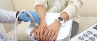
Ultrasound of the lymph nodes of the upper limb (axillary and ulnar) is performed in the following cases:
– suspicion of tumors of the mammary gland or chest organs (lungs, heart, pleura, diaphragm, etc.);
– for the purpose of control after operations to remove the mammary gland;
– signs of a malignant tumor in any organ or HIV infection.
Ultrasound of the lymph nodes of the lower limb (inguinal, popliteal) is targeted in the following cases:
– presence or suspicion of sexually transmitted infections (gonorrhea, syphilis, chlamydia, trichomoniasis, candidiasis, etc.);
– any inflammatory diseases of the pelvic organs and urinary system;
– infectious or inflammatory processes localized on the lower limb (for example, thrombophlebitis, boils, etc.);
- HIV infection;
– presence or suspicion of tumors of the pelvic organs or urinary system.
Diagnosis of the condition of lymph nodes using ultrasound
An ultrasound examination of the lymph nodes is carried out to clarify the diagnosis, as well as monitor the progress of the disease affecting the state of the lymphatic system.
Lymph nodes are a barrier that prevents the spread of bacteria, viruses, toxins, tumor cells and other harmful substances in the blood throughout the body.
These organs of the immune system instantly react to the appearance of any abnormal changes in the body: they become denser, change shape, size, level of mobility, structure, etc.
Their reaction does not always correspond to the severity of the pathology; changes often occur long before the onset of inflammatory processes, thereby helping to promptly prevent the development of the disease. The method is based on the ability of tissues to react differently to the penetration of ultrasonic waves into the body and consists of observing and recording these differences.
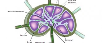
Changes are displayed on the monitor screen and allow the doctor to determine the following parameters:
• Shape of lymph nodes.
• Their sizes.
• Digital ratio of length and width.
• The degree of tissue density in the lymph node.
It must be remembered that ultrasound diagnostics is an auxiliary method of diagnosis; its results must be supplemented by other studies.
Preparation for ultrasound of peripheral lymph nodes
To study peripheral lymph nodes using ultrasound, special patient preparation is not required. The study can be carried out at any time of the day, within any diet and medication regimen.
However, in the case of examination of nodes in the groin area, a preliminary visit to a venereologist or gynecologist is necessary to confirm the absence of sexually transmitted diseases, which allows one to exclude possible infectious causes of changes in nodes and guarantee the reliability of the diagnostic conclusion.
If there is a lot of hair in the area where the lymph nodes are located, you must first remove the hair.
Specifics of echography of lymph nodes
There is no need to carry out any preparatory activities before the study.
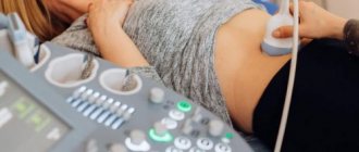
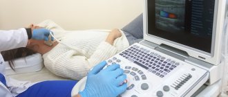
The surface of the ultrasound sensor is covered with gel, the patient exposes a part of the body to which the doctor presses the device. At this time, an image obtained using reflected ultrasonic waves appears on the screen.
A preliminary examination will be required before checking the lymph nodes in the groin area. Since the nature of their changes may be sexually transmitted, consultation with a specialized specialist will be necessary.
The procedure for performing ultrasound of peripheral lymph nodes
The examination takes place on a couch, in a standing or lying position. First, the patient is freed from clothing and jewelry in the part of the body where the necessary lymph nodes are located.
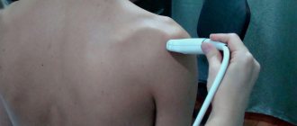
A special gel is applied to the patient’s skin in the area of contact with the sensor to improve ultrasound transmission. The doctor, with light pressure and progressive movements, slowly moves the sensor to the location of each lymph node.
During the examination, the specialist asks the patient to take the necessary position - to examine the axillary lymph nodes, you need to put your hands behind your head, to diagnose the abdominal lymph nodes, you need to take a deep breath while lying down.
The information received by the device is transmitted to the ultrasound machine, displayed on the monitor in the form of an organ, in its two- or three-dimensional projection.
During the diagnostic process, the doctor records the following data:
- location and number of nodes;
- their size and shape;
- the structure of the lymph nodes (absence of cysts and inclusions, smooth contours, normal structure, absence of adhesions of nodes to each other or nearby organs).
Echography of axillary lymph nodes
The study is prescribed in the following situations:
• Suspicion of cancer or HIV (lymph nodes in the groin and neck area increase in size, although the general health status is positive).
• After mastectomy, to exclude the occurrence of metastases. Carried out as planned.
• The doctor suspects the development of tumor-like neoplasms in the breast and mammary glands.
If a woman has had mastitis or toxoplasmosis, a control test will be scheduled no earlier than 2 months after the disease has been eliminated.
Sign up for ultrasound diagnostics
In the medical center, ultrasound examinations are performed using expert-class equipment.
The appointment is conducted by doctors:
– GONTAREV ALEXANDER GENNADIEVICH,
– ZHEVED ELENA SERGEEVNA,
– MAKOVEEVA IRINA SHAUKATOVNA
professional diagnosticians with extensive practical experience.
Ultrasound examinations in the clinic are carried out by appointment.
Phone number for registration.
Address: Sevastopol, Heroes of Stalingrad Avenue, 63
(stop b. Omega)
Opening hours: Mon. - Fri: 07:30 - 17:00, Sat. — Sun: 08:00 — 16:00
The patient should check with the administrators about the need for additional preparation for diagnosis.
The cost of ultrasound of peripheral lymph nodes (for one group) is 800 rubles.
The cost of ultrasound of peripheral lymph nodes (two groups) is 1400 rubles.
How is the ultrasound examination procedure carried out?
The ultrasound diagnostic procedure is carried out quite quickly, it is safe and completely painless. The child is placed on the couch. The skin in the area being examined is lubricated with gel, which is necessary to facilitate the penetration of ultrasonic waves to the surface of the organ. The ultrasound specialist then presses the device's sensor to the skin and uses it to obtain an image displayed on the monitor. From time to time, the doctor may press on certain areas of the body to assess the degree of mobility of the lymph nodes. Examination of the abdominal lymph nodes is carried out in a state of deep inspiration. Therefore, before visiting the clinic, you should practice holding your breath with your child.
Examination of each group of lymph glands usually takes about 10-15 minutes. Most often, lymph nodes located in the neck and abdomen are examined using ultrasound. Changes in the cervical lymph nodes are observed in pathologies of the tonsils, salivary glands, hard palate, hearing organ and thyroid gland. If lymphadenopathy is found in the abdomen or retroperitoneum, this may indicate diseases of the stomach, liver, pancreas or intestines. Lymph nodes are located at different depths from the surface of the body, so it is not always possible to clearly diagnose pathology using ultrasound alone. To clarify the diagnosis, the doctor may prescribe several studies at once; for example, traditional ultrasound diagnostics can be supplemented with MRI, duplex echosonography and biopsy with histological analysis of lymphoid tissue.
If a pathology of any group of lymph nodes is detected, the specialist also evaluates the condition of the organs located next to them in order to find out the cause of lymphadenopathy. If necessary, other laboratory and instrumental studies are prescribed for a more accurate diagnosis.