A kidney biopsy is an invasive diagnostic procedure that examines the cortex and medulla of the organ. The essence of the study is to obtain histological material, with the help of which it is possible to clarify the nature of the pathological process in the tissues of the organ. This study allows you to make a correct diagnosis, resolve the issue of locality or systemicity of the process, and prescribe the necessary treatment. Once a diagnosis has been established, a biopsy makes it possible to monitor the course of the disease and identify the need for organ transplantation or surgical treatment of the identified pathology.
- Types of biopsy
- Indications
- Contraindications
- Preparation
- How the research is carried out
- After the biopsy
- Complications
- The result of the procedure and its interpretation
- Which doctor should I contact?
About the method
Biopsy is an instrumental diagnostic method, the most informative among those available. It is important both to confirm the preliminary diagnosis and to differentiate the presence of tumors and other pathologies in a particular organ. The importance of a biopsy is that with the help of this method the doctor can make an early diagnosis of malignant formations.
Today, biopsy is the only diagnostic method that reliably allows us to understand the nature of various neoplasms. Using this method, the doctor can make an accurate diagnosis for his patient.
A biopsy helps to remove any suspicious cells and tissues of the human body for examination. If a highly qualified specialist takes on the case, the likelihood of error in announcing the diagnosis is minimal.
We have already noted that during a biopsy, a biopsy sample (cells and tissues of the human body) is taken for subsequent laboratory study.
The accuracy of the procedure itself and the study of what is happening behind it depends on a number of factors:
- Doctor's experience;
- Qualification of a laboratory assistant studying elective material;
- Quantities of selected material;
Using the procedure you can:
- Obtain comprehensive information about the specifics of the disease;
- Find out the nature of the neoplasm;
- Confirm/refute the preliminary diagnosis;
- Conduct diagnostics to differentiate between benign and malignant tumors;
- Eliminate the source of inflammation;
- Assess the effectiveness of previously prescribed treatment.
As an invasive diagnostic procedure, biopsy is not dangerous or harmful, but due to its maximum accuracy in making diagnoses, it can hardly be overestimated in oncological practice.
Preparing for a biopsy
Before the procedure, the doctor tells the patient about why and how the procedure will be performed, as well as what complications it may cause. The specialist will also ask the patient questions about existing diseases and general health, and find out whether the patient is allergic to certain medications. If everything is in order, a date for the procedure is set and permission to intervene is signed.
Two weeks before the procedure, the patient needs to stop taking medications that thin the blood and affect blood clotting in order to minimize the risk of bleeding. These medications include anticoagulants, antiplatelet agents, and nonsteroidal anti-inflammatory drugs. Some dietary supplements should also be eliminated from the diet. These are fish oil, garlic, ginkgo. Many medications can only be assessed correctly by a doctor. Therefore, during preparation, list to your doctor all medications that you are taking or have recently taken. Taking these data into account, the doctor will choose the optimal period for the procedure and tell you after what time you can resume taking the medications.
A few days before the biopsy, it is necessary to retake all tests; on the appointed day, you must refuse to eat and drink; the day before, you must cleanse the intestines with an enema or laxatives. A few days before the biopsy, the patient is recommended to have blood and urine tests so that the doctor can assess the condition of the body and its readiness for manipulation.
Indications
Each medical procedure has its own indications and barriers. Biopsy is no exception to the rule
Among the indications:
- Identifying the nature of cell changes, distinguishing benign neoplasms from oncology;
- Determining the effectiveness of treatment previously prescribed by an oncologist;
- Complications after kidney transplantation;
- Kidney failure that develops rapidly;
- Nephrotic syndrome of unknown etiology or suspicion of it;
- Nephrogenic hypertension;
- Infectious diseases of the genitourinary tract;
- Presence of protein and blood in the urine;
- High proportion of creatinine, urea, uric acid and some other cases.
What causes nephroblastoma?
Although there is a clear link between nephroblastoma, birth defects, and genetic mutations, most children with this tumor do not have any birth defects or inherited genetic changes.
The cause of nephroblastoma in children is not yet understood, but significant progress has been made in understanding many of the events that occur during the formation of normal fetal kidneys and how this process is altered to lead to nephroblastoma.
Kidneys develop very early in the womb. Sometimes defects may occur during their development. Some cells that should turn into mature kidney cells do not do this and remain embryonic (fetal, or early). Some clusters of these early kidney cells may exist for a period of time after the baby is born. Typically, these cells mature by the age of 3-4 years. If this does not happen, the cells may begin to grow uncontrollably. The result of this process is the development of nephroblastoma. Having a mutation or deletion of the WT1 or WT2 genes increases the risk that some cells will remain embryonic and lead to the development of nephroblastoma.
Contraindications
Biopsy as a diagnostic method is strictly prohibited or not recommended in the following cases:
- The patient has only one functioning kidney;
- Renal failure;
- Renal mobility;
- Problems with blood clotting;
- Aneurysm and thrombosis of renal vessels;
- Renal tuberculosis;
- Myeloma;
- Suspicion of glomerulosclerosis;
- High blood pressure;
- Vascular atherosclerosis in the final stage;
- Purulent paranephritis and some other pathologies.
Make an appointment
The Sofia Oncology Center is located in the Central Administrative District (CAO) in Moscow, next to several metro stations: Novoslobodskaya, Tverskaya, Chekhovskaya, Belorusskaya and Mayakovskaya.
You can make an appointment for kidney diagnostics and find out the cost of the test by phone or through the online form.
Varieties
Today in medical practice the following types of procedures are common:
- Percutaneous puncture;
- Open;
- Laparoscopic;
- Endoscopic;
- Transjugular.
Percutaneous puncture biopsy has a number of features:
- It is carried out with a long needle, the process is monitored by an X-ray machine;
- If necessary, contrast is injected into the organ;
- The manipulation takes place using local anesthesia.
Open biopsy
The specifics are:
- Carry out in most cases with tumors, as well as on their suspicions;
- It is also indicated for hypertension, if it is impossible to determine the cause of the failure by other means;
- Parenchyma samples taken from several areas are examined under a microscope:
- Before the manipulation, the patient is examined - x-ray of the kidneys, arteriography, ultrasound;
- The procedure requires general anesthesia.
Laparoscopic biopsy
Peculiarities:
- Instruments are inserted into the peritoneum near the location of the kidneys;
- The video camera controls the manipulation of the material capture;
- There is no need to cut the fabric.
Endoscopic biopsy
Features:
- To insert instruments, the kidneys use the urinary tract and bladder;
- There are no punctures or cuts on the body, so they have the least impact on the body.
Transjugular biopsy
The catheter is inserted into the jugular vein, and then into the renal vein.
The technique is used in cases:
- Severe obesity;
- Blood clotting problems;
- Serious respiratory tract diseases;
- When it is not possible to use general anesthesia.
Types of kidney biopsy
Types of biopsy differ in how the sample is taken:
- percutaneous
- taking a sample with a needle under ultrasound guidance; - open
- taken during open surgery, for example, when removing a tumor; - urethroscopic
- performed if stones are found in the ureter or renal pelvis, an anomaly or disease of the upper urinary tract is detected, and also if the kidney has been transplanted. The ureteroscope is passed through the ureter into the renal pelvis. This makes it possible to view the organ and collect tissue. This method is used for children and pregnant women. The manipulation requires spinal anesthesia or anesthesia; - laparoscopic
- performed through several holes in the anterior abdominal wall. It is used for disorders of the blood coagulation system, as well as for patients with one functioning kidney. A camera and illuminator are brought to the kidney through a trocar.
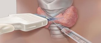
How to do it
Kidney biopsy is performed only in an operating room. Its duration is usually 30-40 minutes.
Most often I use local anesthesia, but general anesthesia is also possible. What actions do the doctor and the patient take? Briefly it looks like this:
- The patient is exposed and lies on the operating table.
- The doctor selects and processes the point of the future puncture (incision).
- The anesthesiologist injects local anesthesia there.
- If the patient is conscious, he is asked not to move, but to retain consciousness.
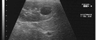
- Under ultrasound guidance, the doctor inserts a special needle and uses it to collect the necessary material.
- There are times when the needle must be inserted several times to collect the required amount of material.
Is early detection of nephroblastoma possible?
Children with birth defects associated with the possibility of nephroblastoma should be monitored and undergo special examination. Ultrasound examination (ultrasound) should be performed every three months until the child reaches 6-7 years of age in order to early recognize a kidney tumor, when it has not yet spread to other organs. It is necessary to inform your doctor about the presence of a relative with nephroblastoma in your family and examine him. all other children of the family with regular ultrasound examinations.
Given the rarity of nephroblastoma, there is no need to recommend ultrasound as a screening method (the use of any test in people without signs and symptoms of the disease), unless, of course, the child has risk factors for developing nephroblastoma.
How is nephroblastoma diagnosed?
Nephroblastoma is sometimes difficult to detect in the early stages because it often reaches large sizes without causing pain in the child. Children with such a tumor may appear quite healthy and lead a normal life. Usually the first sign of the disease is an increase in the size of the abdomen. Parents may notice an enlarged abdomen or tumor formation in it while washing and dressing the child. Up to 25% of people with nephroblastoma may experience abdominal pain, fever, blood in the urine, or increased blood pressure. If your child has any of these symptoms, you should consult a doctor immediately.
After the procedure
After the manipulations associated with collecting the material, it goes to the laboratory for study, and the patient goes to the ward. Bed rest is prescribed for 8-10 hours. A urine test is ordered.
There may be slight soreness in the puncture area. In the vast majority of cases, it does not even require analgesics.
If no complications are identified, the patient leaves the ward the next day. However, he must understand that hard work and physical activity, heavy lifting are prohibited for him for at least two weeks.
Risk factors for nephroblastoma
A risk factor is something that increases your chance of developing cancer. There are different risk factors for different tumors. These may include genetic (inherited) factors, as well as environmental and lifestyle factors. In general, lifestyle factors (smoking, poor diet and physical inactivity) are the most important factors in most adult malignancies. However, they have little, if any, effect on the risk of tumors in children.
Environmental risk factors.
No relationship was found between nephroblastoma and environmental factors either before or after the birth of the child.
Genetic and inherited risk factors.
Cases of nephroblastoma in the family. 1-3% of children with nephroblastoma have one or more relatives who have suffered the same disease. It is assumed that these children inherited an altered gene from one of the parents, which significantly increases the risk of nephroblastoma. Patients with familial cases of nephroblastoma, compared with sporadic tumors (without the presence of relatives with a similar tumor), are more likely to develop bilateral kidney damage.
The presence of certain birth defects . There is a clear relationship between nephroblastoma and certain birth defects. Thus, 15% of children with nephroblastoma have birth defects, most often represented by syndromes. A syndrome is several symptoms, signs, malformations or other disorders that occur in the same patient.
WAGR syndrome : Aniridia (complete or partial absence of the iris, disorders of the genitourinary tract (defects of the kidneys, penis, scrotum, clitoris, testicles or ovaries), mental retardation. Patients with such developmental defects have a 3% chance development of nephroblastoma.
Beckwith-Wiedemann syndrome : Presence of large internal organs, especially the tongue. Hemihypertrophy (an increase in the size of one arm or leg on one side of the body) is possible. These people are more likely to develop nephroblastoma.
Denis-Drash syndrome : Underdevelopment of the penis, testicles and scrotum. For this reason, boys with this syndrome may be mistaken for girls. For some unknown reason, the kidneys in such children begin to function poorly or stop functioning altogether. Nephroblastoma can occur in these altered kidneys.
There are a number of other rarer syndromes associated with nephroblastoma.
Gene and chromosomal changes in children with birth defects and nephroblastoma . Genes determine eye and skin color, blood type, etc. Changes in the DNA of a gene can lead to the development of the disease. Changes in DNA are called mutations. Sometimes these mutations are inherited from parents. This explains the fact that some diseases are more common among members of the same family.
Genes are part of chromosomes, which are giant DNA molecules containing thousands of genes. DNA is contained in 23 pairs of chromosomes. One gene from each pair is inherited from the mother and one from the father.
The previously mentioned syndromes are associated with deletions (missing part of DNA) or mutations of genes on chromosome 11.
Complications
As a rule, complications after a biopsy are not observed. Their most typical manifestation is microhematuria, which can torment the patient for 1-2 days after the procedure. Among the complications:
- Fever;
- Pain;
- Bleeding.
Very severe consequences have been recorded:
- Kidney infarction;
- Perirenal hematoma;
- Infection of the resulting hematoma;
- Damage to neighboring organs.
All this happens due to a violation of basic safety, low qualifications of doctors, and technologies that are lagging behind modern life. At the Onco.Rehab you will not encounter such precedents.
Advantages of Sofia Cancer Center
At the Sofia Cancer Center everything is in one place. In two adjacent buildings with a total area of 35,000 sq.m. there is a hospital, a surgery department, a laboratory, dentistry, an ambulance, a clinic, etc. Patients can receive all the necessary services quickly - we have experienced doctors, leaders of Russian medicine, who provide assistance with any diagnoses.
Advantages of the Sofia Cancer Center:
- impeccable reputation and 30 years of unique experience;
- full staff of specialists: oncologists, nephrologists, surgeons, etc.;
- individual support throughout the clinic and special care for patients;
- increased comfort: modern interior, cozy offices and chambers, a luxurious restaurant with panoramic views, etc.;
- modern operating and manipulation rooms, equipped with everything necessary;
- convenient location (center of Moscow);
- large parking for patients.
Treatment of nephroblastoma
More than 90% of children with nephroblastoma make a full recovery. Advances in treatment have been achieved through the use of surgical, radiation and medicinal methods.
After nephroblastoma is detected and the stage of the disease is clarified, a treatment plan is developed.
Taking into account the rarity of nephroblastoma, it is advisable to be treated in specialized pediatric oncology departments, in which doctors have significant experience. Treatment of the child should not be delayed, since nephroblastoma grows very quickly.
Treatment for nephroblastoma includes surgery, chemotherapy, and sometimes radiation therapy. If there is residual tumor after the first operation, radiation therapy and repeat surgery may be required. The main goal of treatment is to remove the primary tumor, even if there are distant metastases, for example in the lungs.
In some cases, the tumor may be too large and involve vital structures, making surgery impossible at the moment. In some patients, the lesion may be bilateral. In these patients, chemotherapy or radiation therapy, or a combination of the two treatments, is first used to shrink the tumor before removing it.