For anemia
indicators of iron, folic acid, vitamin B12, transferrin, ferritin, bioactive hepcidin, and total protein may change.
Markers of inflammation, rheumatism and rheumatoid arthritis
include total protein, C-reactive protein, procalcitonin, rheumatoid factor, antistreptolysin-O.
Assessment of the content of vitamins and microelements in the body
(A, C, E, K1, B1, B2, B3, B5. B6, B7, B12, D, beta-carotene, etc.) allows you to find out the body’s need for microelements and vitamins.
Abnormal electrolyte levels
(sodium, potassium, magnesium, calcium, chlorine) can detect various metabolic disorders.
Preparation for the procedure
- A biochemical blood test is taken in the morning and strictly on an empty stomach. At least 8 hours should pass between the last meal and blood collection (preferably at least 12 hours). Juice, tea, coffee, especially with sugar, cannot be drunk! You can drink water.
- It is advisable to exclude fatty and/or fried foods and alcohol from the diet 1-2 days before the examination. If there was a feast the day before, the laboratory test should be postponed for 1-2 days.
- One hour before taking blood, you must refrain from smoking.
- It is necessary to exclude factors that influence the research results: active physical activity, emotional overexcitation. Before the procedure, you need to rest for 10-15 minutes and calm down.
- Blood is taken for analysis before starting medications (for example, antibacterial and chemotherapy) or no earlier than 10-14 days after their discontinuation. You must inform your doctor about taking medications.
- Blood should not be donated after radiography, ultrasound or rectal examination, physiotherapeutic procedures, physical therapy, acupuncture (reflexology), massage.
Indications for use
The development of liver diseases is indicated by the following clinical signs:
- heaviness and discomfort in the right hypochondrium;
- bitter taste in the mouth;
- weight loss for no apparent reason;
- darkening of urine;
- stool discoloration;
- yellowing of the skin, eye sclera, mucous membranes;
- dyspepsia;
- fast fatiguability.
If such symptoms appear, you should contact a hepatologist or gastroenterologist and donate blood for a biochemical analysis. Indications for liver tests:
- diagnosis of chronic liver diseases;
- suspicion of the development of cirrhosis;
- diseases accompanied by metabolic disorders in the body, affecting the functioning of the liver (for example, diabetes);
- diagnosis of inflammatory processes in the liver;
- intermediate diagnostics to assess the effectiveness of treatment for confirmed liver pathologies;
- chronic alcoholism;
- changes in the structure and size of the liver identified during ultrasound or other diagnostic methods.
Liver tests are prescribed for patients undergoing treatment with hepatotoxic drugs.
Blood chemistry
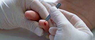
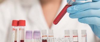
The material for the study is venous blood, which is usually taken from the cubital vein in a volume of 5-8 ml and placed in a clean, dry tube without anticoagulants.
The exception is certain tests (for example, a test for glycated hemoglobin), for which the rules for drawing blood are indicated in the appointment forms. The turnaround time for a basic set of indicators is one to two business days.
However, some tests (eg, serum paraprotein detection) are performed within 11 days.
What may affect the results
Serum protein levels are affected by body position and physical activity.
When changing the horizontal position of the body to a vertical one, the protein content increases by 10% within 30 minutes; with active physical work, the increase is up to 10%. Clamping of blood vessels during blood collection and "hand work" can also cause an increase in total protein levels. When studying bile pigments, the picture of the results is distorted by foods that cause coloration in the blood serum - pumpkin, beets, carrots, citrus fruits. A good piece of roast pork the day before will increase your blood potassium and uric acid levels.
A decrease in blood lipid levels can be caused by drugs that affect this process. Therefore, their use should be stopped 2 weeks before the lipid profile test, in consultation with your doctor.
The level of uric acid is affected by eating offal (liver and kidneys), meat, fish, coffee, tea during the 4-5 days preceding the study.
You can donate blood to determine biochemical parameters at the nearest INVITRO medical office.
A list of offices where biomaterial is accepted for laboratory research is presented in the “Addresses” section. Interpretation of study results contains information for the attending physician and is not a diagnosis. The information in this section should not be used for self-diagnosis or self-treatment. The doctor makes an accurate diagnosis using both the results of this examination and the necessary information from other sources: medical history, results of other examinations, etc.
A little about blood
Human blood is a special tissue that is liquid and adapted to perform many functions. We will not list all the functions; we will focus on just two as an example. One of the main functions of blood is gas exchange. It transports cells enriched with oxygen to organs and tissues, and takes from them the waste product of tissue respiration - carbon dioxide. Blood saturated with carbon dioxide is dark and venous. It rushes into the lungs, into the pulmonary circulation, where it is saturated with oxygen. Oxygenated blood is arterial; it flows through arteries, and gas exchange occurs in capillaries - the thinnest vessels.
The second function is transport. This unique liquid tissue transports a large number of different substances, molecules and compounds. The difference between these two functions is that the first is provided by cells, or red blood cells. And the second function does not require carrier cells. There are simply various dissolved substances in the blood - molecules, proteins, hormones, various ions. If carriers are needed, they are not cells, but special protein molecules that are millions of times smaller than cells.
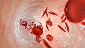
If you take whole blood from a vein (or from a finger, from an ear, from a baby’s heel) and place it in a test tube and spin it for several minutes on a laboratory centrifuge, the blood will separate into two parts unequal in volume. At the top there will be a clear opalescent liquid, or plasma. It does not contain any cellular elements, but only dissolved substances and various compounds.
There will be a dark precipitate at the bottom of the tube. This is a cellular composition: red blood cells, leukocytes and platelets. Accordingly, a general analysis studies the properties of blood as a liquid, its physical characteristics, as well as its cellular composition. When conducting a general analysis, everything that is dissolved in the blood and found in its plasma is not taken into account at all. Let us briefly consider what studies are carried out after taking a standard general or clinical blood test.
Normal values for adults
| Analysis | Men | Women |
| Glucose | 4.1 - 6.0 mmol/l | 4.1 - 6.0 mmol/l |
| Glycated hemoglobin | <6,0% | <6,0% |
| Fructosamine | 170 – 285 µmol/l | 170 – 285 µmol/l |
| Lactate | 0.5 - 2.2 mmol/l | 0.5 - 2.2 mmol/l |
| Total protein 14 – 60 years over 60 years | 64 – 83 g/l 62 – 81 g/l | 64 – 83 g/l 62 – 81 g/l |
| Albumin 14-90 years over 90 years | 35-52 g/l 29-45 g/l | 35-52 g/l 29-45 g/l |
| Homocysteine | 5.46 – 16.20 µmol/l | 4.44 – 13.56 µmol/l |
| Total bilirubin | 3.4-20.5 µmol/l | 3.4-20.5 µmol/l |
| Direct bilirubin | < 8.6 µmol/l | < 8.6 µmol/l |
| Bile acids | < 10 µmol/l | < 10 µmol/l |
| Triglycerides | <1.7 mmol/l | <1.7 mmol/l |
| Total cholesterol | <5.0 mmol/l | <5.0 mmol/l |
| Cholesterol-HDL | >1.0 mmol/l | >1.2 mmol/l |
| LDL cholesterol | 1.3 - 4.5 mmol/l | 1.3 - 4.5 mmol/l |
| AlAT (ALT) | < 41 U/l | < 31 U/l |
| ASAT (AST) | < 37 U/l | < 31 U/l |
| Alpha amylase up to 70 years over 70 years | 25 - 125 U/l 20 - 160 U/l | 25 - 125 U/l 20 - 160 U/l |
| Gamma-glutamyl transpeptidase (GGT) | < 49 U/l | < 32 U/l |
| Cholinesterase | 5800 – 14600 U/l | 5860 – 11800 U/l |
| Lipase | 8 - 78 U/l | 8 - 78 U/l |
| Lactate dehydrogenase (LDH) | 125-220 U/l | 125-220 U/l |
| Alkaline phosphatase | 40-150 U/l | 40-150 U/l |
| Creatine kinase (CPK) | < 190 U/l | < 167 U/l |
| Creatinine | 64-104 µmol/l | 49-90 µmol/l |
| Urea up to 60 years over 60 years | 2.1 - 7.13 mmol/l 2.9 - 8.23 mmol/l | 2.1 - 7.13 mmol/l 2.9 - 8.23 mmol/l |
| Uric acid | 210 – 420 µmol/l | 150 – 350 µmol/l |
| Bilirubin | 5-20 µmol/l | 5-20 µmol/l |
| Potassium | 3.5 - 5.1 mmol/l | 3.5 - 5.1 mmol/l |
| Sodium | 136 – 145 mmol/l | 136 – 145 mmol/l |
| Chlorine | 101 – 110 mmol/l | 101 – 110 mmol/l |
| Total calcium | 2.2 - 2.55 mmol/l | 2.2 - 2.55 mmol/l |
| Magnesium | 0.66 - 1.07 mmol/l | 0.66 - 1.07 mmol/l |
| Phosphorus | 0.74-1.52 mmol/l | 0.74-1.52 mmol/l |
| Iron | 11.6 - 31.3 µmol/l | 9.0 - 30.4 µmol/l |
| Folic acid | 3.1 - 20.5 ng/ml | 3.1 - 20.5 ng/ml |
| Vitamin B12 | 187-883 pg/ml | 187-883 pg/ml |
| Transferrin | 1.9 - 3.75 g/l | 1.9 - 3.75 g/l |
| Latent iron-binding capacity of blood serum | 12.4 – 43.0 µmol/l | 12.5 – 55.5 µmol/l |
| Ferritin | 20 – 250 µg/l | 10 - 120 µg/l |
| Myoglobin | < 70 µg/l | < 70 µg/l |
| C-reactive protein | < 5 mg/l | < 5 mg/l |
Decoding indicators
Decoding a biochemical blood test is a comparison of the results obtained with normal values (reference values).
The norms for women and men for a number of indicators may differ; in addition, some values depend on the age of the patient. Different laboratories may use different research methods and units of measurement. In order for the results to be assessed correctly, the tests should be carried out in the same laboratory, at the same time. Comparing such studies will be more comparable.
What is examined during a general clinical analysis?
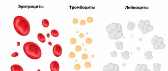
First of all, the total number of all cellular elements, red blood cells, leukocytes and platelets is determined. Red blood cells carry out gas exchange, white blood cells perform a protective function, and platelets regulate coagulation. When studying general analysis, it is very important to know the ratio of the amount of plasma, that is, the colorless part that does not contain cells, to the total volume. This indicator is called blood density, or hematocrit. With dehydration and fluid deficiency it is high, and with a lack of cells it will be low.
Previously, the color index was studied, but now the hemoglobin concentration is directly determined. It is very important for finding the causes of anemia.
The next very important indicator is ESR, or erythrocyte sedimentation rate. ESR is the first and easiest way to diagnose that there is some kind of inflammatory process in the body. The higher the ESR, the heavier the red blood cells and the faster they settle to the bottom of the tube. Their mass increases because they “cling” to their membrane a lot of different molecules, and also form various aggregates among themselves. Various proteins that are responsible for inflammation change the electrostatic parameters of red blood cells and reduce their repulsion from each other. Such cells are attracted to each other and quickly settle to the bottom. Thus, we can tentatively talk about some kind of inflammatory process. Also, a high ESR is characteristic of malignant neoplasms.
Currently, laboratory technology makes it possible to determine many erythrocyte indices. These include the average volume of a blood cell and the determination of its size. Modern laboratory technology makes it possible to determine the amount of hemoglobin in a red blood cell and the hemoglobin concentration. Separately, the processor of the laboratory analyzer will allow you to distribute red blood cells according to their size, or size. All these studies are automated and included in the overall analysis; they do not need to be ordered separately. Red blood cell indices and cell characteristics are very important in the presence of anemia, or a lack of hemoglobin in red blood cells. They help make the correct diagnosis.
Also, when studying the general analysis, attention is paid to what types of white blood cells, or leukocytes that protect us, are represented. In a standard analysis, the leukocyte formula is calculated. Currently, the study is carried out automatically using an analyzer. But still, the most popular is the manual examination of a stained and fixed blood sample under a microscope.
Among leukocytes, 5 subpopulations are distinguished. These are neutrophils that directly destroy foreign microorganisms and engage in phagocytosis, or their absorption. These are eosinophils that take part in allergic reactions and respond to helminthic infestations. These are lymphocytes that take part in immune reactions, these are monocytes, which are often found not only in the blood, but also in tissues as macrophages, as well as basophils. Such names come from the ability of leukocytes to perceive various dyes: neutral, basic and acidic.
A lot can be said from the leukocyte formula. Thus, an increase in white blood cells, regardless of their type, indicates inflammation, intoxication, various injuries, and stress. They increase in malignant neoplasms, including leukemia. A low white blood cell count occurs with anemia, autoimmune diseases, some chronic infections, and an enlarged spleen. Thus, how does a clinical blood test differ from a biochemical one? The study of the cellular composition and general properties of blood as a tissue.
There are some other research methods that also relate to a general or clinical blood test. For example, this is a study of reticulocytes. This is the name given to young red blood cells that have not yet been freed from the remnants of the cell nucleus. Their study is needed to indirectly assess bone marrow function and monitor the course of anemia.
All of the above studies relate to the so-called hematological studies. Hematology is a science that studies blood as a collection of different cells. Hematologists deal with anemia and leukemia, but in any case, the morphological substrate will be various normal or pathological cells, both in the peripheral blood and in the bone marrow. There are, of course, other studies in hematology. This is a determination of blood group and Rh factor, as well as a study of blood clotting ability, or coagulogram. But these methods can no longer be classified as general analysis. How does a biochemical blood test differ from a general blood test?
