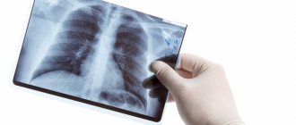
X-ray of the stomach is a diagnostic method for identifying functional disorders and diseases, allowing one to assess the size and shape of the organ and the integrity of the walls. This is an informative study that is easy to follow. The price, compared to other diagnostic methods, is low. It can be used provided there is no risk of developing oncological processes in the upper gastrointestinal tract. If available, it is better to use other methods of imaging the gastric mucosa.
Based on the diagnostic results, you can identify:
- Ulcer;
- Inflammatory processes;
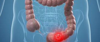
- Stomach cancer.
Complex X-ray examination: radiography and fluoroscopy of the esophagus, stomach, duodenum 1940
+7 (495) 320-43-41
Conditions for the procedure
The Law on Radiation Safety provides for the preliminary conduct of a number of studies: ultrasound, FGDS, laboratory tests. If these methods do not clarify the situation, an X-ray examination of the stomach is performed.
When to take an x-ray of the stomach:
- Suspicion of an ulcer;
- Inflammation;
- Suspicion of cancer;
- Deformations of the walls of the stomach;
- Sharp weight loss;
- Bloody feces;
- Anemia;
- Heartburn;
- Pain in the navel area.
The procedure is suitable for those who have difficulty swallowing endoscopic devices and the gag reflex.
Interpretation of the results of x-ray studies of the stomach
Diagnostic studies make it possible to assess the condition of the stomach, the circular muscle between it and the esophagus and the initial parts of the duodenum and identify any structural changes in them that indicate peptic ulcer disease, protrusions, neoplasms of any nature or stenosis. Their presence is evidenced by the following changes:
- narrowing of the lumen, up to the adjoining walls - stenosis;
- lesions of the orbicularis muscle - gastroesophageal reflux disease;
- Contrast goes beyond the stomach - perforation of its walls.
As for acquired or congenital developmental anomalies, the changes that occur with them are strictly individual. Taking into account the data obtained, the doctor makes a diagnosis and determines further treatment tactics or refers the patient for further diagnostics.
Make an appointment with our specialists by filling out and sending us an application online or by calling us by phone! Trust diagnostics to professionals with over 15 years of experience!
X-ray of the stomach with barium
Plain radiography of the gastrointestinal tract does not show changes, since X-rays easily pass through hollow structures, and they are not displayed on the pictures.
An X-ray of the stomach with contrast aims to identify the area where the movement of the contrast agent is blocked. Places where contrast accumulates indicate a cancerous tumor or pathological narrowing of the intestine.
With the simultaneous introduction of air and barium (double contrast study), it is possible to track changes occurring in pathologies - cancer, gastritis and ulcers.
Before performing the analysis, the doctor asks the patient a number of questions in order to decide on the technique and act purposefully.
Contraindications for X-ray diagnostics of the stomach
| Survey radiography | Contrast radiography |
The procedure is not performed if the patient's condition is such that exposure to X-rays could worsen it. It is contraindicated under the age of 14 years, as well as for:
It is also not performed on persons whose level of radiation exposure over the past 12 months exceeds permissible standards. | Contrast radiography of the stomach is contraindicated if:
|
What does an x-ray of the stomach and esophagus show?
The procedure will make it possible to track changes in the walls of the stomach and additional shadows in nearby tissues.
- Change in lumen. Narrowing occurs with cancer of the stomach or another organ that compresses the stomach from the outside. Enlargement is a sign of a diverticulum - acquired or congenital.
- Displacement of the stomach. May occur with injury or hernia.
- Violation of integrity. The changes are characteristic of peptic ulcer disease. The location of the defect is visualized as a dark area on the x-ray.
- Filling defects. The place where the contrast did not penetrate may be a tumor, polyp or neoplasm.
- Thinning of the folds is possible with atrophic gastritis.
Pathologies of the esophagus:
- Narrowing or protrusion of diverticula;
- Dilatation of veins;
- Foreign bodies.
Stomach diseases:
- Malabsorption;
- Duodenal ulcer.
- Cancer.
Indications for X-ray diagnostics of the stomach
Diagnostics using RG rays is prescribed if the patient suffers from clinical manifestations of diseases of the upper gastrointestinal tract:
- loss of appetite;
- feeling of food retention in the stomach;
- regular nausea and vomiting;
- belching with an unpleasant foreign odor;
- pain symptoms and discomfort in the epigastric region;
- manifestations of early satiety;
- increased gas formation;
- sudden constipation or diarrhea.
The procedure is necessary if the gastroenterologist suspects the presence of the following pathologies:
- stomach ulcer;
- ulcerative colitis of the duodenum;
- narrowing of the pyloric region of the stomach;
- inflammatory-dystrophic changes in the gastric mucosa;
- neoplasms of benign or malignant etiology;
- polypous formations;
- congenital or acquired developmental anomalies.
Diagnostics are carried out after surgical interventions to determine their effectiveness.
Preparation for the procedure
Preparation rules take into account the patient's condition. If the functions of the digestive system are not impaired, no special preparation is required. The only condition is that you should not eat for 6-8 hours before the procedure.
If there are digestive dysfunctions, you need a diet:
- Foods whose consumption causes the formation of gases in the intestines are excluded from the diet;
- You can eat lean meat, fresh eggs, fish, and cereals.
- For constipation, the patient is prescribed an enema or gastric lavage.
How to prepare for a stomach x-ray?
Before undergoing the examination, the patient needs to prepare for an x-ray of the stomach. It is advisable to start preparing a couple of days before the procedure, eliminating foods that contribute to increased gas formation from the diet. This is one of the main requirements on how to prepare for an x-ray of the stomach, because increased gas formation will interfere with obtaining clear images and the examination will be in vain.
The last meal is allowed 8 hours before the examination, since the patient needs to completely cleanse the organ in order to prepare for an x-ray of the stomach. Dinner on the eve of the procedure should consist of easily digestible foods. You should not have breakfast before a stomach x-ray.
There are other restrictions that must be complied with. Otherwise, the survey results will be inaccurate. For example, when preparing a patient for an X-ray of the stomach, experts do not recommend chewing gum or smoking. Increased salivation is a factor that contributes to the secretion of gastric juice, which negatively affects the condition of the mucous membrane and can cause spasms.
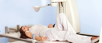
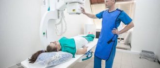
How is the procedure performed?
The study includes examination of the stomach, esophagus and duodenum. The procedure roughly consists of two parts:
- X-ray;
- X-ray of the stomach.
Both parts are performed by a radiologist, who subsequently writes a description.
Procedure algorithm:
- A plain radiograph is performed to evaluate pathology.
- The patient takes barium. This is necessary, since the density of the organs being studied does not differ from those adjacent to them. Taking the contrast leads to its gradual passage, which is monitored by the radiologist. A series of photographs are taken in different positions.
- When barium sulfate completely fills the organ, the doctor can analyze the correct functioning of the gastrointestinal tract.
- The contrast agent is completely removed 1.5 hours after administration. The doctor uses this time to prepare a report.
Where can I get a stomach x-ray?
If you want to have a fluoroscopy of the stomach in Moscow at an affordable price, contact the multidisciplinary clinic CELT. We do not just conduct diagnostic studies in this area - we conduct them as efficiently and efficiently as possible in accordance with international standards.
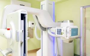
We employ experienced radiologists who are able to identify the slightest pathological changes. In this they are helped by modern equipment that reduces the radiation load on the patient’s body and provides high-quality images: the digital device “SoniavisionVersa” manufactured by “Shiamdzu” (Japan) and mobile X-ray machines “Ziehm Imaging” (Germany), providing three-dimensional images.
Fluoroscopy and radiography of the stomach - 4,800 rubles.
15-20 minutes
(duration of procedure)
You can find out the price of stomach radiography in our clinic in the corresponding section of our website or by calling us: +7 (495) 788-33-88! We regularly update our price list, but to avoid misunderstandings, we still recommend calling us.
Indications for X-ray examination with barium
There are a number of indications that require an X-ray examination of the esophagus with barium. If there are suspicions regarding the disease and diagnosis, this type of diagnosis can also be performed. We list some conditions in which it is recommended to take an X-ray of the esophagus with barium.
Swallowing disorders: feeling of retention of a bolus of food, pain when swallowing food
Various disturbances in the act of swallowing, the appearance of a lump in the throat, pain when swallowing food may indicate an obstacle to the natural flow of food during the act of swallowing. The cause of this pathological condition may be the presence of a foreign body in the esophagus, thickening of its walls, or the growth of neoplasms of various origins.
An X-ray of the esophagus with barium will clearly show the location of the obstruction in a certain part of the esophagus, and will also make it possible to assess its size and shape, which will help specialists in making a reliable diagnosis and selecting rational treatment.
Chest pain not associated with pathology of the respiratory system and cardiovascular system
If there is a history of pain in the chest and, with the help of differential diagnosis, it was found that they are in no way related to the cardiovascular and respiratory systems, then it is possible to assume a pathology of the esophagus. Since the esophagus is located directly behind the trachea and descends all the way to the diaphragm and stomach, the cause of pain can very likely be localized there.
Diseases such as hiatal hernia, esophagitis and peptic ulcers can be accompanied by pain and are detected using contrast-enhanced radiography of the esophagus.
Repeated vomiting, heartburn, belching
If the clinical picture contains symptoms such as repeated heartburn, vomiting and belching, then this may indicate a violation of the motor function of the esophagus, as well as its anatomical sphincters or the presence of various pathologies of the walls. Thus, this symptomatology can occur with diverticula of the esophagus, dyskinesia and achalasia. All these diseases can be detected using a barium X-ray of the esophagus.
In what cases is the study indicated?
The method allows you to visualize the area of the esophagus, assess its internal state and find most anomalies and pathologies. Thanks to what an x-ray of the esophagus shows, a specialist can make a diagnosis for almost any disease. Therefore, examination is prescribed for a large number of different symptoms.
Unmotivated, repeated vomiting
In most cases, vomiting is of visceral origin (caused by irritation of stomach receptors). However, the symptom can also appear with a malignant neoplasm in the esophagus. Then vomiting will recur for no apparent reason. Blood clots may appear in it.
To diagnose a tumor, the patient is sent for a contrast-enhanced x-ray of the esophagus. The method allows you to detect a tumor, localize it and make an assumption about its nature.
Belching that accompanies the patient constantly
Belching often occurs when swallowing function or esophageal motility is impaired. The patient complains of a lump in the throat and heartburn, vomiting and other symptoms. The problem may lie in achalasia cardia, scleroderma or other diseases.
It is almost impossible to make an accurate diagnosis without a detailed examination of the esophagus. Therefore, in diagnosis, the specialist relies on what an x-ray of the esophagus shows.
Suspicion of reflux
Reflux is the reflux of stomach contents up the gastrointestinal tract into the esophagus. This occurs due to insufficiency or damage to the lower esophageal sphincter, which is supposed to act as a valve.
With reflux, stomach contents rich in hydrochloric acid and pepsin (a protein-breaking enzyme) enter the esophagus. In this case, the patient experiences heartburn and discomfort. If reflux occurs regularly, erosion and peptic ulcers will appear in the lower part of the organ. If the disease is not detected in time by X-ray of the esophagus and not treated, severe bleeding caused by perforation of the esophageal wall and other dangerous complications are possible.
Pain syndrome behind the sternum
The pain is caused by stretching of the walls when food passes through. Often the sensation is spasmodic in nature, pain radiates to the jaw, ear, neck and back.
Pain may appear due to irritation of the mucous membrane of the organ due to esophagitis, reflux or malignant neoplasm. To make an accurate diagnosis, a specialist needs to conduct a series of examinations, including an x-ray of the esophagus.
Conditions for obtaining reliable diagnostic results
To avoid distorted results of hardware diagnostics of the stomach and intestines, patients are advised to follow a simple diet the day before. Alcohol should be discontinued one day before the test, tobacco and medications that can affect intestinal motility – 12 hours before.
to undergo fluoroscopy of the intestines on an empty stomach, after a three-day slag-free diet. There should be no metal objects in the clothes or on the patient.
The results can be distorted by:
- increased gas exchange;
- full stomach (metabolic disorders, in this case an enema is given a few hours before fluoroscopy of the stomach and intestines);
- metal on the patient (jewelry, crowns, prosthetic plates).
At the end of the study, barium suspension will be released for another 2-3 days. It can cause constipation or mechanical obstruction of the canal, which will aggravate the disease, if any. Therefore, if you experience serious discomfort (bloating, pain, nausea) during this period and (especially) after 72 hours, you should contact your doctor.
X-ray procedure
Before starting the procedure, the patient must remove all metal jewelry that interferes with the correct passage of x-ray radiation and interferes with obtaining a contrast shadow. The procedure begins with a standing X-ray, which helps to identify primary gross disturbances in the functioning of the gastrointestinal tract.
Before radiography with contrast, the patient drinks a 200 ml barium solution in portions (has the taste of chalk and the consistency of sour cream, does not cause discomfort). Next, the study is performed in several body positions so that the contrast is evenly distributed over the walls of the gastrointestinal tract. In total, the patient has to change about 6 positions, including standing, lying on his side, back, stomach, etc. The procedure takes 20-40 minutes. After the examination, to quickly remove the contrast, you should drink 1-2 liters of water. X-ray results during the procedure at NEARMEDIC will be ready in 20 minutes.
Pros and cons of fluoroscopy
The positive aspects of the procedure include the fact that X-ray examination of the esophagus is almost harmless and does not require special preparation. The specialist receives the data in real time and can not only localize the pathology and assess the degree of its development, but also draw a conclusion about the functioning of the esophagus as a whole.
Meanwhile, the examination is carried out using ionizing radiation. Therefore, the patient receives a certain amount of radiation (0.2-0.5 mSv at a rate of 5 mSv per year). Because of this, the procedure is not recommended for pregnant women, children and patients in serious condition.