In cases of pathological lesions of the lung tissue, bronchial tree or trachea, a secretion called sputum is formed. Sputum production accompanies a lot of pathologies of both the human respiratory system and the cardiovascular system.
Microscopic examination of sputum, or rather its fixed, stained and native preparations, indicates the cellular composition of this secretion.
The cellular composition, in turn, reflects the nature of the pathological processes of the lungs and bronchi, its activity, and also helps to distinguish between crystalline and fibrous formations in the secretion (important in the diagnosis of various diseases), to evaluate bacterioscopic indicators (the state of the microflora) in the respiratory organs.
general characteristics
Cytological examination of sputum helps to establish the nature of the pathological process in the respiratory organs, and in some cases, determine its cause. Cytological examination of sputum (five times) can detect cancer cells in 50 - 85% of patients with central lung cancer and in 30 - 60% of patients with peripheral lung cancer. Particularly valuable is sputum examination obtained after bronchoscopy. It also allows you to establish a diagnosis of bronchial asthma, suggest the presence of pulmonary mycoses, tracheobronchial bleeding, dysplasia of the mucous membrane of the bronchial tree, and identify siderophages - “cells of heart defects” (in case of cardiovascular failure, rheumatic heart defects). According to the literature, initial cancer can be detected earlier by this method compared to other diagnostic methods. The absence of cancer elements in sputum does not provide grounds to deny a lung tumor. The availability of the cytological method for examining sputum makes it possible to use it when examining individuals at “high risk” for cancer, and during mass preventive examinations of the population.
Cells and elastic fibers in the analysis results
Content:
- Cells and elastic fibers in the analysis results
- Spirals and crystals in microscopic studies
Stained and native sputum preparations serve as the basis for microscopic examinations. If it is necessary to identify microflora, then sputum smears are stained with Gram, Romanovsky-Giemsa, and if it is necessary to detect tuberculosis, they are stained with Ziehl-Neelsen.
When pneumonia occurs, epithelial cells, leukocytes, erythrocytes and alveolar macrophages can be observed in the examined sputum.
Epithelial cells in large numbers indicate a poor and low-quality sample of the secretion that was taken for research.
The fact is that when the indicator of 10 epithelial cells in the field of view during microscopic examination is exceeded, laboratory assistants conclude that the analytical data contains samples from the nasopharynx and oral cavity, which have no diagnostic value. Simply put, the laboratory received for analysis not sputum, but the patient’s saliva.
Large reticulohistiocytic cells with an eccentric nucleus and numerous cytoplasmic inclusions, slightly present in sputum, are called alveolar macrophages. Inclusions in the cytoplasm contained in them may contain dust cells, leukocytes, etc. In the presence of an inflammatory process in the respiratory tract and pulmonary parenchyma, in particular with pneumonia, the number of alveolar macrophages in the sputum increases.
The cylindrical ciliated epithelium is located in the body in the mucous membranes of the larynx, bronchi and trachea. These are elongated cells with an extension of one end with a nucleus and cilia. Ciliated epithelial cells are present in any sputum, but if their number exceeds the permissible maximum, this may indicate acute bronchitis, tracheitis, laryngitis, bronchiectasis and other damage to the mucous membrane of the trachea and bronchi.
There is nothing unusual in the number of 2-5 leukocytes per field of view on microscopic examination. This concentration is present in absolutely every secret.
However, a significant increase in their number indicates inflammatory processes in the tissues of the lungs, mucous membranes of the trachea and bronchi.
If you stain sputum preparations using the Romanovsky-Giemsa method, it becomes possible to differentiate leukocytes, which in some cases plays a decisive role in the diagnosis of diseases.
A general increased number of neutrophilic leukocytes and their number in a degenerative form with fragments of nuclei and destroyed cytoplasm may indicate inflammation of the bronchial mucosa or lung tissue. A high concentration of degenerative leukocytes always indicates the activity of inflammation and a severe prognosis during the course of the disease.
In any sputum, single red blood cells are usually present, but if their number is too high in microscopic examination, this indicates disturbances in vascular permeability in patients suffering from pneumonia, the process of destruction of pulmonary or bronchial tissue, the presence of stagnation in the pulmonary circulation, or pulmonary infarction. With hemoptysis of any origin, red blood cells are always present in the sputum.
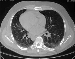
With various destruction of lung tissue as a result of abscesses, tuberculosis, disintegrating lung cancer in sputum, experts detect plastic fibers in the form of double-circuit thin and twisted threads with a dichotomous division at the ends that is unique to them.
In pneumonia, the appearance of such fibers in the sputum indicates an incipient complication - an abscess of the lung tissue. In this case, elastic fibers can arise at the start of a complication even before its X-ray visualization.
Often, when tuberculosis, actinomycosis, pneumonia, or obstructive bronchitis occurs, the finest fibrin fibers begin to separate in the sputum.

Coral-shaped fibers characterize the occurrence of cavernous tuberculosis, decalcified elastic fibers (with calcium salts) characterize the decomposition of petrificate in tuberculosis. So, with a rapid inflammatory process in the lung tissue, experts see in sputum tests:
- mucopurulent or purulent nature of the examined sputum;
- an increased number of neutrophils in the biomaterial, as well as their degenerative forms;
- increased number of alveolar macrophages in the study material;
- the appearance of elastic fibers, which signals the process of destruction of lung tissue.
Patient preparation rules
Discharge of the lungs and upper respiratory tract Standard conditions:
Self-collection of BM: sputum is collected before eating or no earlier than 2 hours into a clean, dry container. The BM is delivered to the first branch of “ML “Dila”” within 2 hours at the address:
st. Podvysotskogo, 6A,
Obolonsky Ave., 49.
Attention! New branch: Yu. Gagarin Ave., 9.
Important:
Avoid taking local antibacterial drugs for 24 hours (if the patient’s condition allows it).
Possible:
During business hours DC.
You can add this study to your cart on this page
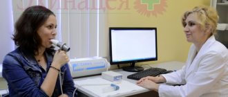
How to properly collect sputum
Sputum is collected in a medical facility, or independently at home. After collection, it must be delivered to the laboratory as soon as possible (1–2 hours). You must first purchase a sterile, sealed container.
Before collecting sputum, you need to brush your teeth and rinse your mouth thoroughly. Cough and collect the secretions in a container. It is necessary to minimize the entry of saliva into the material.
To make phlegm come out easier:
- on the eve of the analysis, it is recommended to drink plenty of warm drinks;
- the analysis is given in the morning;
- you need to take three deep breathing movements and then cough;
- in case of unsuccessful attempts, inhalation over water vapor with the addition of table salt and baking soda for 5–7 minutes is effective.
Collection of biomaterial
Sputum for culture is collected in the morning before the first meal in a special sterile container with a tight-fitting lid. To ensure that impurities of saliva and mucus do not get into the biomaterial, the oral cavity and nasopharynx are thoroughly cleaned before collecting a sample for research.
Only fresh sputum isolated as a result of prolonged coughing can be analyzed.
Collecting sputum for culture and determining sensitivity to antibiotics is carried out before starting treatment with antibacterial drugs. Otherwise, the result may be false negative.
results
Examination of a patient's sputum to determine antibacterial sensitivity includes:
- microscopy of sputum smear and preliminary identification of the type of pathogen;
- sowing the material on a nutrient medium;
- cultivation of microorganisms in individually selected conditions;
- repeated microscopy with detailed identification of grown colonies and determination of the basic properties of microorganisms;
- determination of sensitivity to antibiotics.
Using various methods for determining sensitivity, it is possible not only to identify a spectrum of antibacterial drugs that have a detrimental effect on identified microorganisms, but also to establish the minimum acceptable concentration of substances for a positive therapeutic effect.
In what situations is sputum culture necessary?
This study is indicated for all diseases of the middle and lower respiratory tract of infectious origin: acute and chronic bronchitis, pleurisy, pneumonia. It is necessary to carry out in case of complications of these pathologies, for example, with a lung abscess. Repeated analysis should be carried out at the slightest suspicion of tuberculosis
or be carried out as a control study during its therapy. In some cases, sputum culture is prescribed for lung cancer, to clarify the diagnosis in case of possible combinations with infectious pathology. It must be taken into account that the respiratory tract contains normal microflora, some types of which can become pathogenic if the immune system is weakened. The most common causative agents of inflammatory diseases of the respiratory tract are Staphylococcus aureus, Haemophilus influenzae, Klebsiella, Streptococcus, Enterobacteriaceae, and microscopic fungi. In tuberculosis, Koch's bacillus is detected.
How the research is carried out
Sputum collection should be done before starting antibiotics and is carried out in 3 ways: by expectoration, aspiration or using bronchoscopy. Before this, the patient should brush his teeth and rinse his mouth. The resulting sample is hermetically packaged and sent to the laboratory, where within 2 hours it must be inoculated into Petri dishes on a nutrient medium. This method is called bacteriological.
When colonies of microorganisms form, they are identified.
Detection of settlements created by pathogenic bacteria is an important diagnostic criterion. The next stage of the study is to determine the sensitivity of microbes to antibiotics.
For this purpose, the disk method is more often used: small disks soaked in solutions of various antibacterial drugs are placed into bacterial colonies. Next, the rate of death of the colony around them is monitored. The faster this happens, the more effective the antibiotic.
“Who are” alveolar macrophages?
This is the name given to the cells that are found in lung tissue. They are located in the alveoli, where gas exchange occurs. These cells belong to tissue macrophages, that is, they are a component of the immune system. Macrophages have a changing shape, are capable of movement, among their organelles they have a nucleus and a large number of lysosomes containing a variety of enzymes.
All these features indicate that alveolar macrophages are not just structural components of lung tissue. Indeed, their role is different: to protect the respiratory system from foreign objects.
When we take a breath, the air is first cleared of dust in the cavity and sinuses of the nose, then dirt and other foreign particles settle on the walls of the trachea. Then air filtration continues in the bronchi, where microscopic foreign fragments “sink” in the mucus secreted by special cells and are “swept out” from the respiratory organs by “cilia” - the long processes of these cells. To cleanse the bronchi from impurities and microbes, the “cilia” make up to 1000 beats per second. So that readers can imagine this, let’s draw a parallel: a mosquito flaps its wings with approximately the same frequency when flying.
Even after passing through all these “filters,” when entering the lungs, the air is not 100% clean: there are still germs, microscopic dust particles, and so on. It is from these contaminants that the air is purified by macrophages (it’s not for nothing that they are also called dust cells). They devour and digest harmful objects.
Unfortunately, macrophages cannot withstand a heavy load: having carried out phagocytosis, they die. Before they stop functioning, they move to the base of the respiratory bronchioles (the smallest bronchi leading to the alveoli). There, the cells leave the walls of the respiratory tract, enter the mucus and remain there. This is how alveolar macrophages end up in sputum.