Oncologist
Lobov
Mikhail Yurievich
Experience 27 years
Oncologist
Make an appointment
Lung cancer is a neoplasm that develops from pathologically altered epithelial cells lining the internal surfaces of the bronchi and bronchial glands. Under the influence of certain conditions, epithelial cells cease to perform their function. An uncontrolled, disorderly process of division is launched, as a result of which the pathological tissue quickly grows, penetrates into nearby and then into distant organs. The respiratory function of the lungs and other affected organs gradually decreases, intoxication with decay products increases, and in the absence of treatment, inevitable death occurs.
Kinds
In accordance with histological characteristics, lung oncology is divided into three groups of tumors:
- small cell – accounting for approximately a fifth of all clinical cases and characterized by an extremely rapid aggressive course;
- non-small cell - this includes adenocarcinomas, which make up half of all cases, squamous cell carcinomas (20-30%) with a slow progression and undifferentiated cancer;
- other types - bronchial carcinoids, neoplasms from surrounding tissues and metastases from other organs.
Based on location, lung cancer is classified into central, peripheral and disseminated lung cancer.
Choosing treatment tactics for lung cancer
If PET/CT reveals lymph nodes suspicious for metastatic lesions, a biopsy is necessary - transbronchial / transesophageal puncture, mediastinoscopy or thoracoscopy at the discretion of the thoracic surgeon.
After completing all these studies, patients are divided into four groups:
- Group 1 should first receive surgical treatment. The extent of the operation is determined by the thoracic surgeon. After surgery, patients with stage IIA and certain risk factors should receive chemotherapy. Also, postoperative chemotherapy is mandatory for patients with stage IIIA.
- Group 2 patients (with stage IIIA - N2) receive 2-4 cycles of chemotherapy as the first stage, followed by a decision on surgical treatment. From the end of chemotherapy to surgery, 3 to 8 weeks should pass.
- Patients of group 3 with initially inoperable stage IIIA and IIIB receive chemoradiotherapy. If the patient’s physical condition allows, then at the same time. If not, then sequentially - first chemotherapy, then radiation therapy.
- Group 4 patients with stage IV - with pleurisy, pleural invasion or detection of distant metastases - should receive drug antitumor therapy.
Symptoms
Most patients mistake the first signs of lung cancer for a common cold, since at the initial stage of tumor growth there are no specific manifestations. As the pathological tumor grows, the patient develops:
- feeling of general malaise, decreased performance;
- increased body temperature;
- wet cough with expectoration of sputum;
- difficult, noisy breathing;
- swelling of the lymph nodes;
- frequent respiratory tract infections.
The symptoms of lung cancer in men and women are practically the same. Over time, the listed manifestations intensify, adding to them:
- chest pain that gets worse when coughing and taking deep breaths;
- shortness of breath with minor exertion or at rest;
- blood streaks or brown clots in expectorated sputum;
- headaches, bone pain;
- dizziness, periodic loss of consciousness;
- causeless sudden weight loss, loss of appetite.
If a cough, fever, or increased sputum production continues for more than two to three weeks, you should definitely contact an oncologist for examination.
Reasons for appearance
Factors in the development of lung cancer can be grouped into three groups: genetic, exogenous (external) and endogenous (internal). Genetic causes include:
- The familial nature of the disease is three or more cases of lung cancer in close relatives.
- The patient has a history of another malignant tumor.
External factors that influence the development of this neoplasm:
- Smoking. It is addiction to this habit that is the main reason for the development of lung cancer. Moreover, not only active but also passive smoking can lead to the appearance of a tumor.
- Exposure to carcinogens: exhaust gases; some organic substances (for example, arsenic, cadmium); tars and cokes that appear in the air as a result of coal and oil processing.
- Occupational hazards: work in uranium mines; industrial processing of steel, wood, metal. Also, the risk of lung cancer is increased among those workers who come into contact with stone dust, asbestos, aluminum and nickel. The combination of these factors with smoking further increases the likelihood of developing the disease.
Endogenous factors for the development of central lung cancer include:
- Age over 50 years.
- Chronic inflammatory processes in the bronchi.
- Presence of endocrine diseases.
- Immunodeficiency conditions (HIV infection, therapy with cytostatic drugs).
Under the influence of unfavorable factors, precancerous changes are formed in the bronchi. They play an important role in the pathogenesis of this disease.

Causes and risk factors
Although the causes of lung cancer and the mechanisms of malignant cell degeneration have not yet been precisely established, oncologists have well studied the factors contributing to the development of the disease:
- smoking, including passive smoking;
- work in hazardous production;
- inherited predisposition to lung cancer;
- long-term exposure to radon on the respiratory system;
- chronic respiratory diseases, including tuberculosis;
- age over 40 years.
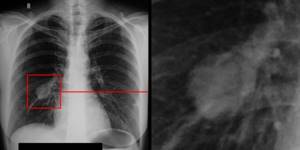
Men get sick 5-6 times more often than women, since heavy smokers and workers in hazardous industries with high levels of dust or gas pollution are much more common among them.
Preparing for chemotherapy
The use of targeted and immunotherapy does not require a special regimen or diet - adequate sleep, regular healthy eating, and sometimes taking multivitamins are recommended.
When chemotherapy is carried out, healthy cells are also affected; “building material” is needed to replenish protein losses. Therefore, the diet should actively include legumes, lean meat and fish, and poultry. It is also advisable to take additional vitamins of group B. Any types of raw and boiled vegetables, salads and fruits are useful, and especially those with a high content of vitamin C - citrus fruits, apples, currants. It is also advisable to increase the amount of fluid by consuming vegetable, fruit and berry juices. The feasibility of this increases significantly when treated with platinum drugs. Carrot, beet, tomato, raspberry and lingonberry juices are especially useful.
Stages
In oncology, it is customary to distinguish the stages of lung cancer by the size of the tumor and the degree of its growth and spread to other organs. There are four main stages of the disease, depending on which the treatment method is determined.
- The neoplasm, measuring no more than 3 cm, is located compactly, without penetration into the pleura. Lymph nodes are not affected, there are no metastases.
- The tumor enlarges and clogs the lumen of the bronchus or grows, forming a compaction within one lobe of the lung. One or two metastases may appear in regional lymph nodes.
- The dimensions of the neoplasm exceed 6 cm, the pathology affects the chest wall, and can reach the area of separation of the main bronchi or the diaphragm. Metastases penetrate to distant lymph nodes on their half of the body.
- The tumor grows into several neighboring organs, affects the esophagus, heart, stomach, gives multiple metastases to distant lymph nodes on both sides and to other organs.
For small cell tumors, a separate classification is often used, consisting of only two stages - localized within one lung and spread beyond the original lung, including to other organs.
Diagnostics
In the early stages, the disease is usually diagnosed accidentally, when studying fluorographic images. When contacted regarding symptoms of lung cancer, a pulmonologist or oncologist usually prescribes the following diagnostic procedures:
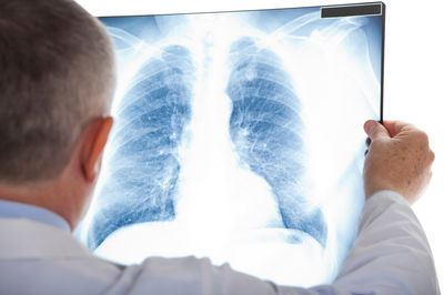
- X-ray of the lungs in several projections for the initial detection of a tumor;
- CT scan of the chest to identify a tumor, accurately determine its size, stage, extent, etc.;
- MSCT of the chest to determine the exact location, size of the tumor and other data;
- fibrobronchoscopy – examination of the bronchi using an endoscope with a biopsy of tumor tissue;
- transthoracic tumor biopsy performed with a puncture of the chest wall;
- histological examination of a biopsy to determine the type of cancer cells and their degree of malignancy;
- blood test for tumor markers CEA, NSE, CYFRA21-1;
- cytological analysis of expectorated sputum to identify malignant cells;
- Ultrasound of the abdominal cavity to detect metastases in other organs.
Attention!
You can receive free medical care at JSC “Medicine” (clinic of Academician Roitberg) under the program of State guarantees of compulsory medical insurance (Compulsory health insurance) and high-tech medical care.
To find out more, please call +7, or you can read more details here...
1.What is “central lung cancer”?
Lung cancer is an oncological disease that occurs as a result of mutations in the epithelial cells lining the bronchi. Further development of the disease leads to the involvement of various lobes of the lung in the process. Central cancer is diagnosed when the primary focus is localized in the proximal sections of the bronchi, that is, pathology occurs on the mucous membranes of its large and medium segments. The cancerous tumor is most often squamous cell. The neoplasm can be endobronchial or peribronchial. As the malignant tissue grows, it spreads and involves the peripheral parts of the lungs, pleura and neighboring organs in the pathological process, and also metastasizes by moving cancer cells through the lymphatic system.
Statistically, men are eight times more likely to get this type of cancer.
This can be associated with smoking and professions associated with the effects of toxins, radiation and other oncogenic factors. There is also a hereditary predisposition in the presence of close relatives with a history of central cancer.
Sign up for a consultation
A must read! Help with hospitalization and treatment!
Treatment
Early detection is critical to effective treatment of lung cancer. In this case, after removal of the malignant tissue, a complete recovery and return to normal life is possible.
- Surgery. If there is no growth into other organs or damage to the lymph nodes, part or the entire lung is removed during the operation. Surgical treatment is possible for patients with the first or second, partly with the third stage of the tumor. If the pathology spreads to other vital organs or if the patient’s general condition is severe, surgery is impossible.
- Radiation therapy. Targeted ionized radiation, which destroys malignant cells, is used in combination with surgery, chemotherapy and other methods at all stages: for non-surgical tumor removal, destruction of residual cells and metastases after surgery, to prevent relapse or to improve the condition of inoperable patients.
- Chemotherapy. Drugs that destroy tumor cells are prescribed in combination with radiation treatment before and after surgery, as well as in inoperable clinical cases.
- Targeted therapy. Modern targeted drugs are used to inhibit the growth of strictly defined cell types. Targeted drugs do not destroy the tumor, but inhibit its growth, thereby improving the symptoms of lung cancer in women and men, and extending the life of patients at the inoperable stage of the disease.
What types/types of lung cancer are there?
Based on the location of the tumor, they are traditionally classified as:
- Central lung cancer
- Peripheral lung cancer
Central lung cancer is characterized by tumor growth in the bronchus. In peripheral lung cancer, the tumor is located directly in the lung tissue (at the periphery).
At the present stage of development of medicine, high-quality treatment of lung cancer is only possible if the morphological type of lung cancer is known. The type of cancer can only be determined by a biopsy (intravital sampling of cells or tissues from the body) and examination of a piece of the tumor under a microscope. Treatment is fundamentally different for the two types of cancer:
- Non-small cell lung cancer.
- Small cell lung cancer.
Non-small cell lung cancer is the most common type of lung cancer, and these tumors do not grow as quickly as small cell lung cancer. Non-small cell lung cancer includes the following types of tumors: squamous cell carcinoma, adenocarcinoma, large cell and mixed cancer.
Small cell lung cancer is a type of cancer in which the tumor grows quite quickly. This is one of the most malignant lung tumors. It is characterized by a latent and rapid course, early metastasis (spread to other organs) and a poor prognosis.
Diagnosis and treatment of lung cancer in Moscow
If you need a high-quality diagnosis of lung cancer, contact the Meditsina clinic about this. At your service are modern tomography and X-ray equipment, a well-equipped laboratory, and diagnostic radiologists of the highest category. Consultation and treatment of patients is carried out by experienced oncologists and pulmonologists. Consultations with highly qualified endocrinologists, infectious disease specialists and doctors of other specialties are possible. Treatment is carried out in our own comfortable hospital.
Questions and answers
How long do they live with lung cancer?
A patient's life expectancy depends on many factors, but early detection and the type of cells that make up the tumor are critical. For non-small cell cancer of the first or second stage, the possibility of complete recovery reaches 85%. Therefore, it is necessary to undergo an annual examination in order to detect and treat a serious disease in time.
How do you know if you have lung cancer?
If a cough with sputum and a slight fever, general malaise and weakness continue for more than two to three weeks, this is a significant cause for concern. It is necessary to contact a pulmonologist as soon as possible for a lung examination.
How to make breathing easier with lung cancer?
To make breathing easier, try to keep your back straight. When going to bed, place several pillows under your back and head, lie mostly on your side in a semi-sitting position. Learn diaphragmatic breathing and other breathing exercises. To eliminate dryness, constantly rinse your mouth with water or a weak soda solution, drink more ordinary or mineral water.
Attention! You can cure this disease for free and receive medical care at JSC "Medicine" (clinic of Academician Roitberg) under the State Guarantees program of Compulsory Medical Insurance (Compulsory Medical Insurance) and High-Tech Medical Care. To find out more, please call +7(495) 775-73-60, or on the VMP page for compulsory medical insurance
How is the examination carried out?
The procedure is carried out using a tomograph, which is a device in the form of a large capsule with a retractable table. On the table, the patient is fixed in a motionless position using belts and pillows. During the procedure, your arms should be raised up. During the X-ray scan, medical personnel leave the room in which the tomograph is located. The patient is monitored through a special window and voice communication is provided. The whole procedure takes no more than 30 minutes.
Important! The frequency of examinations must correspond to the calculated x-ray dose. Violation of the permissible radiation dose is possible only in critical cases.

