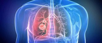
Lung cancer is a tumor that develops from the tissue lining the inside of the bronchi, bronchioles and mucous bronchial glands - the epithelium. In most cases, lung cancer develops in men after 60 years of age. This disease is the most common of all cancers: there are more than 1 million new cases of lung cancer every year, and this number is growing.
Molecular tumor analysis in lung cancer is a new step in the treatment of particularly aggressive tumors. We provide the opportunity to test tumors for sensitivity to chemotherapy. Based on the results of the study, we receive an effective plan to combat lung cancer, giving the most favorable chance of recovery.
- Symptoms of lung cancer
- Pancoast tumor symptoms
- Symptoms of peripheral lung cancer
- Types of lung cancer
- Central and peripheral cancer
- Small cell lung cancer
- Stages of lung cancer
- How to recognize lung cancer at an early stage?
- What increases the risk of developing lung cancer?
- Is it possible to reduce the risk?
- Metastases to the lungs
- Treatment of lung cancer at different stages
Symptoms of lung cancer
After clinical examination in our country became much less popular, malignant neoplasms of the lungs increasingly began to be detected at later stages. In this case, lung cancer is often diagnosed accidentally - during an X-ray of the lungs during an examination for another disease.
If you do chest fluorography every year, in most cases the diagnosis can be made at earlier stages, when the prognosis looks most optimistic.

This is especially important because there are practically no symptoms due to which one can suspect oncology at an early stage. How lung cancer manifests depends on various factors: the stage of development of the tumor, its location in the lung, the disorders it causes, etc. Lung cancer can masquerade as various diseases. For example, sometimes it looks like long-term pneumonia that is difficult to treat. A growing tumor can cause coughing, secretion of mucous or mucopurulent sputum. Sometimes there is an admixture of blood in the sputum - this happens when the mucous membrane of the affected bronchus becomes inflamed. Such patients experience chest pain, shortness of breath, and hoarseness.
Due to the fact that the tumor releases harmful substances, other symptoms of lung cancer arise, which can be mistaken for manifestations of completely different diseases. These are fatigue, decreased ability to work, weakness, fever (usually slight), and weight loss.
Pancoast tumor symptoms
Cancer of the apex of the lung (Pancoast tumor), due to the close proximity of other organs and nerve trunks, often causes more pronounced symptoms: weakness of the muscles of the hand, pain in the shoulder girdle, along the front surface of the chest and between the shoulder blades, and discomfort also occurs in the area of the hand or forearm or sensitivity is impaired, muscles atrophy. Unfortunately, such symptoms often disorient the doctor, and the person wastes time treating non-existent cervicothoracic osteochondrosis.
Can this be avoided? Yes. It is enough to pay attention to Horner's syndrome. Due to damage to the cervical sympathetic ganglia, the upper eyelid of one eye droops (ptosis), one pupil narrows (miosis), and sweating significantly decreases on only one side of the face (anhidrosis). A person's voice may also become hoarse due to damage to the recurrent laryngeal nerve.
Symptoms of peripheral lung cancer
Peripheral lung cancer can be mistaken for sluggish pneumonia, which cannot be controlled with antibiotics, and the temperature usually rises to low levels.
In rare cases, paraneoplastic syndrome may indicate that a tumor has appeared or has begun to appear in the body. This is a complex of various manifestations that arise not due to the direct effect of the tumor on the tissue, but through the production of various substances by cancer cells. In lung cancer, more often than in any other cancer, paraneoplastic syndrome affects the nervous system. This, in particular, is manifested by difficulties in walking, lack of coordination, problems with maintaining balance, such a person becomes difficult to swallow, and his speech is slurred. In addition, deterioration in memory, sleep, vision, etc. may occur.
Paraneoplastic syndrome sometimes involves elevated calcium levels. In some cases, the tumor can even produce hormones that in a healthy body are synthesized by the pancreas and parathyroid glands, pituitary gland and hypothalamus. That's why your doctor should pay special attention to warning signs in order to conduct an in-depth examination and possibly detect lung cancer at an early stage.

Usually the first symptom of the disease is a persistent dry cough. If it bothers you for several weeks and is not associated with allergic or infectious diseases, you need to visit a doctor and get examined. Another early sign is an increase in body temperature. Usually it is insignificant, up to 37.5 degrees, but persists for a long time. Nonspecific symptoms such as increased fatigue, weakness, and frequent ailments are of concern. Often these manifestations are attributed to bronchitis and pneumonia.
Whistling sounds occur during breathing, and the voice becomes hoarse. Over time, the cough becomes more and more painful, during which sputum is released, in which an admixture of blood can be seen. This is definitely a reason to be screened for lung cancer.
There are no pain receptors in lung tissue. Therefore, in the early stages there will be practically no pain. They occur periodically and pass quickly. Later, when the tumor has time to grow significantly and spread into the pleura and intercostal nerves, long-term painful pain in the chest occurs. They can spread to the shoulder, the outer part of the arm.
The appearance of shortness of breath indicates that the tumor affects a large amount of lung tissue, due to which the respiratory surface is reduced. This symptom may be associated with exudative pleurisy.
Write to the oncologist
Diagnostic methods
After a visual examination and study of the medical history, the doctor prescribes a comprehensive examination to establish an accurate diagnosis. To identify oncology, various diagnostic methods are used . They determine the presence of a tumor process, help identify the degree of its prevalence, and differentiate it from other diseases.
Radiography
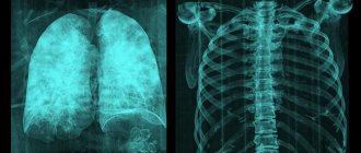
Many people doubt whether the tumor is visible on an x-ray. The information content of this method is 80%. Lung cancer is not always detected early on X-rays. The small formation grows gradually, involving the lymph nodes and other organs in the process. Later, an x-ray makes it possible to see lung cancer very clearly. This becomes a reason for further examination, which gives a more accurate result.
If with central lung cancer the x-ray shows a cloudy area with an expanded vascular network, then with peripheral oncology, such as bronchioloalveolar cancer, clear shadows with ribbon processes going to the pulmonary root are visible. Spreading, metastases affect the lymph nodes of the mediastinum, regional lymph nodes, and penetrate through the blood into the brain, liver, and bones. Lung cancer is only diagnosed by X-ray. Determining the nature of the tumor is much more difficult . Other techniques are used for this purpose.
Fluorography
This is one of the most accessible ways to examine the lungs. The procedure must be done regularly. Disputes about whether fluorography shows lung cancer are pointless. An experienced radiologist will easily detect any pathological changes. Another thing is that other diseases can be mistaken for lung cancer on fluorography, for example, calcification of lung tissue or hamartoma. To diagnose the problem at an early stage, it is recommended to take pictures in several projections. This allows you to identify any suspicious areas . Therefore, doubts about whether lung cancer is visible on fluorography are groundless.
Magnetic resonance imaging
The principle of the technique that helps to see what lung cancer looks like is a complex of magnetic resonance and software capable of processing the data obtained. The diagnosis is safe - there is no radiation exposure or any side effects. MRI provides high-resolution images of lung cancer. This method helps solve the issue of how to identify the slightest structural abnormalities of tissues, including in the lymph nodes.
A contraindication for screening examination methods is the presence of a metal implant.
CT scan
When an X-ray picture or fluorographic image does not clearly show the state of the tumor process, computed axial tomography is used. Lung cancer screening identifies all foci of tumors. Images taken in different projections provide an opportunity to more carefully examine lung cancer on a CT scan.
Computed tomography allows one to recognize the smallest formations, including tumor metastases that penetrate the lymph nodes . If necessary, a three-dimensional image of the organs is taken. A similar diagnostic picture of such a disease, for example, is impossible with radiology. To obtain a better image, the patient is given contrast agents to help detect lung cancer.
Bronchoscopic diagnosis
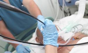
Fiberoptic bronchoscopy (FBS) is one of the main ways to detect lung cancer. The procedure allows you to visually examine the airways using fiber optic probing. Considering the possible discomfort during the procedure, the patient is given an anesthetic drug. Bronchoscopy of the lungs allows you to examine the bronchi and trachea, which gives a more accurate picture of the developing pathological process.
As a result, the disease is determined by the following characteristics:
- The pattern of cartilage tissue is blurred.
- The lumen of the bronchus is narrowed.
- An infiltrate is visible on the mucous membrane, which is a slight elevation.
- Tumor formations of different colors and sizes are detected.
- Tracheobronchial lymph nodes are enlarged.
Bronchoscopic examinations are mandatory when performing a biopsy of tumor-affected tissue.
Cytological examination of sputum
One of the simplest and safest ways to recognize lung cancer in the early stages. The sputum secreted by the patient is examined under a special microscope. The presence of atypical fractions in it is an indicator of existing oncology. The limitation of this technique is that sometimes, even in the presence of a malignant tumor, pathogenic cells in the sputum may be absent. In addition, in the presence of an inflammatory process, there is a possibility of deformation of benign cells.
Video
Video - cancer symptoms
Biopsy
One of the methods of histology is a procedure for studying lung tissue under a microscope, known as a biopsy. Performed if pathological changes are suspected. Preparing for a biopsy includes restricting food intake for at least 6 hours before the biopsy. On the question of whether you can take medications, you need to ask your doctor. You should definitely avoid using non-steroidal drugs before the procedure. The day before, you need to do an X-ray or CT scan of the chest and donate blood for analysis.
In total, 4 methods of obtaining biomaterial are used:
- Lung biopsy with a bronchoscope. The biomaterial is obtained using bronchoscopy by inserting a probe into the patient's respiratory tract.
- Percutaneous biopsy, when a puncture is taken from a suspicious area using a long thin needle. The procedure is accompanied by X-ray control.
- Open method. A piece of lung tissue is taken through an incision made in the area of the respiratory organ. The operation is performed under general anesthesia.
- Video thoracoscopic biopsy. A painless procedure using a camera, performed by most modern medical clinics. A biopsy is prescribed only when the pathological process has affected the pleura.
Bronchoalveolar lavage may be prescribed to identify histological changes in carcinoma. The procedure helps the cells of the lung tissues located deep.
Blood analysis
It is unlikely to detect signs of lung cancer at an early stage using this method. Specific changes such as eosinophilia, leukocytosis, low hemoglobin in cancer and other blood parameters are diagnosed later, when metastasis reaches the bone marrow. A general blood test for lung cancer determines the deficiency of a number of enzymes, which may indicate the development of metastases.
Tumor markers
Lung cancer markers are a new development by scientists based on the identification of certain proteins. The body of a sick person produces them in response to a tumor.
Tumor markers for lung cancer help:
- Detect a tumor at the earliest stage of development.
- Determine whether the formation is malignant or benign.
- Detect metastases in time.
- Monitor and test the effectiveness of cancer treatment.
- Carry out preventive work to prevent the disease.
However, it should be taken into account that healthy cells are also capable of producing similar proteins in other conditions of the body that are not related to oncology. Therefore, based on tumor marker tests alone, lung cancer is not recognized.
Signs of malignant lesions of the respiratory system are often similar to the symptoms of pneumonia, tuberculosis, abscesses and other pathologies. Only differential diagnosis of lung cancer helps to exclude the likelihood of these diseases.
It is not easy to distinguish any type of cancer. The process is complicated by the absence of pronounced symptoms in the initial stages of the disease. The result of the diagnosis is timely detection of lung cancer, treatment of which at an early stage increases the chances of recovery.
Types of lung cancer
To determine the prognosis and choose a treatment method, the doctor needs to know the histological structure of the tumor. To do this, a biopsy of the bronchi or lung is performed, that is, a small piece of tissue is taken during bronchoscopy (examination of the inner surface of the trachea and bronchi using special optical instruments) or thoracoscopy (examination of the pleural cavity through a puncture of the chest wall). After this, a specially processed and stained tissue sample is examined under a microscope and the type of tumor is determined. It is important to have “glasses” and “blocks” with tissue samples with you if you decide to seek a consultation at another clinic, so that they can look at them themselves and form their own opinion.
Depending on the type of cells there are:
- small cell carcinoma (oat cell, combined oat cell, intermediate cell);
- squamous cell or epidermal lung cancer (poorly differentiated, keratinizing, non-keratinizing);
- lung adenocarcinoma (acinar, papillary, bronchiolo-alveolar cancer, solid cancer with mucin formation);
- large cell carcinoma (giant cell, clear cell);
- glandular squamous cell carcinoma;
- cancer of the bronchial glands (adenocystic, mucoepidermoid, etc.).
There are also other, rarer types of lung cancer - there are at least twenty of them in total. One tumor can contain different types of cells. If there are metastases in the lungs, then the cells that make up them will look like the cells of the mother’s tumor.
In 40% of cases, malignant lung tumors are represented by adenocarcinomas, which are formed from cells that produce mucus. Lung adenocarcinomas predominantly occur in smokers or in people who once smoked. However, it is also the most common type of lung cancer in non-smokers. It is also one of the most common types of malignant tumors in young people.>
Adenocarcinoma is a relatively slow-growing malignant tumor. The chances of detecting it in the early stages are quite high. However, this is individual; in some patients this cancer behaves more aggressively.
To select the optimal treatment regimen for a patient with an atypical course of the disease, we use international databases, including cases from leading oncology clinics, results of medical research, and scientific articles. If doubts arise regarding the assessment of the histological analysis performed, we turn to our foreign colleagues: we scan images of histological sections and send them to a partner clinic. Within 2-5 days, we receive a second expert opinion with a histological report and a treatment plan option.
Thanks to scientific advances, it is possible to determine the sensitivity of a tumor to chemotherapy even before the start of treatment. Molecular analysis allows us to develop a more effective plan to combat lung cancer. At Euroonko we use exactly this approach: it gives the best possible chance for treatment success.

Types of radiography
The types of research are determined by the characteristics of the anatomical zone being studied and the process of taking the image.
Standard radiography is a survey, that is, a picture that gives an idea of the condition of the entire organ, or rather pictures in two mutually perpendicular projections - anterior and lateral.
In dentistry and for oral tumors, a version of the overview image is known as orthopantomography, but in radiography technology it is a panoramic image, during which the beam emanating from the apparatus passes along a wide arc, appearing on film in the form of the upper and lower jaw, and not an individual tooth.
The opposite of the survey view, targeted radiography allows you to “photograph” a specific area, for example, the upper mediastinum or the root of the lung altered by a tumor. This type is performed only after a survey x-ray.
Essentially the same targeted contact radiography is used in dentistry, when a film is placed in the mouth and only the diseased tooth is removed. In oncology, intraoral technique is used for cancer of the oral mucosa and oropharynx to assess the involvement of bone in the tumor conglomerate.
Similar to sighting is close-focus radiography, usually of small structures and at a close distance from the beam tube. In a small focus, the pathology is seen more clearly.
The introduction or intake of a contrast agent visualizes the cavitary organs of the gastrointestinal tract, urinary system and vascular network - contrast study of the intestine - irrigoscopy, bile ducts - cholecystography, urinary tract - urography, fistulas - fistulography.
X-ray according to Vogt is not used in oncology; a photograph of the eye without skull bones is necessary for injuries. For malignant eye processes, CT is used.
There is also rarely a need for x-rays with functional tests when the patient is filmed in a certain position, for example, with spinal tumors.
Soft tissue radiography is also not a widely used method, but may be useful in sarcomas.
Central and peripheral cancer
But when choosing a treatment, it is very important to consider not only the cell type: the location of the tumor is also of great importance. There are central and peripheral lung cancer. In central cancer, large bronchi are affected (main, lobar and segmental), while in peripheral cancer, smaller bronchi are affected.
In turn, there are four types of peripheral lung carcinoma:
- Subpleural node – this option also includes Pancoast tumor.
- Intralobar node.
- Diffuse and miliary forms.
- Cavity form.
In addition, mediastinal cancer is distinguished separately - these malignant tumors in the lungs are usually small in size, but quickly metastasize to the lymph nodes of the mediastinum.
Where the tumor grows is also an important factor when determining treatment tactics. If it grows into the lumen of the bronchus (exophytic cancer), it can partially or completely block the lumen. Then the development of secondary pneumonia is extremely likely. If the tumor grows into the thickness of the lung tissue (endophytic cancer), this does not affect the patency of the bronchial tube for quite a long time. Branched cancer is also found - the tumor is located around the bronchus and evenly narrows the lumen. To finally get an idea of the nature of tumor growth, you need to remove the tissue surgically and study it.
Peripheral cancer comes in three main types:
- round or nodular tumor;
- pneumonia-like cancer - has no clear boundaries and symptoms resemble pneumonia;
- cancer of the apex of the lung (Pancoast tumor).
There are also atypical forms that have different features of metastasis. To estimate the prevalence of lung cancer, the TNM classification system is used worldwide. Thanks to it, it is possible to systematize various clinical situations, determine treatment tactics, and also make a prognosis for the development of the disease - all this based on the anatomical characteristics of the tumor.
Small cell lung cancer
Small cell lung cancer is the most malignant of all. Such a tumor not only grows quickly, but also actively metastasizes. There are a number of other unpleasant features of this type of cancer.
- If the tumor grows inside the bronchus, it can cause atelectasis (“collapse” of part of the lung) and obstructive pneumonitis (inflammation) with cough, fever and chest pain.
- When the tumor disintegrates, there is a danger of pneumothorax - when air enters the pleural cavity (the sealed cavity between the layers of pleura that cover the surface of the lungs).
- If in the later stages the tumor, spreading, involves the pleura, this can cause hemothorax. In this condition, blood accumulates in the pleural cavity.
These situations are often extremely life-threatening, and it is often impossible to do without urgent surgical help.

Stages of lung cancer
The stage of malignant lung tumors is determined in accordance with the generally accepted TNM classification:
- The letter T indicates the size of the primary tumor. Next to it there may be symbols is, 1, 2, 3 and 4. Tis is a very small tumor that is located in the uppermost layers of the mucous membrane of the respiratory tract. T4 is a cancer that grows into neighboring organs.
- N – cancer has spread to nearby lymph nodes. N0 means that there are no lesions in the lymph nodes. Numbers 1, 2 and 3 characterize different degrees of lymph node involvement.
- M – presence of distant metastases. One of two numbers can be assigned to this letter: 0 – no distant metastases, 1 – distant metastases detected.
Stage classification differs slightly between non-small cell and small cell lung cancer. In NSCLC, depending on the characteristics of T, N and M, five stages are distinguished, they are designated by Roman numerals:
- Stage 0
: “cancer in situ”, with no lymph nodes involved and no distant metastases. This is the most favorable situation, characterized by the best prognosis for the patient. - Stage I
: the tumor is located only in the lungs, does not invade neighboring organs, lymph nodes are not affected, and there are no distant metastases. - Stage II
: Along with the primary tumor in the lungs, there are lesions in nearby lymph nodes. - Stage III
lesions in the lymph nodes of the mediastinum. This type of cancer is called locally advanced. - Stage IV
: The cancer has spread to both lungs, cancer cells are found in the fluid in the pleural cavity and pericardial cavity (the sac around the heart), or distant metastases are found.
For small cell lung cancer, the TMN system is also used, but in clinical practice the division into two stages is more important:
- Locally advanced
: the tumor is found on only one side, affecting only one part of the lung and nearby lymph nodes. - Extensive
: cancer has spread to other organs and there are distant metastases.
CT scan
For lung cancer, x-rays do not provide as accurate information as computed tomography. This is another x-ray study. Only it takes layer-by-layer photographs of tissues.
Can lung cancer be seen on an X-ray? Undoubtedly, the tumor can be detected. But in the future, clarification of its location and size is required. It is necessary to clarify the shape of the tumor and assess the condition of the lymph nodes. This is why a CT scan is performed. Cancer on a CT scan usually appears as a round, sometimes oblong, nodular formation.
How to recognize lung cancer at an early stage?
When central lung cancer is at an early stage, it is very difficult to recognize. Examination by a therapist and x-ray examinations are ineffective. If such a person undergoes bronchoscopy with a biopsy, then the correct diagnosis can be made. Sometimes computed tomography helps to recognize the disease in the early stages.
If the cancer is peripheral, then it is impossible to do a biopsy, since it is simply not possible to get to the suspicious place using bronchoscopy. Therefore, a transthoracic needle biopsy is performed, that is, a piece of tissue is taken through a puncture in the chest wall. If lesions are in the mediastinum (the part of the chest cavity located between the sternum, spine, diaphragm, pleura and surfaces of the lungs), mediastinoscopy is performed (examination for the purpose of biopsy through an incision in the neck). Sometimes it is impossible to do without diagnostic thoracoscopy and thoracotomy (opening the chest cavity). To clarify how widespread the tumor is, a variety of diagnostic methods are used: ultrasound, bronchoscopy, multislice computed tomography, magnetic resonance and positron emission tomography, as well as radionuclide studies. Without this, it is impossible to choose the best approach to treating a particular patient.
Make an appointment with an oncologist
What is radiography?
Radiography is an image of a specific part of the body made using ionizing radiation directed from a machine.
Fluoroscopy is an X-ray examination lasting several minutes, when the doctor, through a special screen, visually monitors what is happening inside the patient’s body, occasionally photographing what is happening.
Recording the internal state of an organ on film in real time - an x-ray or radiograph.
You can take an x-ray of any part of the body and get a true image; in fact, it is a black and white “photograph” of all layers of the anatomical area of interest.
The reliability of the image can be increased by adjusting the shutter speed and power of the X-ray tube. Tissues and organs filled with air will be dark in the image, bones will be light, that is, the greater the density of the tissue, the lighter it will be on the x-ray.
Radiography provides an image fixed at a point in time; several images in different positions of the patient will help to get an idea of the deviations of processes from the norm in dynamics, but the idea will be somewhat incomplete without the introduction of contrasting solutions into the vascular bed, hollow organ or ducts of the studied area.
Modern X-ray machines have not become completely harmless to the subject, but the dose of radiation received during the study is minimal and is offset by the importance of the diagnostic information obtained. Analogue devices are gradually becoming a thing of the past, giving way to digital ones, where everything is calculated by a computer program, the image is displayed on the monitor without the need to develop the film, and the pictures taken during the process are stored on electronic media.
What increases the risk of developing lung cancer?
At the moment, the connection between lung cancer and smoking is not in any doubt. This is especially true for central squamous cell and large cell cancer: in 70–95 percent of cases, such patients smoked or smoke. The International Agency for Research on Cancer has concluded that smokers are 10 times more likely to develop lung cancer. Tobacco smoke contains a lot of carcinogenic substances. These are, in particular, polonium-210, polyaromatic hydrocarbons (naphthylamine, 2-toluidine, benzopyrene, 4-aminobiphenyl), nickel, a number of N-nitroso compounds, etc. The longer a person smokes, the higher his risks. In addition to smoking, some occupational factors also negatively affect the likelihood of developing lung cancer: for example, prolonged contact with asbestos and other hazardous substances. The likelihood of lung cancer also depends on air pollution with carcinogenic substances.
Research technique
Before diagnosis, the patient must undress, since all tissues repel some of the X-rays; this rebound creates secondary radiation, which reduces the clarity of the image. Attenuation of ricochet rays from the skin, soft tissues, and organs is provided, and each X-ray machine has its own correction table.
Metal decorations also get in the way, creating artifacts; they are removed.
I place the patient on the table in a certain position that allows optimal visualization of the pathological area. The X-ray room staff goes into a room with special protection, from where they communicate with the patient via speaker, offering to freeze at a certain moment to take the image. This is followed by developing the film and describing the x-ray picture.
Metastases to the lungs
Approximately every fifth patient with metastases in the lungs experiences cough, hemoptysis, shortness of breath, chest pain, low-grade fever, and weight loss. Often the occurrence of these symptoms indicates that the process has gone quite far. In most cases, metastases can be detected by X-ray examination, which is performed twice a year after treatment of the primary tumor. If metastases are found, a number of important studies must be carried out to choose the most appropriate treatment method. We are talking about computer, magnetic resonance and positron emission tomography of the chest organs, as well as bronchoscopy.