Down syndrome is a relatively common congenital disorder caused by the presence of an extra chromosome in pair 21. Down syndrome is also called trisomy 21 pairs of chromosomes. Of all the chromosomal abnormalities, this pathology is the most well-known to a wide range of people.
Down syndrome is associated with many significant developmental defects. Such as, for example, congenital heart defects, duodenal atresia (a condition where part of the small intestine does not develop), and an increased risk of developing acute leukemia (leukemia). About 50% of children with this condition are born with heart defects, the most common of which is a ventricular septal defect. Another disease that is often observed in such children is Hirschsprung's disease, an aganglionic area of the large intestine that leads to the development of intestinal obstruction.
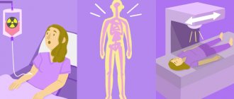
In order to assess the risk of having a child with Down syndrome, screening tests are used during pregnancy. These tests are painless and non-invasive (do not cause damage to the tissues of the pregnant woman and the fetus), but do not accurately determine the presence or absence of congenital pathology. Nevertheless, their widespread use makes it possible to suspect risks and recommend invasive procedures. Invasive diagnostic methods include interventions performed under ultrasound guidance - amniocentesis, chordocentesis and chorionic villus biopsy.
The mechanism of development of Down syndrome
In the majority of cases, a chromosomal abnormality is caused by an oocyte (female germ cell), which at the time of fusion with the male germ cell contains not 23, but 24 chromosomes.
Since the sperm involved in fertilization has 23 chromosomes, one of the oocyte's chromosomes remains extra. Only in 10% of cases the extra chromosome is passed on by the father. Why is an extra chromosome formed? To answer this question, we should recall the process of cell division. In one of the phases of division (meiosis), a pair of chromosomes is stretched to the poles of the dividing cell, so that as a result both daughter cells receive a chromosome. The process of chromosome stretching involves a motor protein with microtubules for transport. If the microtubule weakens or thins on one side, both chromosomes are pulled to one pole, after which a shell is formed around them. This is how a sex cell is formed with a set of 24 chromosomes. If she manages to take part in fertilization, the fetus will develop Down syndrome.
Risk factors for having a child with Down syndrome
The mechanism of development of the disease determines its genetic randomness. The causes of random mutation and trisomy on chromosome 21 cannot be explained by the peculiarities of lifestyle and nutrition, place of residence, ecology, or race.
As a factor of heredity, this anomaly can manifest itself only in the rarest case if both the mother and father are carriers of the mutation of the 21st chromosome. The number of such couples does not exceed 2% of registered cases of birth of children with Down syndrome.
The only reliably established factor that can cause the birth of a sick child associated with trisomy 21 is the age of the mother. Based on static data, for women of different age groups, the birth of a child with Down syndrome occurs on average per 700 healthy newborns, while for women over 40 years of age this ratio is 1:19.
The older the pregnant mother was at birth, especially if the expectant mother herself is at a late fertile age, the higher the risk of a chromosomal abnormality.
An early age of a pregnant woman (up to 18 years) can also lead to a chromosomal mutation during the period of oogenesis, since the mechanism of egg maturation in young girls has not yet formed.
Conditionally, the risk group may include a married couple whose family history includes cases of the birth of children with Down syndrome. The risk increases if the couple is closely related.
Main ultrasound markers of Down syndrome
Ultrasound markers are ultrasound measurements that are most likely to be associated with a high chance of a fetus having Down syndrome. None of these markers are specific, that is, unique to this pathology. The following indicators are taken into account: thickness of the nuchal translucency (75% sensitivity), absence of nasal bone (58%), heart defects, short femurs and humeri, hyperechoic intestine, choroid plexus cysts, echogenic foci in the heart, signs of duodenal atresia . Of course, none of these markers is an absolute sign of a chromosomal abnormality.
Measuring the thickness of the collar area
The collar space (neck fold) is the area between the folds of fetal tissue that is transparent to ultrasound and located below the back of the head in the area where the neck is formed. In children with chromosomal abnormalities such as Down syndrome or trisomy 18, fluid accumulates in this area. This space is measured from 11 to 14 weeks during an ultrasound scan of pregnancy in the first trimester. Measurements obtained at this time are the most significant for predicting the individual risk of developing chromosomal abnormalities in the fetus. A nuchal translucency thickness of more than 3 mm is perceived as an indicator of an increased likelihood of genetic defects in the fetus. An increase in the nuchal space does not mean that the fetus necessarily has Down syndrome, but if we take into account the gestational age of the fetus at which the measurements were taken, the age of the mother and biochemical screening indicators, the probability of a correct diagnosis becomes above 80%. Although nuchal translucency thickness screening is one of the most valuable tests for identifying chromosomal abnormalities, there are uncertainties. The error arises due to violations of measurement techniques. This applies not only to measurements of the collar space, but also to the nasal bone. Errors occur due to human factors. The correct measurement technique and the experience of an ultrasound specialist are very important.
Measurements of the fetal pelvis and brain structures
In fetuses with trisomy 21 pairs of chromosomes, ultrasound examination may reveal cerebellar hypoplasia (reduction in size) and a reduction in the frontal lobe. These facts are used to assess the likelihood of Down syndrome. The combination of a shortened transverse dimension of the cerebellum and a decrease in the frontothalamic distance is more important for the diagnosis of this chromosomal abnormality than measuring each distance of intracerebral structures separately.
In a fetus with Down syndrome, when performing an ultrasound, the length of the iliac bones is clearly reduced and the angle between these bones is increased. This can be most clearly measured at the level of the middle of the sacrum in cross section.
Signs of Down syndrome
Phenotypic signs of Down syndrome are common to almost all people with this syndrome:
- Mongoloid eye shape is characteristic of 90% of patients;
- flat face due to a poorly defined bridge of the nose and shallow-set eyes;
- altered shape of the skull – flat occiput, brachycephaly (width to length ratio of 80% and above);
- hypermobile joints;
- muscle hypotonicity;
- a fold of skin in the inner corner of the eye, overlying the lacrimal tubercle (epicanthus);
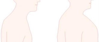
- skin fold of the neck;
- shortened length of legs and arms;
- the shape of the outer ear in the form of an almost perfect semicircle;
- a shortened fifth finger (“little finger”) turned inside the palm;
- short fingers in general due to underdeveloped middle phalanges;
- single transverse palm fold;
- splayed big toe;
- skin folds of the foot;
- constantly slightly open mouth;
- large tongue with numerous deep grooves;
- speech defects.
People with Down syndrome have a number of diseases characteristic of this chromosomal pathology.
Down syndrome – a sentence or hope?
Down syndrome is the most common genetic abnormality caused by trisomy 21 pairs of chromosomes. According to statistics, approximately 1 in 700 children are born with Down syndrome. This ratio is the same in different countries, climate zones, and social strata. It does not depend on the parents’ lifestyle, their health, bad habits, nutrition, wealth, education, skin color or nationality. The likelihood of having such a child increases with the age of the mother, but since younger mothers give birth more often, all age categories are equally represented among parents of children with Down syndrome.
This syndrome was described by John Langdon Down in 1866 as a mental retardation associated with a number of characteristic external signs. And in 1959, Jerome Lejeune revealed its genetic nature.
By 1964, three main types of chromosomal abnormalities in Down syndrome became known:
- Standard trisomy (94%) - tripling of chromosome 21 is present in all cells and occurs as a result of a violation of the process of meiosis.
- The mosaic form (2%) is caused by disturbances in the mitosis process in one of the cells at the blastula or gastrula stage; tripling of chromosome 21 is present only in the derivatives of this cell. In these forms, the parents have a normal genotype.
- Translocation form (4%) - the arm of one 21st chromosome is attached to another chromosome and during meiosis moves into the resulting cell along with it. Before the birth of their next child, parents must undergo genetic testing.
A presumptive diagnosis, as a rule, is made immediately after the birth of a child based on a number of external signs: “flat” face (90%), brachycephaly (81%), skin fold on the neck in newborns (81%), anti-Mongoloid eye shape (80%), epicanthus (80%), joint hypermobility (80%), muscle hypotonia (80%), short limbs (70%), arched palate (58%), etc. Transverse palmar fold, considered a universal sign of Down syndrome, occurs in 45% of cases, and Brushfield spots (pigmented spots along the edge of the iris) in 19% of cases. Usually, a newborn with Down syndrome has some of the known signs, sometimes some signs are also found in ordinary children. The final diagnosis is made after receiving the results of the karyotype analysis.
Down syndrome is often accompanied by somatic diseases.
Many children are often born with signs of morphofunctional immaturity. Congenital heart defects occur in 40–60% of cases. A common disorder is sleep apnea (up to 50%), which occurs due to the structural features of the nasopharynx, and obstruction of the oropharynx by the root of the tongue. Acquired forms of hypothyroidism can account for up to 35% and require special attention, since the clinical signs of hypothyroidism are masked by manifestations of Down syndrome. Pathology of the musculoskeletal system (including muscle hypotonia and hyperelasticity of ligaments) is present in almost all children. Ophthalmological problems occur in 45%, hearing loss in 38–78%, and gastrointestinal abnormalities in 12% of cases.
Developing prenatal diagnostics make it possible to better diagnose Down syndrome and become more accessible to women in Russia. However, this method does not help resolve the question of whether the family is ready to raise a child with Down syndrome, but only postpones it to the period before the birth of the child. More and more families are turning to our Center in which the parents knew about Down syndrome before the birth of the child and decided to continue the pregnancy.
For many years, Down syndrome was considered a medical problem, as reflected by its old name "Down disease", and since this chromosomal abnormality cannot be cured, such patients were usually institutionalized from birth, where they were given general care . Comorbidities were also considered part of the syndrome and often went untreated. Children with Down syndrome in such conditions demonstrated a low level of psychophysical development with an average life expectancy of about 10 years.
The situation began to change about 50 years ago. Against the backdrop of the humanization of society in developed countries, it was recognized that the conditions of keeping people in closed institutions are not satisfactory, their rights are often violated, and long-term stay in inpatient conditions has an adverse effect on their condition. In addition, keeping people with intellectual disabilities in hospitals required unjustified financial costs. At the same time, psychological and educational research has shown the importance of emotional attachment to an adult and a stimulating environment for the development of a child. Parents of patients with developmental disabilities also advocated for the right of their children to live at home and receive all the care they need in the community. These requirements have found wide support among pediatricians. All this led to the beginning of the process of deinstitutionalization, that is, the dissolution of the boarding school system and the creation of local help services. These changes are reflected in UN documents defining the rights of children with developmental disabilities to life in a family, education and integration into society.
In Russia, the majority of children with Down syndrome continued to be in government institutions and only about 10% of children were raised at home. Attitudes towards people with Down syndrome have begun to change since the early 90s. Parent associations and public organizations played a big role in this. In 1997, a charitable foundation was created in Moscow, the purpose of which is to improve the quality of life of children with Down syndrome in Russia. In 1998, on the basis of this organization, an Early Help Center was opened, providing free psychological and pedagogical assistance to children from birth to 8 years. Until this time, families raising their children at home found it difficult to find professional support, and the developmental level of such children was not studied. All examinations of the level of development of children with Down syndrome were carried out in boarding schools, where for many children the factor of deprivation, that is, lack of parental love and care, turns out to be almost the leading factor in the genesis of their disorders. On the other hand, the development of a system of assistance to children living in families has made it possible to achieve good results in their rehabilitation and changed the idea of their capabilities. The data below is borrowed from foreign literature and confirmed by the experience of the Early Help Center (
.).
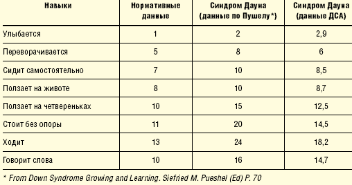
Thus, children with Down syndrome go through the same stages of development as ordinary children, and with special education, although somewhat later, they master the same skills.
Today, more than 1,000 families from Moscow and Russian regions receive information support, they are regularly treated and provided with advisory assistance. Such programs are becoming increasingly widespread in our country and neighboring countries.
The modern approach to teaching children with Down syndrome is based on a family-centered model of early assistance, which includes psychological and pedagogical support for parents and is focused on helping parents in raising their children. Much attention is paid to the development of young children (from 0 to 3 years), the prevention of secondary disorders that arise due to concomitant diseases or uneven development of the child. From the first weeks, teachers’ attention is focused on the interaction of parents with the child, motor development, cognitive processes, and the development of communication. From the age of 1.5 years, children begin to attend group classes aimed at socialization and preparation for kindergarten. By age 3, children typically enter kindergarten while continuing to receive additional special education activities. More and more kindergartens are organizing integrative groups, where children with Down syndrome are together with ordinary peers, and celebrate their positive influence on each other. By the age of 8, children enter schools selected according to their level of development. The majority of children study in specialized schools, but there are isolated examples of children with Down syndrome being taught in general education schools.
Of course, only interdepartmental interaction, including health, social protection and education authorities, makes it possible to comprehensively solve the problems of a child with developmental disorders and his family.
Since 2002, active work has been carried out aimed at preventing social orphanhood of children with Down syndrome, as well as changing attitudes towards this category of children in the professional medical environment and in the public consciousness.
The main components of the program are:
- Providing doctors in maternity hospitals with up-to-date knowledge about the developmental characteristics of children with Down syndrome living in families, the possibilities of pedagogical assistance for such children, and tactics for communicating the diagnosis. Information about the program and printed materials about Down syndrome are provided free of charge to maternity care facilities.
- Providing crisis psychological assistance and social support to parents who are at the stage of making a decision about the fate of the child.
- Inclusion of the family in the early assistance service literally from the first days of the child’s life, psychological and pedagogical support in the preschool period and upon admission to school.
The result of the implementation of this program is a steady increase in the number of children with Down syndrome raised in families. Thus, in 2002 in Moscow, approximately 15% of children remained in families, the same as the Russian average, and after 4 years of the program - in 2006 - 50% of children.
Let's talk about this project in more detail.
Many parents remember the words they heard in the maternity hospital for many years. Because Down syndrome is diagnosed immediately after a child is born, parents learn the diagnosis before their relationship with the child has formed, and often even before they see the child for the first time. The way in which the diagnosis is communicated greatly influences the subsequent development of the parent-child relationship and the acceptance of the child, even if the child remains in the family.
The message about Down syndrome destroys the image of the child that parents dreamed of during pregnancy, their plans and ideas about themselves as parents. The significance of this loss determines the intensity and duration of the experience. This process has several phases and ultimately aims to reorganize life in accordance with the new situation.
The first phase (shock) can be described with the words: “This cannot be, this is not happening to me.” It can last from a few minutes to several days. Then comes the reaction phase, which manifests itself in feelings of anger, conflict, distrust, and the search for someone to blame. These feelings often fall on those who happen to be nearby and report the diagnosis. It is important to understand that such behavior is a normal manifestation of this stage of grief, and to try to emotionally support the parents. This phase can last from several days to several weeks.
At the next stage, the adaptive phase begins. Parents gradually accept the fact that their child has Down syndrome, their anxiety noticeably decreases, and they begin to think about immediate needs. This phase can last up to a year. After this, the orientation or reorganization phase begins, when the family masters the role of parents of an unusual child, takes a constructive approach to solving their problems, seeks help from the appropriate services, makes plans for the future, and establishes new relationships.
Unfortunately, parents usually have to make a decision about the fate of the child while he is in the maternity hospital, that is, within the first few days after his birth. At this time, they are in a shock or reactive phase, and in this state it is almost impossible to make an informed decision. The ability to delay a choice gives them the time they need to adapt. The Early Help Center has recorded about 30 cases in which parents took their child home several days or months after abandonment.
The doctor in the maternity hospital who reports the diagnosis is also under stress caused by the failure of the newborn to meet his expectations, the need to convey unpleasant news, the expected reaction, etc. In addition, he turns out to be the only person to whom parents can turn, and he has to answer the question: “What does this mean?”, presenting pedagogical information about the development and socialization of the child, and also provide psychological support to the mother, answering the question: “What to do now?”
Without sufficient psychological preparation, doctors often act at the expense of their mental strength, “trying the situation on themselves.” Often it is difficult for a doctor to determine the boundaries of his responsibility, and he, sincerely wanting to help parents, advises them to abandon the child, based on their own ideas and their psychological resources. In this case, it turns out that the doctor advises to abandon a child who is not his own, and the parents have to abandon their own.
The mother's reaction, confusion, tears, and aggressive behavior also cause a desire to help her, and she is often prescribed medications that reduce the intensity of grief, but slow down its experience.
Understanding the limits of one’s capabilities and responsibilities, and the opportunity to invite a psychologist, teacher or representative of the parent association make the task facing them easier for doctors.
Let's take a closer look at what is important to do in the maternity hospital:
- congratulate the parents on the birth of a child, list the usual positive things in such cases: gender, height, weight, etc.;
- talk about his problems, examination plan and necessary medical measures;
- bring the baby to the mother, try to maintain breastfeeding, since this is extremely important for the child’s health, improving his contact with the mother and the development of speech in the future;
- provide parents with general information about Down syndrome, avoiding predictions, since the development of a newborn child cannot be predicted;
- support any decision of the parents about the future fate of the child;
- It is not permissible to agitate parents to make this or that decision, since we do not fully understand the circumstances of the life of this particular family and cannot share with them the responsibility for the jointly made decision;
- By kindly questioning parents about their decision, we bring them back to their feelings, their life circumstances, and their own motivations and responsibilities. This leads to the fact that they make a more natural decision - not to abandon the child and subsequently take a more active parental position;
- give time to make a decision about the fate of the child, since the condition of the parents after reporting the diagnosis often does not allow them to make an informed choice. Arrange, if necessary, a meeting with other family members or a psychologist;
- provide information about organizations where the family can receive support in the future. If the child still has medical problems, he should be sent to a medical institution; if he has developmental problems, he should be sent to a psychological or pedagogical institution. These could be educational services, parent mutual support groups, social services, etc. The best option, of course, is the early intervention service, since the family-centered model of early intervention allows parents to receive psychological support when contacting about the child's development.
Starting work with the family at the stage of the maternity hospital, including the family in the early intervention service, with the subsequent transition of the child to kindergarten, school and employment projects for adults, ensures the development, training and socialization of children, and also gives their parents the opportunity to work and lead a normal life life.
T. P. Medvedeva , Moscow
Diseases associated with Down syndrome
A feature and one of the signs of a genetic abnormality on chromosome 21 is the presence of a large number of diseases of various organs and systems in Down syndrome. This is explained by a decrease in muscle tone, weakened immunity at the cellular level, metabolic disorders and other pathologies. As a result of these disorders, children and adults with Down syndrome experience:
- persistent infectious diseases affecting the pharynx, bronchi and trachea;
- congenital heart defects in almost half of patients;
- vision pathologies (myopia, cataracts, increased intraocular pressure, etc.) due to congenital eye abnormalities and infections - more than 60% of patients are affected;
- narrowing or complete obstruction of the duodenum is observed in 8% of newborns;
- reflux of food into the esophagus from the stomach (reflux);
- seizures with signs of epilepsy;
- hypofunction of the thyroid glands, leading to metabolic disorders, obesity, dermatological problems - up to 15% of patients;
- congenital hip dislocation;
- stopping breathing during sleep due to weak tone of the muscles of the pharynx and large tongue;
- hearing pathologies due to frequent inflammation and structural anomalies of the middle ear;
- congenital blood cancer (leukemia) with hemorrhages in internal organs, mucous membranes, skin;
- mild to moderate mental retardation, systemic speech underdevelopment, underdeveloped motor skills;
- early onset of Alzheimer's disease and other mental illnesses associated with senile dementia.
Signs of Down syndrome during ultrasound in the second trimester of pregnancy
When performing an ultrasound of the fetus in the second trimester of pregnancy, it is possible to detect such abnormalities as a violation of the formation of skeletal bones, expansion of the nuchal translucency space by more than 5 mm, the presence of heart defects, expansion of the renal pelvis (pyelectasia), echogenicity of the intestine, and cysts of the choroid plexus of the brain. Moreover, only heart defects, disorders of skeletal formation and expansion of the collar space are independent risk factors.
Using only one of the measurements in assessing the likelihood of fetal chromosomal abnormalities, for example, determining the thickness of the nuchal translucency or measuring the iliac angle, is less accurate than taking into account the totality of measurements.
How is Down syndrome diagnosed during pregnancy in NEARMEDIC
If the embryo is affected by Down syndrome, there is a high risk of spontaneous abortion at approximately 6-8 weeks of pregnancy, which is observed in almost a third of women. If such miscarriages recur in the next pregnancy, it is necessary to undergo prenatal screening to identify Down syndrome in the early stages of fetal development.
Prenatal diagnosis of Down syndrome in the multidisciplinary network of NEARMEDIC clinics is carried out as part of the examination of pregnant women in each trimester. The gynecologist who is seeing the expectant mother refers her for prenatal diagnosis.
For prevention and if Down syndrome is suspected during the diagnostic process, various tests and procedures can be performed according to indications at different stages of pregnancy.
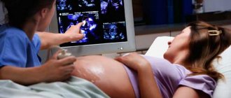
Ultrasound examination
This non-invasive examination method is carried out in the gynecological departments of the NEARMEDIC clinic network in the first trimester (11-13 weeks) and again at 24 and 34 weeks. Ultrasound diagnostics at different stages of pregnancy can reveal the following signs indicating possible Down syndrome in the fetus:
- shortened hip and shoulder bones;
- cysts in the brain;
- abnormalities of heart development;
- absence or underdevelopment of nasal bones;
- absence of one of the umbilical arteries;
- the increased space between the neck bone and the skin is filled with fluid, etc.
The simultaneous presence of these pathologies makes it possible to establish Down syndrome with 90% certainty. One symptom allows one to suspect the disease and place the woman at risk.
Blood chemistry
The blood volume of certain proteins and hormones that are produced in the body of a pregnant woman is checked.
It is carried out in the 1st (double test - examination of blood serum for hCG and PAPP-A) and 2nd trimesters of pregnancy (triple test - examination of the level of hCG, AFP and free estriol).
The ability to detect Down syndrome in a fetus through a biochemical blood test is about 70%. Its presence may be indicated by a reduced content of the listed proteins and hormones.
Study of amniotic fluid
If the above non-invasive tests cause doctors to suspect a chromosomal abnormality, more highly accurate methods are used. One of them is amniocentesis, a study of amniotic fluid, or amniotic fluid, which is performed at 8-14 weeks of pregnancy.
This invasive method involves withdrawing fluid through the abdomen using a video-guided needle, followed by genetic analysis of fetal cells in the amniotic fluid. The presence of three chromosomes 21 in the cells 99% confirms the preliminary diagnosis.
Cord blood studies
This study is called cordocentesis, and is carried out from the 18th week of pregnancy, since in earlier stages the umbilical cord vessels are too thin and do not allow blood to be taken with a special needle. A puncture under the control of an ultrasound machine is made in the abdominal wall or cervix. The optimal period for cordocentesis is 22-24 weeks.
Genetic research of fetal cells contained in umbilical cord blood makes it possible to diagnose Down syndrome with an accuracy of 98-99% when three twenty-first chromosomes are detected.
Examination of the villi of the outer membrane of the embryo
A chorionic villus biopsy (the so-called fuzzy outer membrane of the fetus) is performed at 10-12 weeks. A biopsy needle is used to puncture and remove a tissue sample from the small finger-like projections on the placenta or using a flexible probe if the procedure is performed through the cervix.
The chromosomes contained in the chorion cells are similar to the cells of the fetus. If, during a genetic study, chorion cells contain three chromosomes 21, we can talk about Down syndrome with 99% accuracy.
The NEARMEDIC Genetics Department conducts a rare study of the genotype of future parents for gene translocation that causes a chromosomal mutation.
After all the examinations and the final diagnosis of the presence of Down syndrome in the fetus, the expectant mother must independently decide whether to terminate the pregnancy or give birth. At the same time, she receives reliable information that this chromosomal abnormality is incurable, and what difficulties she has to overcome. But modern medical science and the capabilities of the NEARMEDIC genetic department make it possible to support such children and ensure their vital functions at all stages of life, providing adequate medical care and counseling parents. In Western Europe, “sunny” people, as those with an extra chromosome are called, are no longer treated as a burden, having appreciated their empathic qualities, gentle and affectionate disposition. Among such people, the first athletes, artists, and painters have already appeared, since they are highly trainable, as John Down spoke about.
Make an appointment with a gynecologist or geneticist using the feedback form on the website, or call our contact center at the specified phone number.
Fetal ultrasound is the main way to diagnose the likelihood of Down syndrome
Ultrasound of the fetus to detect developmental abnormalities is called ultrasound screening. Most often, ultrasound screening is combined with biochemical blood tests of a pregnant woman, and this research is called combined screening. Depending on the choice of criteria, the probability of identifying Down syndrome in the second trimester is 60-91%. The use of color Doppler technology for ultrasound screening makes it possible to detect fetal heart defects and increases the accuracy of diagnosis.