HE4 (human epididymal secretory protein 4)
– serum protein secreted by epithelial cells of the respiratory and reproductive systems. It is a marker of malignant tumors of the endometrium and ovaries. The study is used to diagnose cancer, the result allows you to differentiate malignant neoplasms. To monitor the course of cancer pathology, the analysis is prescribed to patients with negative CA125 test data as a way to assess the effectiveness of treatment and identify relapses. Blood is taken from the cubital vein. The method for determining a tumor marker is ICL. The normal level for premenopausal women is up to 70 pmol/l, for postmenopausal women - up to 140 pmol/l. The duration of the procedure is 1 day.
- Indications
- Preparing for analysis
- Normal values
- Increasing the indicator
- Treatment of abnormalities
- Prices in Moscow
HE4 is a protein with a small molecular weight. It was first identified in the epithelium of the distal ducts of the appendages. These ducts are called epididymal, hence the expanded name of the protein - secretory protein 4 of the human epididymis. Later it was discovered in the glandular epithelium of the mammary gland, reproductive organs, distal renal tubules, large intestine, and salivary glands. Presumably this protein ensures the normal functioning of epithelial tissue and supports the innate immunity of the oral cavity, trachea, and lungs. Its synthesis increases with the development of malignant tumors of the female reproductive glands, lungs, kidneys, uterus, mammary glands, and gastrointestinal tract. HE4 promotes the progression of cancer and is its laboratory marker.
Norm CA 125 in women with cysts and other conditions
This tumor marker is a glycoprotein produced by epithelial cells of the digestive tract, kidneys, and pancreas.
But the maximum concentration is found in the ovaries, which allows the antigen to be used as a marker of diseases of the female reproductive system. The maximum permissible value is 35 U/ml; venous blood is taken for research. The specificity of CA 125 is low, since its level can increase in tumors of various locations:
- ovaries;
- uterus;
- pancreas;
- intestines and stomach;
- lungs.
Concentration indicators also increase in benign formations - ovarian cysts, fibroids. A physiological increase in the norm is typical for the first trimester of pregnancy and menstruation.
To diagnose ovarian cancer in women of fertile age, CA 125 is rarely used because it is characterized by low sensitivity. In addition, in the first stages, when there are no metastases yet, the amount of glycoprotein increases slightly. To suggest adenocarcinoma, additional tests for antigens HE4, CA 15-3, CA 72-4, CEA and others are required. The tumor marker is of practical value at the stages of monitoring treatment and after it, for predicting relapses.
The information content of the analysis increases in the postmenopausal period, in women over 50. It is for this age category that CA 125 is recommended as the main tumor marker for ovarian cancer. In addition, specific values make it possible to judge the presence of certain diseases.
Thus, the norms of CA 125 in women do not differ by age, and the table shows the concentrations of the antigen in the blood under certain conditions and pathologies:
| Patient's condition | Analysis results, U/ml |
| Healthy woman | 10-15 |
| Pregnancy, 1st trimester | Upper limit of normal (35) |
| Cyst | 35-60 |
| Endometriosis, fibroids | up to 110 |
| Ovarian cancer | From 100 and above, most often five times higher than the maximum permissible value |
Ovarian cyst
This benign formation is diagnosed in women of different ages. With an ovarian cyst, which is a cavity with fluid or tissue proliferation, the CA 125 value rises to 60 U/ml. This indicator is normal, but it is necessary to constantly monitor the antigen level, since the tumor can degenerate into malignant. More information about the characteristics of the disease can be found in the video about tumor markers for cysts.
Endometriosis
A fairly common pathology in women of reproductive age. It is a growth of the muscular layer (endometrium) of the uterus. It may be accompanied by painful sensations, inter-cycle bleeding, and in advanced cases – an increase in abdominal volume. The values of the CA 125 marker reach 100 U/ml.
After additional diagnostics that exclude cancer, hormonal therapy is prescribed, and surgery is also indicated. In this case, systematic monitoring of the antigen level is mandatory, since malignant degeneration of endometrial cells is possible.
Myoma
Against the background of hormonal imbalances, benign tumors of the uterus - fibroids - often arise. They can be localized both in the thickness of the muscular wall of the organ and in the cavity (connecting to the uterus with the help of the so-called stalk). CA 125 values in this case are higher than with ovarian cysts, reaching 110 U/ml. Myoma itself is not life-threatening, but the cells of such a formation are prone to degeneration into malignant ones. Therefore, it is recommended to take hormonal drugs to reduce the tumor or undergo surgery to remove it.
Pregnancy
Changes in hormonal levels also occur in the initial stages of pregnancy, which affects the increase in the level of antigen in a woman’s blood. In addition, the glycoprotein is also synthesized by fetal cells, also entering the bloodstream. Thus, in the first trimester, an increase in CA 125 above the permissible 35 U/ml is a physiological norm, and should be alarming only if it is significantly exceeded.
When and who to get tested
In the modern world, the incidence of cancer has increased significantly not only in people of older age categories, but also in the younger population.
Oncological markers make it possible to determine the presence and level of a tumor at the very beginning of its development or at the time of its development without obvious symptoms. Despite this, it is not necessary for every person to get tested.
First of all, the risk group consists of those who are prone to cancer. These are people whose blood relatives had malignant tumors of different stages. For such people, screening tests are required 1-2 times a year. Also, 1-2 times a year, such tests should be carried out by people who have been identified as having benign formations (for example, fibroids), or in the presence of tumor-like formations (for example, a cyst). People who have been in situations that could activate the development of neoplasms, in unfavorable environmental conditions, or have experienced severe mental shock or other illnesses should undergo tests once every two to three years.
There is a special scheme for taking samples for tumor markers for people who have undergone successful cancer treatment. To exclude the possibility of relapse, the following tests must be taken for 3 years after treatment:
- after 1 year - monthly;
- after 2 years - once every two months;
- after 3 years - 1 time per trimester.
After 3 years, it is recommended to take samples every six months to a year to detect relapse in the early stages and prompt treatment.
Tests must be taken regularly over several years.
People suspected of having a malignant neoplasm should immediately undergo testing in order to refute or confirm their fears in the early stages.
It is also worth taking a sample if you have complaints about:
- unsatisfactory health for no obvious reason;
- pain in the pelvic area;
- disruptions in the menstrual cycle;
- weight loss;
- decreased appetite.
Let's summarize who should undergo testing for cancer markers:
- people whose relatives had cancer;
- people who have undergone cancer treatment;
- people suffering from precancerous diseases;
- people working in hazardous industries or in hazardous environmental conditions;
- if you notice unrelated symptoms.
ROMA Index
The Roma tumor marker is an algorithm that is calculated based on the results of tests of two protein markers CA-125 and HE-4. The ROMA index is a marker of the pelvic organs, the most accurate way to calculate the risk of ovarian cancer of epithelial origin in women.
A blood test for the Roma index divides patients into two groups for the likelihood of ovarian cancer:
- high probability;
- low probability.
The ROMA index study is prescribed to the patient by a gynecologist, oncologist, or carried out on the woman’s initiative. An analysis to determine the level of tumor markers is necessarily prescribed in the following cases:
- hereditary tendency to form cancer tumors in the body;
- a history of an ovarian or pelvic tumor removed by surgery.
Also, the main indications for getting tested for the ROMA index are:
- detection of ovarian cancer in the early stages;
- determining the degree of malignancy of the tumor;
- monitoring the effectiveness of surgical treatment of cancer.
Multiple studies using the ROMA index may be required. This is necessary to detect relapses and determine the spread of metastases. The ROMA index is calculated using special formulas and depends on the period of a woman’s hormonal activity. In premenopause, the ROMA1 index values are used, in postmenopause – ROMA2.
There are no special requirements for preparing for this study, but you still need to adhere to some restrictions:
- donate blood in the morning on an empty stomach or 5-8 hours after eating;
- if possible, do not take medications 3 days before the test;
- stop drinking alcoholic beverages within a few days;
- 5-7 days before the analysis, do not eat fried, fatty and spicy foods, smoked foods;
- eliminate physical and emotional stress in a few days;
- do not conduct instrumental studies (ultrasound, X-ray, MRI, etc.) for several days;
- stop smoking 3-4 hours before the test;
- Before the analysis, you can only drink clean water without gas.
Don't waste your time searching for inaccurate cancer treatment prices
*Only upon receipt of information about the patient’s disease, a representative of the clinic will be able to calculate the exact price for treatment.
The test is deciphered by a laboratory diagnostician and an oncologist using a table of norms and pathologies of ovarian cancer markers. Usually the analysis result is ready the next day. The ROMA index test is not performed for patients under 18 years of age and for patients who have previously undergone treatment for oncology.
The normal value of the ROMA1 index result in premenopause is less than 7.4%. This result indicates the absence of cancer processes in the body and the minimal likelihood of their occurrence. In cases where the rate is 7.4% or higher, ovarian cancer is suspected.
The norm for the ROMA2 index in postmenopause is 24.7%. If the indicator is higher than normal, then the risk of a malignant tumor is high. In cases where the index value is elevated, but does not decrease or increase over time, and remains at the same level, this may indicate a benign tumor in the body.
Features of the analysis
Venous blood is required for the study. The accuracy of the results largely depends on proper preparation for the analysis:
- Blood sampling should occur in the first half of the day, preferably in the morning.
- Before donating blood, you should not eat breakfast or drink any drinks other than water.
- 12-24 hours before donating blood, you should completely stop smoking, drinking alcohol and avoid increased physical and emotional stress.
In addition, it is important to stop taking any medications 7-10 days before blood collection. If for some reason this is not possible - for example, you are undergoing a course of drug treatment that cannot be interrupted - be sure to tell your doctor
The names of all medications taken and their dosage should be listed. Such information will allow you to adjust the analysis results taking into account new circumstances and avoid incorrect interpretation of the test.
What the analysis result shows depends on several factors
Any other diagnostic measures should be prescribed after the test, as they can potentially distort the blood picture, which will give the doctor false information.
What does the SCC tumor marker show?
Normally, a minimal amount of this glycopeptide is produced in the epithelial cells of each person, however, its diffusion into the intercellular space does not occur.
For the first time, the scc molecule, which is classified as a tumor marker, was isolated from mutant squamous epithelial cells of the cervix in 1977. According to statistics, the specificity of this marker reaches 80% for stages 3-4 of oncological pathology.
With the development of squamous cell carcinoma in the patient's body, the secretion of the tumor marker SCC increases significantly. This fact is due to the reaction of the immune system in response to the development of abnormal cells in the human body. Presumably, an increase in the value of the antigen, indicating squamous cell carcinoma, creates optimal conditions for the implementation of the mechanism of invasion (spread) of mutant cells throughout the body with the help of metastases.
When is an SCC test ordered?
A study to determine the level of squamous cell carcinoma antigen SCC is an important diagnostic criterion and is prescribed for:
- large-scale screening of a person with suspected cancer pathology in combination with establishing the spread of metastases throughout the body;
- prescribing a treatment regimen as a result of a reliably confirmed diagnosis of squamous cell carcinoma;
- the need to monitor the effectiveness of the prescribed course of treatment for oncology;
- determining the severity of the disease and predicting the outcome.
Indications for choosing this type of diagnosis are:
- suspicion of cancer of the mucous epithelium of the esophagus, cervix, lungs and other organs;
- drawing up a course of therapy for people at the initial stage of the disease and, if necessary, transferring the patient to more aggressive methods of fighting cancer;
- control over possible recurrent tumors of various organs;
- annual routine examination of patients who have undergone removal of malignant tumors.
High scc level
A correlation has been established between the size of the antigen, indicating squamous cell carcinoma, and the severity of the pathology, the size of the malignant neoplasm, its growth rate, as well as the penetration of metastases into neighboring organs.
The squamous cell carcinoma antigen is detected in more than 50% of women with cervical cancer at any stage of the disease. However, the sensitivity of the method varies from 10% (stage 1) to 80% (stage 4).
In this case, the absence of a decrease or its increase indicates a relapse of the pathology and the need for a second course of therapy. Carrying out control tests makes it possible to establish the occurrence of repeated neoplasms in more than 90% of cases, long before the first clinical signs of disease manifestation.
By what method and how is the analysis carried out?
To check the HE 4 tumor marker (what it shows), the ECLIA method is the best way to help - an immunological method for the qualitative and/or quantitative identification of all kinds of low-molecular compounds.
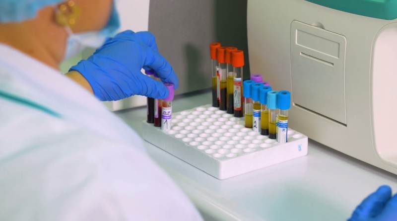
It is carried out in laboratory conditions using special equipment and manual work. To begin with, venous blood is collected from the patient in the traditional way, after which it is transferred to a transport medium, where the serum is separated from the blood plasma. The resulting serum serves as a sample for testing, which is done manually.
The laboratory technician injects antibodies into the resulting sample. Based on the results of their interaction, a reaction is set - the presence or absence of an increase in the norm of HE4 tumor marker in the body.
As a rule, the results are ready within a day. But if you need them “for yesterday,” you can do an express analysis, which is carried out in modern laboratories equipped with high-tech equipment.
If an increase in pathological proteins is detected, additional examination is prescribed: ultrasound (ultrasound), magnetic resonance imaging (MRI), radiography, computed tomography (CT) and a blood test for related tumor markers. The best combination is HE-4+CA125.
Explanation of the analysis and reasons for deviation of indicators from the norm
Deviations from the norm in the analysis of the he4 marker can occur in both cancer and benign diseases. These are mainly diseases of the pelvic organs in women. The most possible include ovarian cancer and cysts, uterine fibroids, and endometriosis.
Ovarian cancer
The he4 marker is highly informative in the presence of epithelial ovarian cancer. In this case, there is a significant deviation from

norms, and the indicator increases with tumor growth.
Its detection is very important, since this type of cancer manifests itself clinically in late stages, when treatment is palliative and cannot result in recovery. Symptoms include:
Symptoms include:
- Menstruation disorders
- Pelvic pain
- Impaired urination and defecation (in later stages when the tumor puts pressure on nearby organs)
Uterine fibroids
Occurs when the function of sex hormones is impaired. Refers to benign oncological diseases. It is the growth of muscle tissue (myometrium). It may be asymptomatic or, with pronounced myomatous nodes, manifest itself:
- Pain in the lower abdomen.
- Uterine bleeding.
- Anemia.
For reference. Determination of a tumor marker in this case is not the basis for diagnosis. A gynecological examination and ultrasound of the uterus are much more informative.
Ovarian cyst
A cyst is a cavity with clear boundaries and is a benign disease. He4 may be normal or slightly elevated. Symptoms include
- Abdominal pain
- Heavy periods
- Cycle disruption
- Disruption of the pelvic organs.
However, it is worth keeping in mind that most often this pathology is asymptomatic.
Endometriosis
A disease characterized by the proliferation of endometrial cells beyond their anatomically normal location. Considering that the endometrium contains receptors for sex hormones, abnormally located cells also exhibit cyclic activity - bleeding occurs.
Endometrial cells can grow both in the genital organs (ovaries, vagina, fallopian tubes) and extragenitally - in the large and small intestines, navel, bladder.
For reference. He4 increases slightly in endometriosis.
Indications for the purpose of analysis
Doctors rarely prescribe an analysis to detect the HE4 tumor marker. Such a study is recommended for patients with unfavorable heredity for pelvic cancer, women who have chronic pathologies of the genital organs, and those who work in hazardous industries. Diagnostics is indicated for the following complaints about health:
- the menstrual cycle is irregular;
- frequent painful sensations appear in the lower abdomen;
- difficulties arise associated with emptying the bladder - difficulty urinating or urinary incontinence;
- weight decreases sharply;
- there is general weakness.

The study is also prescribed in the following cases:
- suspicion of an oncological process in the ovaries. Determination of tumor marker levels is carried out as part of early screening of the disease. It is for this purpose that an analysis is performed to determine the level of this protein;
- when it is necessary to determine whether a neoplasm is malignant or not;
- if necessary, find out how effective the prescribed treatment was for ovarian cancer;
- if it is necessary to make a prognosis for further treatment;
- to prevent tumor relapses, as a preventive screening measure;
- to detect metastases.
What does carcinoembryonic antigen (CAA) mean and show in the results of a blood test?
Often, patients whom the doctor sent for this type of study ask the question - what is CEA and what does it show?
Carcinoembryonic antigen is a tumor marker first isolated in 1965 by S. Friedman and colleagues from colon cancer cells of a sick person. The molecular weight of a glycoprotein molecule varies from 180 to 200 thousand Daltons. A distinctive feature is the predominance of the carbohydrate part over the protein part (about 60%).
Deciphering the chemical structure made it possible to establish that CEA is a protein molecule connected by covalent bonds to heterooligosaccharides.
Currently, its function in adults has not been established. It is known that during the ontogenesis of a child, CEA molecules are actively produced by epithelial cells of the gastrointestinal tract and indirectly controls the work of cell division, enhancing it.
The name of the criterion under consideration largely characterizes its properties that are important for laboratory diagnostics, and also partly its biological nature. The term “cancerous” determines its primary importance for identifying oncology
The word “embryonic” emphasizes the importance of the molecule at the stage of intrauterine development of the child. In turn, “antigen” means that it can be detected using immunochemical reactions
The essence of the technique is the binding between an antigen molecule and specific antibodies.
It is necessary to answer that in the human body, the PEA tumor marker does not exhibit antigenic properties, and therefore does not trigger the mechanisms of the immune system. This means that protective antibodies are not produced directly in the patient’s body.
What is REA used for?
A blood test for CEA is used to diagnose oncopathologies and differentiate malignant neoplasms from benign ones. At the same time, the maximum diagnostic value of the CEA tumor marker is observed when detecting cancer of the intestinal tract (rectal and colon). In the case of the development of malignant oncopathology in a patient, the concentration of cancer embryonic antigen sharply increases to critical values.
It should be noted that using a blood test for carcinoembryonic antigen, the presence of a number of other pathologies can also be determined. Thus, the value of the CEA antigen tumor marker indicates the development in the patient’s body of autoimmune diseases, inflammatory processes, as well as diseases of etiology other than oncology.
The dynamics of the tumor marker rea determines the effectiveness of the chosen treatment regimen for a malignant tumor. So, after surgical removal of the tumor, the antigen value should return to normal. The absence of a decrease in the level indicates the ineffectiveness of the therapy, and after removal - a relapse of the disease or the spread of metastases to neighboring organs and tissues.
In this case, correction of the course of treatment is necessary. If the estimated risks of the consequences of a malignant tumor exceed the magnitude of side effects from more aggressive methods, then the possibility of deciding to transfer the patient to them cannot be ruled out.
Deciphering the finished results
On average, the transcript for tumor marker n4 is ready in a couple of days.
Important! The level of the defining blood tumor marker (by itself) does not indicate the exact presence or absence of cancer. To make this diagnosis, it is necessary to use he4 together with data from other antigens; it is also worth going through various types of research methods: from instrumental to laboratory. Chemiluminescent analysis allows you to determine the current level of he4 antigen in the blood
The essence is this: the desired 4th protein enters into a certain reaction with the labeled compounds, after which these compounds begin to change all their inherent properties. In the presence of this catalyst, they begin to glow. With instrumental help, the determination and calculation of the he4 level then begins
Chemiluminescent analysis allows you to determine the current level of he4 antigen in the blood. The essence is this: the desired 4th protein enters into a certain reaction with the labeled compounds, after which these compounds begin to change all their inherent properties. In the presence of this catalyst, they begin to glow. With instrumental help, the determination and calculation of the he4 level then begins.
“Recognize” a tumor, regardless of gender

Many “female” tumor markers can recognize other locations of cancer, but there are antigens associated with tumors that are little or not attracted to the genitals, and they choose a place for growth somewhere in the intestines, liver, and gall bladder. Basically, the gender of the patient does not play a role for them if the pathology does not concern the reproductive sphere, because in women the list of diseases that may be indicated by an increased concentration of a tumor marker is much wider, as the reader can see for himself:
AFP (alpha fetoprotein)
It became one of the first markers that began to be called tumor markers (Tatarinov Yu. S., 1964). This glycoprotein is normally produced in the fetus during intrauterine development, entering the blood of a pregnant woman, it gives a positive result, which is quite understandable.
The appearance of alpha-fetoprotein in other people in concentrations above 10 IU/ml may indicate problems in the liver (hepatitis, cirrhosis, hepatocellular carcinoma, hepatoblastosis), gastrointestinal tract (ulcerative colitis, gastrointestinal tumors), as well as malignant forms of leukemia, cancer breast and lungs. The normal values for men and women differ slightly; AFP increases significantly during pregnancy, so the normal for such women is determined using a special table.
CEA (CEA, carcinoembryonic antigen)
Its concentration should not exceed 5 ng/ml, however, this norm does not apply to pregnant women. In non-pregnant patients, CEA increases with cancer of the ovary, uterus, and breast.
If this indicator increases, cancer of the colon, liver, or pancreas can also be suspected, but it should be borne in mind that, like other tumor markers, CEA also increases with benign processes in the gastrointestinal tract (Crohn's disease, Meckel's diverticulum, peptic ulcer of the duodenum and stomach ), as well as for pancreatitis and cirrhosis. In smokers, the level of CEA in the blood serum also increases markedly.
SA-19-9
An antigen associated with tumors of the pancreas, liver, gallbladder and biliary tract, stomach, lower intestine (rectum and sigmoid), that is, to some extent it is considered a tumor marker of the gastrointestinal tract. However, in addition, the concentration of CA-19-9 increases in cancer of the breast, ovary, uterus and in metastases of carcinomas of various localizations to the liver.
The normal level of a tumor marker is up to 10 U/ml, an increase in level to 1000 U/ml or more indicates that the malignant process has reached the lymphatic system, but the tumor can still be removed (in 5% of patients), an increase in concentration over 10,000 U/ml ml indicates hematogenous dissemination.
Tumor marker 19-9 is not suitable for screening studies and does not detect tumors well in the early stages of development, therefore it is mainly used to monitor the progress of treatment in combination with other tumor-associated antigens (CA-125, CEA, HE4, AFP). When deciphering the results regarding CA-19-9, one should remember and take into account the fact that it is rare in some blood groups (A/B in the Lewis system), when it is simply not produced, regardless of whether the body is healthy or sick.
SA-242
A tumor marker for gastrointestinal diseases, detected in cases similar to CA-19-9, however, it is more sensitive and can be used to diagnose a malignant process at an early stage of its development. In addition, it is often found in increased concentrations (the norm is up to 30 IU/ml) in benign lesions of the stomach and intestines.
SA-72-4
This glycoprotein is expressed by various carcinomas localized in the mammary and pancreas, stomach, large intestine, lungs, ovaries, and endometrium. The marker is often used in combination with CA-125 and CEA to monitor cancer therapy.
Obviously, when diagnosing tumors, preference is given to one antigen that is more sensitive to a certain type of tumor, which is called the main one (CA-15-3, PSA, HE4), while others are of secondary importance and are designed to assist the main ones in carrying out their tasks (often REA). In addition, some tumor-associated antigens are able to detect the disease at the earliest stages (HE4, AFP, PSA), when others serve to monitor the effectiveness of therapy (CA-125, CA-19-9, SCC). Meanwhile, tumor markers sometimes change places, that is, the secondary one becomes the main one in relation to a particular pathology, while in other cases the main one solves a secondary problem (CA-125).
How to get tested
To conduct the study, blood is drawn from a vein. To get the most accurate results, you must follow the rules of preparation for the procedure.
Where can I do this?
The described examination can be completed in many medical centers or private laboratories where various blood tests are performed. It is possible to undergo diagnostics at any of the oncology centers.
Preparation
Compliance with the rules for preparing for research and submitting biological material is the most important factor in obtaining objective results, which is important for making a diagnosis and making a doctor’s decision on treatment tactics, monitoring the disease, and the need for further diagnostics. Preparation for analysis includes the following rules:
Preparation for analysis includes the following rules:
- quit smoking for at least half an hour before blood sampling;
- refusal to eat and drink, with the exception of clean drinking water, 8 hours before the procedure;
- You must stop taking medications two days in advance, but only if stopping the medications will not cause harm to your health;
- It is recommended to avoid sexual intercourse two days before the study;
- drinking alcohol in any form is contraindicated 7 days before the test;
- physical activity is not recommended;
- it is necessary to avoid stress as much as possible;
- maintaining a proper diet - for this purpose, limiting the consumption of fatty, fried and spicy foods.

The results of the examination can be obtained within the next two days.
Technique
In the laboratory, the necessary reagents and catalysts are added to the obtained blood samples, and the analyzed protein “glows.” This is how the level of this glycoprotein in the collected biological material is calculated.
Features of testing for children, pregnant and lactating women
In some cases, testing for the HE4 tumor marker is also prescribed for children. To obtain a reliable result, the same preparation is required as for adults. In this case, the girl can drink a glass of boiled water half an hour before taking the test.
To make an accurate diagnosis, it is necessary to use other diagnostic methods, such as ultrasound, MRI, and undergo an examination by a pediatric gynecologist. Only a comprehensive examination shows the complete clinical picture of the disease.
For pregnant and lactating women, such an analysis is not very effective and the only possible diagnostic method. Features of biochemical processes in the female body during periods of gestation and lactation often lead to false positive results. This determines the need for additional examinations.
Normal values of tumor marker HE-4
The concept of norm in the case of HE-4 analysis does not have a clear framework. The laboratory can only give reference values, that is, the average value characteristic of a healthy person of a certain age group. The value of reference values largely depends on the equipment on which the study is carried out and the test systems.
Important
In different laboratories, the normal values for the level of the tumor marker being determined may differ slightly.
If we take average figures, then the content of the HE-4 tumor marker in the blood of women should be as follows:
- up to 40 years – no more than 60 pmol/l;
- in premenopause – no more than 70 pmol/l;
- in postmenopause – no more than 140 pmol/l.
In addition, correct interpretation of the results of any blood test is impossible without knowledge of other data about the patient (age, weight, disturbing pathological symptoms, presence of concomitant diseases, treatment taken, results of other tests, etc.). In this regard, a competent interpretation of the result of the test for HE-4 can only be given by the attending physician, who knows all the anamnestic and clinical information about the patient being examined.
Symptoms of cancer
Since women are more susceptible to this problem, they need to carefully and attentively listen to their body and all disturbances in its functioning must be taken into account and react to them properly.
If there is a disturbance in the functioning of the ovaries due to tumors, the symptoms are subtle and practically do not bother the patient. In this condition, diagnosis takes place almost at the very end of the disease at the last stage, when treatment already produces less results.
This test should be performed for the following symptoms and associated indications:
- disruption of the ovaries and problems with menstruation in women;
- acute pain syndrome in the pelvic area;
- deterioration of general condition, accompanied by a decrease or complete absence of appetite;
- sudden and causeless weight loss;
- apathy.
General description of glycoprotein CA 125 and HE 4
Ovarian tumor marker CA 125 is the main test that makes it possible to determine the presence of a malignant disease. It is produced by epithelial tissue cells. Moreover, it is produced not only by the ovary, but also by the pancreas, gall bladder, stomach, bronchi and intestines. However, tests are most often used to diagnose cancer of the reproductive organs. That is, the specificity of tumor markers is important. To achieve the most accurate result, the study is combined.
This tumor marker can be detected not only in endometrial cells, but also in the serous uterine fluid. The analysis does not always indicate the presence of an oncological process in the ovaries. Often, a change in the level of tumor marker CA 125 for ovarian cancer is observed in the presence of normal inflammatory processes in the body.
It is detected mainly in those patients who develop oncological pathology of the serous type. Such a study is mandatory in women with a complicated medical history. The fact is that malignant tumors in a large number of cases are caused by genetic predisposition.
This tumor marker has certain features:
- Low sensitivity. That is, in the early stages of the disease it is practically undetectable. Its level increases approximately 8 months before diagnosis.
- Not very good specificity. That is, protein is produced by the epithelium of both the ovaries and other organs.
For the most accurate result, it is necessary to analyze two tumor markers simultaneously: CA 125 and HE 4. The latter is considered more sensitive precisely at the early stage of pathology development, and increases sharply, even if there are no symptoms. Its level changes 3 years before the final diagnosis. An additional advantage of such a tumor marker is its high specificity. It does not respond to inflammatory processes in the uterus or ovaries. That is, with its help, it is the malignant process that is determined.
Marker HE 4 in combination with CA 125 makes it possible to detect a malignant tumor with 90% accuracy.
What is HE-4 analysis
HE-4 is a protein compound that is considered specific to the epithelial tissue of the reproductive organs, respiratory tract and pancreas, so it can normally be present in the blood of women and men.
But an increased amount of it is most often found in epithelial cancer tumors of the ovaries and endometrium, because in them, under the influence of certain genes, this protein is synthesized in large quantities. The peculiarity of the HE-4 protein is that it begins to “go off scale” already at the early stage of cancer.
note
Numerous studies have shown that approximately 75% of women with asymptomatic preclinical ovarian or endometrial cancer test positive for HE-4. In addition, the test for HE-4 practically does not give false-positive results for benign neoplasms of the female reproductive organs, that is, it is highly specific.
For a more accurate diagnosis of malignant epithelial ovarian tumors, oncologists recommend simultaneously determining two tumor markers: HE-4 and CA-125 - this is the so-called ROMA test (link).
Cyfra 21 1 tumor marker - interpretation and norm
This antigen is characterized by low specificity, that is, it appears in the blood in various types of cancer tumors. The exact location of the lesion is determined during combined tests. However, if the diagnosis has already been confirmed, the stage of the disease and the size of the tumor can be determined based on the test results and the amount in the blood.
The norm for the Cyfra tumor marker (in other words, the reference value) ranges from 0 to 3.3 nanograms per ml of blood. Deviations towards the larger side indicate a pathological process, which allows not only to assume the presence of cancer, but also to monitor its progress. Regular tests during treatment provide information about its effectiveness, since a reduced level of cyfra 21.1 after surgery is an indicator of successful therapy.
Cyfra 21.1 is a tumor marker that is found in cancer:
- Lungs (non-small cell)
- Bladder
- Esophagus
- Cervix
- Colon
A positive result may also be a consequence of metastasis of a cancer tumor to the thyroid and mammary glands.
However, the analysis does not guarantee 100% accuracy. For example, the sensitivity of the antigen for non-small cell lung cancer (the main disease that is detected using this tumor marker) is no more than 65%. As the tumor grows, it increases to 75%, but in the early stages the result may be negative. Therefore, in case of suspicion of lung carcinoma, a combined test is performed, with additional detection of antigens NSE, SCC, CEA and others.
In addition, increased values are observed not only in cancer. Indicators of the Cyfra 21 1 tumor marker above normal are characteristic of such conditions and pathologies as:
- tuberculosis;
- chronic inflammation of lung tissue;
- obstruction (obstruction) of the pulmonary tract;
- hemodialysis;
- inflammatory processes affecting the serous membranes of organs (pericarditis, pleurisy);
- renal failure;
- pulmonary fibrosis;
- benign tumors localized in the neck and head;
- cirrhosis of the liver.
What to do if the test for the number 21 1 is positive?
First of all, do not make diagnoses yourself and do not panic. As written above, the level of antigen increases not only in the presence of cancerous tumors. Therefore, deciphering the lung tumor marker cyfra 21-1 is a matter for the attending physician who wrote out the referral for analysis. Only a specialist can correctly compare clinical symptoms with laboratory data and draw adequate conclusions. In addition, the diagnosis is never made only by analyzing tumor markers: a full examination is required to confirm their results.
If you have been diagnosed
If you experience any painful symptoms, you should consult a specialist.
After the woman has completed all the prescribed examinations, been tested several times for oncological markers, the doctors have assessed the dynamics of growth, and they have no doubts about the diagnosis, they develop an individual set of procedures. First of all, chemotherapy is prescribed in most cases. If the growth of the He4 tumor marker does not stop during chemotherapy and the treatment does not produce the desired effect, doctors prescribe surgery and further postoperative therapy.
If treatment is prescribed correctly, tumor markers should fall and reach normal levels. If the indicators do not return to normal, this may lead to a relapse. In this case, the doctor should prescribe alternative treatments.
Important! In the initial stages of ovarian cancer, doctors give more than 90% probability of a positive treatment result. But even if the treatment gave a positive result, the woman must undergo tests periodically in order to determine it at the initial stage in case of relapse. If you donate blood for the He4 tumor marker and it shows an increased result, do not panic - this does not mean that you have cancer
As practice shows, any analysis, including cancer markers, can give a false result due to the presence of certain factors. The error may be caused by either faulty equipment or a laboratory technician’s mistake, or non-compliance with the rules for taking the test and third-party diseases.
If you donated blood for the He4 tumor marker and it showed an increased result, do not panic - this does not mean that you have cancer. As practice shows, any analysis, including cancer markers, can give a false result due to the presence of certain factors. The error may be caused by either faulty equipment or a laboratory technician’s mistake, or non-compliance with the rules for taking the test and third-party diseases.
Among third-party diseases that may cause an increase in the growth of markers are the following:
- inflammatory processes;
- endometriosis;
- kidney diseases;
- fibroids and uterine fibroids, etc.
Many women have a question: does the He4 tumor marker increase with a cyst? According to the observations of doctors, indeed, with a cyst, the indicators may increase.
If, after taking all the tests and undergoing examinations, you are still diagnosed, you should follow the doctor’s recommendations - this will lead to a positive result. Cancer is not a death sentence, especially in the initial stages.
How to prepare for a blood test?
The result of a laboratory test is influenced by numerous factors that can distort it either upward or downward. In order for the tumor marker values to be as reliable as possible, you need to properly prepare for taking a blood sample:
- For three days before the planned laboratory visit, adhere to certain nutritional principles, excluding spicy, fried and fatty foods, as well as alcohol from the diet.
- Do not take any medications, including vitamin and mineral complexes, during the same time period.
- Donate blood on an empty stomach. Samples to detect the Cyfra tumor marker are taken from a vein in the first half of the day, so the last meal should be the night before.
- On the morning of the test, do not drink tea or coffee, do not smoke, or play sports.
- Directly in the laboratory, allow the nervous system to calm down - sit for 15 minutes before the analysis.
The result is usually ready in two days (a two-hour express test can be done for an additional fee). Regardless of the results of the study, you should not draw conclusions about your own health. Only a doctor can correctly decipher and compare the data obtained, and the final diagnosis is made based on the results of a full examination.
What you need to know before the procedure
Before carrying out the analysis, you must be aware of the recommendations of doctors and contraindications to it. Below are some of the most important steps you should avoid to ensure your test results are accurate. Preparation for analysis is as follows:
It is not recommended to donate blood during the menstrual cycle in women or during inflammatory processes. Changes affecting hormones can distort the results obtained. Three days before blood sampling, you should not drink alcoholic beverages. Ethanol quickly penetrates the blood, changing its composition. This will compromise the accuracy of the test results, producing an elevated antigen reading. The examination must be carried out in the first half of the day. At this time, the level of antigen concentration is high, so the test performance increases. It is recommended to donate blood on an empty stomach, when processes associated with digesting food are not occurring in the body. The blood does not contain impurities or additional substances and is normal. In the morning you are allowed to drink several glasses of water. This will muffle the feeling of hunger, and a feeling of fullness will appear in the stomach. Two to three days before the procedure, you should carefully review your diet. Excessive amounts of protein foods will skew the results
It is also important to minimize the consumption of fatty and smoked foods seasoned with a lot of spices.

- You should stop smoking and drinking alcohol. This will affect the accuracy of the indicators.
- Exhaustive physical activity is strongly not recommended during the examination period. It is better to avoid visiting sports complexes, fitness clubs, and playing sports during the test. This will have a beneficial effect on the accuracy of the study.
- Taking medications distorts the examination results. Therefore, it is recommended to stop taking medications three days before blood collection. If stopping medications is not possible, then you should inform your doctor about this.
- If the procedure is performed on a child, it is advised to drink water for half an hour to replenish the volume of fluid in the body.
The results of the analysis are prepared within ten days, depending on the clinic and the complexity of the disease. If a repeat procedure is required, the attending physician will inform the patient. Tumor marker testing is carried out monthly to exclude relapse after therapy, as well as to study the effectiveness and efficiency of the chosen treatment.
Five years after the course of therapy, examinations are carried out once a year. You should donate blood for protein pigment regularly in case of metastases. If high levels are detected, doctors perform a repeat analysis. The diagnosis is not established after a single blood draw. If there are signals of pathology, a comprehensive examination is carried out in order to identify the true cause.
Female tumor markers
Tumor markers in gynecology are often used not to diagnose primary tumors, but to evaluate the effectiveness of the chosen method of therapy and to identify the occurrence of a secondary tumor in a woman. What tumor markers there are for women, as well as what diseases they occur with, can be found out from any oncologist, but still, it is better for women to know the main types of tumor markers in advance.
CA 125
Tumor marker CA 125 is a carbohydrate antigen (glycoprotein) and most often increases in the blood in the presence of malignant tumors in the ovaries. The norm is considered to be 15 – 35 units/ml of blood. This antigen is almost never tested independently; in combination with it, a woman’s blood is also tested for other markers, the concentration of which is increased in ovarian cancer, uterine and endometrial cancer, breast cancer, as well as the digestive and respiratory system.
SA 15-3
The tumor marker CA 15-3, like the previous one, belongs to glycoprotein proteins, but more often the concentration of CA 15-3 increases when metastases occur in the mammary glands. The positive point is that in most cases we have to talk about benign neoplasms. In some cases, an increase in this antigen occurs with cirrhosis of the liver.
HE-4
This antigen has high specificity for epithelial ovarian or endometrial cancer and makes it possible to detect pathology at an early stage, when a woman does not yet have any symptoms. Its increase can indicate pathology three years before the first signs appear. Before the onset of menopause, the level of the tumor marker should not exceed 70, and during menopause in a healthy woman it can increase to 140. Such a tumor marker for benign tumors has a specificity higher than the tumor marker CA 125.
SCC (SCCA) squamous cell carcinoma antigen
This tumor marker cannot accurately identify pathology, but it is well suited for determining the effectiveness of therapy against cervical cancer. By examining a woman’s blood for SCC, it is also possible to diagnose a relapse of oncological pathology, since it increases when metastases spread not only to the abdominal organs, but also to the lungs, liver, and stomach.
Tumor markers
Tumor markers are substances secreted by a tumor that appear in the blood of a cancer patient. A tumor marker consists of hormones, proteins, antigens and metabolic products. This is a kind of indicator that shows the likelihood of cancer. The tumor marker also determines the tumor structure and stage of development.
Using this test, you can determine treatment methods not only for cancer, but also for inflammatory processes. Elevated blood test results for the level of tumor markers indicate the likelihood of disorders in the ovaries or other pelvic organs with a suspected oncological genesis.
To identify a malignant neoplasm, the following is usually used:
- the main ones are CA-125 and CA 19-9 (normal result is from 9 to 37 units/ml.);
- secondary - HE-4;
- additional – AFP and CEA (CEA).
Tumor marker CA-125
CA-125 increases in the initial stages of disease development only in 50% of cases. Using this tumor marker or in combination with other markers, the presence of cancer in the mammary gland, uterine mucosa, respiratory organs, rectum, liver, and stomach is determined.
For example, if there is no pathology, the analysis will show a CA-125 level of 10 to 15 units/ml. The normal level of this tumor marker in women in the absence of cancer is allowed up to 35 units/ml. Exceeding this CA-125 value indicates the presence of a cancerous tumor in the body.
A high level of this marker in the blood can also occur in some other diseases:
- ovarian cyst;
- pleurisy of the lungs;
- inflammatory or infectious diseases of the female reproductive system;
- liver diseases;
- purulent appendicitis;
- pregnancy;
- bleeding;
- dysfunction of the thyroid gland;
- endometriosis.
Therefore, an increase in CA-125 levels does not always accurately indicate a malignant neoplasm in the ovaries. But a comprehensive examination will help to accurately determine the cause of the high level of tumor marker in the blood.
Tumor marker HE-4
Among other tumor markers, HE-4 is most suitable for identifying cancerous tumors in the ovaries. A high level of this tumor marker in the blood at the very initial stage of cancer development is observed in more than 95% of cases, and its sensitivity to malignant cells is 85%.
An increase in HE-4 levels confirms the appearance of a malignant tumor in the ovaries, uterine and breast cancer. In some cases, the reason for the elevated levels of this marker is not related to cancer.
False-positive test results for HE-4 determination occur in the following diseases:
- benign tumor in the ovaries;
- uterine fibroids;
- inflammatory process in the female genital organs;
- cystic fibrosis;
- hepatitis;
- renal dysfunction;
- cirrhosis of the liver.
The main advantage of the HE-4 tumor marker is that its level in the blood increases two to three years before a cancer diagnosis is made. HE-4 does not detect mucinous tumor. In the presence of such a neoplasm in the body, the content of this marker in the blood does not increase.
HE-4 levels depend on the menstrual cycle or menopause and are calculated in picomoles per liter. In patients under 45 years of age, the blood level of this tumor marker is normally 60 pm/l. In women aged 50 years and older, the level of indicators is up to 121 pm/l.
What does CEA mean as a result of a blood test?
The CEA marker is a protein formed in the tissues of the embryo during its development. It stimulates the formation of new cells. In an adult, active cell proliferation is observed during the growth of a malignant tumor. Therefore, the detection of CEA, a cancer embryonic antigen, in an adult indicates a possible cancer.
This substance is present in the tissues of a healthy adult, but its amount is very small. The growth of the marker shows the active proliferation of cells of a particular organ. And this is the first suspicion of a malignant tumor.
On the blood test form, the fetal antigen marker may be negative or positive. When this tumor marker is detected, the laboratory technician indicates the quantity by which the doctor will judge the presence or absence of a tumor.
What it is?
Tumor markers are substances that contain protein. Indirectly, they indicate the occurrence of some kind of cancer process. Using markers, you can understand the degree of localization of the tumor. Also, specialists are able to analyze how effective the therapy or surgery performed was.
The production of tumor markers occurs in response to the growth of the tumor. If a person is healthy, then protein molecules are present in the blood in small quantities. When a malignant neoplasm forms, their content increases.
Innovative technologies are used in oncology. The effectiveness of tumor markers in identifying pathology is very high.
Decoding and normal values of CA 19 9 tumor marker
No criterion can be used as an isolated indicator to make a final diagnosis. The interpretation of the results obtained should be carried out exclusively by a specialist. Self-diagnosis followed by independent choice of treatment tactics significantly aggravates the course of the disease and worsens the prognosis, including death.
Ca 19 9 norm (reference values): from 0 to 34 U/ml.
A blood test for CA 19 9 within normal limits means:
- absence of cancer;
- the effectiveness of the prescribed course of treatment;
- low concentration of antigen 19 9, characteristic of the initial stages of oncological pathologies.
In women over 50 years of age, the normal concentration of the tumor marker ca 19 9 is similar to the reference values presented above. A slight deviation from the norm is allowed (no more than 1-5 U/ml), a twofold excess is considered critical and requires immediate registration with an oncology clinic.
Increased level
A correlation has been established between the value of the criterion under consideration and the stage of the oncological disease. Thus, in the early stages, the value of ca 19 9 is minimal, often even insufficient for detection by instruments. Moreover, the higher the level, the more severe the degree of pathology. Critical deviations from the norm are characteristic of the stage of extensive spread of metastases in the body.
In addition, increased values are observed in gastrointestinal diseases:
- cancer of the intestinal tract, liver, stomach, gallbladder or bile ducts, ovaries;
- liver cirrhosis due to chronic hepatitis;
- stones in the bile ducts;
- pancreatitis;
- cystic fibrosis.
In addition, this type of examination does not allow us to establish the exact location of the abnormal cells of the epithelial tissue.
How dangerous is pancreatic cancer?
Statistical data indicate an annual increase in detected cases of oncology of the organ in question. The disease is in 6th place in terms of frequency of occurrence among the total number of patients with cancer pathologies. It has been noted that people over 60 years of age are more susceptible to the disease, with no difference between genders.
The prognosis for the outcome is considered conditionally unfavorable. Modern methods of surgical removal have reduced the mortality rate by 5%. Moreover, the average life expectancy of more than 5 years after surgical removal of the tumor is about 20%. If it is impossible to remove the tumor, metastases develop in neighboring organs. Such a small percentage of survival allows us to classify the pathology in question as one of the most dangerous for humans.
This fact necessitates timely qualified diagnosis and prescription of adequate treatment.
What to do if sa 19 9 is elevated?
Initially, it is necessary to establish the exact cause of this condition. It should be remembered that false results are possible if the patient is not properly prepared for the delivery of biomaterial or if the technological process of the analysis is not followed. Initially, it is necessary to consistently exclude other gastrointestinal diseases using additional diagnostic methods using ultrasound and additional tests.
It is noted that the diagnostic value of examination for CA 19 9 increases significantly in combination with determining the level of other indicators:
- CEA is a cancer embryonic antigen, recorded mainly in malignant neoplasms. Normal values should not exceed 3.8 ng/ml for non-smokers and 5.5 ng/ml for smokers. Minimal deviations from the norm indicate benign diseases, however, with a multiple increase, a malignant tumor is highly likely to be diagnosed. When metastasis spreads, CEA increases tenfold;
- Ca 242 is an antigen produced by the mucous epithelium during oncology of the gastrointestinal tract. It is used for the purpose of differential diagnosis of malignant and benign neoplasms, since ca 242 is practically not produced in benign tumors. Permissible concentration is from 0 to 20 IU/ml;
- Ca 72-4 - preferential synthesis is observed in stomach or ovarian cancer. In a healthy person, the value of ca 72-4 does not exceed 6.9 U/ml.
What does tumor marker ca 242 mean?
Antigen ca 242 is synthesized in large quantities by the mucous epithelium lining the digestive organs: the rectum and colon, stomach, and pancreas.
Characteristics of tumor marker ca 242: high molecular weight protein covalently linked to oligosaccharides. However, the final chemical structure of the molecule has not yet been deciphered, despite the fact that the marker was discovered in the early 90s.
It has been established that ca 242 is synthesized by mutant tumor cells of epithelial tissue, after which it diffuses in large quantities into the bloodstream. This fact determines the effectiveness and indicativeness of the criterion under consideration even in the early stages of the disease. In addition, using the ca-242 tumor marker, the doctor can make a preliminary prognosis (six months in advance) for the recurrence of pancreatic, rectal and colon cancer.
It is noted that the value of this indicator increases in the majority of patients with tumors of the digestive tract. In this case, the maximum concentration is recorded in the case of pancreatic, rectal and colon cancer.
Indications for testing on CA 242
The study is not planned and mandatory for all patients. Its purpose is appropriate when:
- suspected pancreatic cancer. In this case, the patient is characterized by severe pain in the abdominal area, nausea, periodic bouts of vomiting, uncontrolled weight loss or jaundice, symptoms of diabetes mellitus (thirst, itchy skin, frequent urination, decreased immunity);
- an initial increase in the values of tumor markers ca 19-9 and 242, followed by a diagnosis of pancreatic cancer. This type of examination is necessary for the purpose of periodic regular monitoring of the effectiveness of the chosen treatment tactics, as well as for early detection of the possibility of tumor recurrence;
- the need to confirm the development of oncopathologies affecting the intestinal tract.
How to prepare for the test?
The reliability of the results obtained depends not only on the correctness of the analysis technique. It also depends on the correctness of taking the biomaterial and preparing the patient himself. The material for the study is venous blood, which is collected from a vein in the bend of the elbow.
This rule is due to the fact that food intake can significantly affect the reliability of the results obtained. You are allowed to drink unlimited amounts of still water. The day before the analysis, in order to exclude false positive results, it is recommended to exclude fried, smoked and fatty foods from the diet. These products create an increased load on the digestive organs, activating the production of molecules similar to tumor markers in chemical structure.
Half an hour before donating blood, it is important to limit the patient’s physical and emotional stress. During physical or emotional stress, a microscopic change in the structure of mucosal epithelial cells is possible, so the values of the tumor marker ca 242 can be significantly distorted. In agreement with your doctor, it is advisable to eliminate all medications you are taking 2 days in advance.
If it is impossible to limit the intake of vital medications, it is necessary to notify the laboratory
In agreement with the attending physician, it is advisable to eliminate all medications taken 2 days in advance. If it is impossible to limit the intake of vital medications, the laboratory must be notified.
Method and timing of the study
To perform the analysis, the enzyme-linked immunosorbent method (ELISA) is used. The essence of the technique is a highly specific reaction that occurs between antibodies (produced by the human immune system) with antigens (synthesized by mutant cells).
The price of analysis for private clinics starts from 800 rubles, the turnaround time is up to 9 days, not counting the day of taking the biomaterial.


