From time to time, women are advised to examine their mammary glands so as not to miss the appearance of tumors. There are two ways to do this - mammography and ultrasound. Patients often ask the question: mammography or ultrasound of the mammary glands - which is better? Which of these methods is more effective and safe, and what criteria do doctors use when prescribing them?
Ultrasound of the mammary glands
This is a harmless and painless procedure that is carried out within a few minutes. It is done using a special ultrasound scanner.
The patient lies down on the couch, undressed to the waist, and the doctor uses an ultrasound probe to examine her mammary glands, supra- and subclavian areas and axillary lymph nodes.
The sensor emits ultrasound, the waves of which are reflected and returned to the receiver, then converted into an image, and it is displayed on the screen. If a tumor is suspected, the doctor gives a referral to the oncology clinic.
On what day of the cycle should I do a breast ultrasound?
For both breast ultrasound and mammography, the best time to conduct the examination will be days 5-14 of the cycle (the first day of the onset of bleeding is taken as the beginning of the cycle). During this period, the mammary glands have a homogeneous structure, false cysts do not form in them, they have good echogenicity, etc.
Menopausal women can undergo screening at any time.
If you delve into the process of preparing for an ultrasound or mammography, then a large preparation procedure is not required. If possible, the patient should refuse deodorant, cream or body lotion in the chest and armpit area on the day of the examination. These products, due to some of their components, can impede the passage of ultrasonic waves, and also appear on the image in the form of darkening and spots that will be incorrectly deciphered.
Also, if the procedure is not being performed for the first time, then it is worth providing the doctor with the results of past examinations in order to assess your physiological characteristics and changes in tissue structure.
Mammography
Today, Russian clinics use analogue or digital mammography. The first method involves taking a picture on film, and with the second, the image is processed on a computer and recorded on a disk or flash card. Modern mammographs take better images, which minimizes misdiagnoses. Selenium is used as the emitting substance, which gives less radiation exposure than in conventional x-rays.
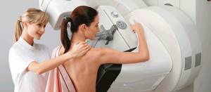
During operation of the mammograph, the breast is placed on a stand and pressed on top with a transparent plate. The mammary gland is compressed to allow images to be taken from different angles, but this may cause some discomfort to the patient. The radiating tube is designed so that X-ray radiation reaches only the mammary gland.
A procedure that includes two photographs in different projections, after which the patient is given a result, on the basis of which the mammologist makes a preliminary diagnosis.
Mammography studies
This type of examination can be carried out by both healthy women for preventive purposes, and those who have been diagnosed with various diseases of the mammary glands. Mammography allows you to determine pathology in the breast even at the beginning of its development.
According to the recommendations of oncologists, after reaching 30 years of age, a woman should undergo basic mammography for preventive purposes. After 40 years, such studies are recommended every 2 years, and after 50 years – annually.
Indications for mandatory examination are:
- Mammalgia;
- Seals in the form of nodules in the chest area;
- Nipple discharge;
- Suspicion of cancer.
When diagnosing cancer, research involves determining the stage of the disease, accurately identifying the area of the breast for puncture, and monitoring after surgery.
Before the examination, the woman should see a gynecologist. To take a high-quality picture, before the examination you should not use deodorants, apply talc or any cosmetics to the body, as they form a film on the body that does not allow you to take a high-quality picture.
The procedure is similar to an ultrasound examination. The chest is placed between the plates. To reduce the degree of radiation exposure and obtain an informative image, the procedure is performed with low compression. The examination may be accompanied by minor and short-term painful sensations. To complete the research, photographs are taken from two angles - straight and oblique. The photo is developed on a mammograph.
How to prepare for the procedure
The manipulation does not require special preparation. The reliability of the study does not depend on the functioning of the digestive system or blood composition. Accordingly, there are no recommendations prohibiting eating or drinking before the procedure.
On the day of a mammogram, it is advisable for a woman to refrain from using antiperspirants, talcs, and other antiperspirants. On photographs, their remains are sometimes displayed as dark spots, which the oncologist may mistakenly mistake for malignant formations. Before the procedure, the girl must remove any jewelry from her neck and raise her hair.
The doctor or laboratory assistant will first interview the patient, make sure that the woman is not pregnant, and clarify the phase of the menstrual cycle. To obtain the most correct interpretation of the received data, you need to bring the results of previously conducted examinations (ultrasound, MRI, and others). It is permissible to undergo other x-ray procedures at least every other day so as not to exceed the radiation dose to the body.
Price category of the examination
Many patients ask the doctor: “How much does a mammogram cost?” The price of the procedure can vary greatly depending on the region in which the manipulation is performed. Mammography is provided free of charge in government institutions. In this case, it is necessary to have appropriate indications.
Most often, a woman does an ultrasound, based on the results of which the doctor prescribes this procedure. Mammography is performed free of charge only upon referral. In this case, there may be a preliminary registration (queuing) for the examination.
How much does a mammogram cost on average? The price category in private clinics ranges from 3,000 to 7,000 rubles. It all depends on the device and the qualifications of the specialist who conducts the examination.
conclusions
When prescribing an examination, the doctor will be guided by your symptoms and other indicators, since some types of formations can only be examined with a certain type of examination: cysts are easier to detect on an ultrasound examination, and microcalcifications are well amenable to X-rays. For these reasons, it is impossible to say which is better - a mammogram or an ultrasound of the breast and mammary glands.
To summarize, it is worth saying that both methods are complementary and are often prescribed together in order to obtain the clearest and most clear picture of the condition of the mammary glands.
Brief summary
Now you know how mammography is performed (what it is and what it is needed for is described in the article). Choose the appropriate examination method yourself, but do not forget to listen to the advice of a specialist.
All results obtained must be provided to the doctor for evaluation and diagnosis. It is worth noting that mammography is an accurate and reliable examination method. However, in some cases, it is necessary to perform a biopsy of the tumor after it. Most often, this procedure is prescribed when a controversial situation arises regarding the nature of the tumor. In most cases, mammography can provide the specialist with all the necessary information to make a diagnosis and prescribe treatment.
Examine your mammary glands regularly, especially if there are indications for this. In this case, the pathology can be detected at the very initial stages of its development.
Comparison of methods
Taking into account all of the above, ultrasound:
- safely;
- informative;
- provides guaranteed results.
In turn, mammography:
- accompanied by x-ray radiation;
- causes discomfort;
- provides high image detail;
- in some cases it may not be reliable enough.
Thus, in the vast majority of cases, ultrasound is recommended. For some categories of patients, taking into account their characteristics, mammography is indicated.
At what age should you have regular breast health checks?
Girls and women over 18 years of age are referred for an ultrasound of the mammary glands in case of lumps or painful sensations in order to identify the nature of the pathology. The presence of the described symptoms is not necessary. Every year, women of childbearing age should undergo a preventive examination.
Frequency of ultrasound diagnostics
The described method for assessing the health of a woman’s breasts is painless and safe. The frequency of examination of patients is determined by the doctor. For preventive purposes, obstetricians-gynecologists recommend doing an ultrasound once every 2 years for women under 30 years old, and once for teenagers. Pregnancy and its planning are a reason for an unscheduled visit. With age, the frequency of diagnostics increases (30–45 years old - once a year, after 46 - every 6 months).
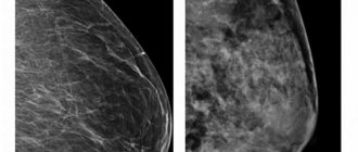
What does a mammogram show?
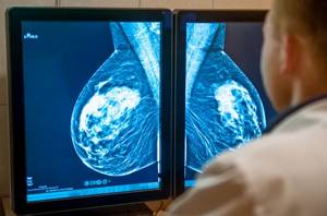
Studying the image, the doctor analyzes all possible changes in breast tissue. Pays special attention to tumor formations, as well as calcifications - bright white spots. It is not possible to find them using ultrasound.
Oncologists distinguish two types of calcifications:
- macrocalcifications
- large deposits of the element are usually formed in women over 50 years old and are associated with age-related changes in the body, injuries, and inflammatory processes. The formations are benign and do not require additional examinations; - microcalcifications
- tiny accumulations of mineral carry the risk of malignancy. Malignant inclusions differ from the safe form by localization. A biopsy is usually not required, but the patient will most likely need additional testing.
Mammography reveals benign formations:
- mastopathy of various forms - fibrocystic non-malignant changes of a hormone-dependent nature with or without atypical cells. Conditions require constant monitoring by specialists, tracking cell changes, treatment;
- Cysts are capsules filled with fluid and can be single or multiple. Simple ones do not pose a threat. If multi-chamber, large formations are detected, it may be necessary to clarify their status and analyze the contents of the cavities using a biopsy;
- papillomas inside the milk ducts are noncancerous growths. They are dangerous because they can ulcerate, bleed, and create favorable conditions for the onset of the inflammatory process;
- fibroadenoma is a benign tumor formed from healthy breast cells. When the formation reaches a large size, it is cut off surgically.
The main advantage of the mammographic method of hardware diagnosis of breast cancer is the ability to visually assess the number and structure of malignant cells. The procedure is often the starting point in identifying breast diseases and helps determine cancer treatment tactics.
What to choose, ultrasound or mammography?
Which is better, ultrasound or mammography of the breast
? At first glance, it may seem that ultrasound diagnostics is a more expedient and safer method of research. However, the order for the procedure is made by the attending physician. The specialist analyzes the data obtained during the examination and survey and selects a more appropriate diagnostic technique.
It is worth noting that over the age of 40, all women should undergo annual mammography for preventive purposes.
Pros and cons of procedures
Like all other diagnostic techniques, ultrasound and mammography have advantages and disadvantages.
Features and disadvantages of mammography
Mammography
involves x-ray examination of breast tissue. During the procedure, the patient receives dosed x-ray radiation through the examined area, which is retained in the tissues, which facilitates the visualization of information on x-ray film. Frequent use of this diagnostic method is contraindicated. Experts recommend conducting such a study no more than once a year unless absolutely necessary.
The advantages of this research method are:
- the ability to determine the development of benign and malignant neoplasms even of small size;
- identifying the condition of the mammary gland ducts;
- the ability to achieve good visualization of calcifications if they are present.
Flaws:
- Carrying out a mammography requires the woman to be forced to sit in front of the machine for some time, which can cause inconvenience.
- During the examination, slight pressure is applied to the chest (for better visualization of data), which causes discomfort.
- During diagnostics, although insignificant, X-ray exposure occurs. That is why frequent mammography is contraindicated.
- The results obtained are not always reliable, especially if the age of the subject is less than 35 years.
Features and disadvantages of ultrasound
Ultrasound examination of the mammary glands
carried out through exposure to ultra-high frequency sound waves. It is absolutely harmless and safe.
Advantages of ultrasound diagnostics:
- widespread;
- cheapness;
- high information content;
- harmlessness;
- Convenience for both the specialist and the patient.
The disadvantages include the short period in which it is advisable to carry out diagnostics.
What is more accurate, breast ultrasound or mammography? These two research methods are indicative, and the attending physician chooses one of them.
Types of research and equipment used
The X-ray examination method is highly accurate, specific, and informative. The results of mammography of the mammary glands form the basis for diagnosing organ pathologies.
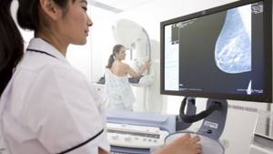
The set of equipment for the procedure consists of:
- mammograph;
- laboratory assistant workstation;
- digital detector;
- printer;
- doctor's workstation.
Devices are constantly being improved; modern equipment makes it possible to detect breast cancer at the very beginning of tumor formation from altered organ cells. Today, doctors detect up to 96% of breast pathologies in the early stages, compared to 80% diagnosed 10 years ago.
Radiologists use two types of mammographs:
- analogue (film)
- images of organs obtained by passing X-rays through tissue are transferred to film. Such devices are cheaper, but exclude the possibility of editing the image, transferring it to various media, or transferring it over the Internet for consultations with specialized oncologists; - digital mammographs
- display a “picture” on a computer monitor. It is saved as a graphic file. The devices operate at a reduced dose of radiation and allow you to quickly transfer images of the organ to the attending physician. It is convenient to work with them, zoom in or zoom in on the problem area, and make changes to the description. However, the clarity of a digital image is somewhat inferior to an analog photograph.
Tumor diagnosis is carried out in several ways and, if necessary, combined with other methods. For breast cancer, the doctor can use the following types of mammographic examination of the breast:
- with tomosynthesis – produces a layer-by-layer image of tissues, makes it possible to evaluate contours and comparative analysis of different projections of the “picture”;
- projection optical procedure - identifies malignant tumors, determines the type of their cells due to the effects of infrared radiation, provides two projections of images;
- with a biopsy attachment - designed for targeted examination of changes in the formation with the collection of suspicious tissues for histological analysis;
- tomographic optical - provides a three-dimensional image of the organ, outlines the specific location of the tumor, the main characteristics of oncopathology;
- luminescent procedure - involves the use of special marker substances that color pathological structures, which improves the visualization of malignant formations, as well as metastases;
- MRI mammography of the breast is considered one of the most informative methods for diagnosing breast cancer;
- radiothermometry - uses the penetrating ability of microwaves, the condition of cells is assessed based on temperature indicators of pathological tumor tissues. Together with the classical technique, radiothermometry detects breast cancer at any stage of the formation of the primary focus.
Mammographic diagnosis of breast cancer cannot be called completely safe, but modern devices allow the procedure to be carried out with minimal radiation exposure to the body. If you follow safety rules and frequency of manipulations, they will not cause harm to health. The diagnostic value of the analysis is very high.