“The results of an ultrasound of the mammary glands revealed a formation suspicious for oncology,” “the results of mammography do not clearly exclude the presence of a malignant formation,” “a biopsy showed the presence of cancerous changes in the mammary gland and an extensive examination is required.” These are just a few of the worst words a woman can hear from her doctor.
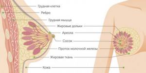
The mammary glands consist of three main types of tissue - adipose, connective and glandular. Breast cancer (BC) is a malignant tumor of the mammary gland that develops from glandular tissue cells. Contrary to popular belief, breast cancer affects both women and men, but it is approximately 100 times more common in women.
- How does breast cancer occur?
- Types of Breast Cancer
- Causes and risk factors
- Symptoms
- Self-diagnosis
- Diagnosis by an oncologist-mammologist
- Stages of breast cancer
- Breast cancer treatment
- Prognosis for breast cancer
How does breast cancer occur?
Breast cancer develops in the same way as any other malignant tumor. One or more cells of glandular tissue, as a result of a mutation that has occurred in them, begin to divide abnormally quickly. They form a tumor that can grow into neighboring tissues and create secondary tumor foci - metastases.
Mutations that lead to breast cancer can be hereditary or acquired.
Common hereditary genetic causes are mutations in the BRCA1 and BRCA2 genes. BRCA1 mutation carriers have a 55–65% risk of developing breast cancer, and BRCA2 carriers have a 45% risk of developing breast cancer. Such genetic defects are inherited from parents to children and cause cancer in approximately 15% of cases.
Much more often, breast cancer occurs due to acquired mutations: they arise in the cells of the gland and are not inherited. For example, in 20% of cases, the number of copies of the gene encoding HER2, a receptor protein that is located on the surface of cells and stimulates their reproduction, is increased.
The “molecular genetic portrait” of a breast tumor is important when choosing the optimal treatment.
Heredity
If a blood relative has ever been diagnosed with breast cancer, the girl is also considered to have an increased risk of the disease. At the end of the last century, researchers discovered genes that are responsible for the inheritance of breast cancer: BRCA1 and BRCA2.
With hereditary cancer, the disease is detected when a woman is only 40-45 years old, or even earlier. There is a high chance that cancer will affect both the right and left breast. In such patients, tumors are also detected in other organs and tissues, and not just in the chest. Also a characteristic feature is the identification of many lesions in the chest.
Prevention of cancer due to mutations of the BRCA1 and BRCA2 genes:
- oophorectomy surgery
- surgery to remove both breasts
- taking anti-estrogen medications such as raloxifene or tamoxifen
Types of Breast Cancer
Types of breast cancer are divided into two groups: ductal and glandular. Ductal cancer is more common. It can be intraepithelial (in situ) and invasive. Intracellular ductal breast cancer has a more favorable prognosis, it rarely metastasizes and is curable in 98% of cases. The invasive version of the tumor is prone to uncontrolled growth and generalization of the process.
Glandular cancer can be lobular (invasive lobular carcinoma) or grow from other cells of the glandular tissue. Lobular cancer is often characterized by multicentric growth. The rate of increase in size and timing of metastasis of forms of nodular breast cancer depend on the degree of differentiation of the breast tumor.
Prevention
Since the causes of the disease are not known, but only the risk factors, there is no 100% protection against breast cancer. But you can provide prevention, which will reduce your risk of illness. Women over 40 years of age need to have a mammogram twice a year. After age 50, breast examinations should be performed every 12 months. If there have been cases of breast cancer in your immediate family, then from the age of 35 you need to be screened for cancer every year.
Girls and women of any age need to regularly examine their breasts on their own. If you notice one or more of the symptoms described above, immediately make an appointment with a doctor, do not try to “cure yourself on the Internet.” You need to carefully plan your pregnancy, feed the baby naturally, and not from a bottle, as much as needed.
Also, preventive measures include monitoring your own weight. If you have diseases of the endocrine system, including diabetes, it needs to be treated on time. It is necessary to carefully monitor the breast health of those patients who have relatives diagnosed with ovarian and breast cancer.
You cannot take oral contraceptives on your own; you need to consult a gynecologist. Hormone replacement therapy is also prescribed by a doctor, because the dosage and regimen must be purely individual. If the patient is confirmed to have hereditary breast cancer, she should undergo a prophylactic mastectomy, surgically remove the ovaries, or take tamoxifen. But the decision, again, must be made by the doctor. Self-medication can only cause harm and significantly worsen the prognosis.
Causes and risk factors
Unfortunately, scientists do not yet have complete information about the causes of breast cancer. There is a list of risk factors that affect the likelihood of cancer, but some people are diagnosed with the disease in the absence of these factors, while others remain healthy even if many of them are present. Nevertheless, scientists still associate the occurrence of malignant breast tumors with certain circumstances that most often precede its appearance. These include:
- Age. The majority of breast cancer cases occur in women aged 55 years and older.
- Heredity. If breast cancer is diagnosed in one of your close relatives, the risk doubles.
- History of breast cancer.
- Increased breast tissue density as determined by mammography.
- Some benign neoplasms in the mammary gland.
- Early onset of menstruation - before 12 years of age.
- Late menopause - after 55 years.
- Lack of children or late (after 35 years) first birth.
- Exposure to radiation, such as from radiation therapy given to treat another type of cancer.
- Smoking and drinking alcohol. If a woman consumes 28–42 g of ethyl alcohol daily, her risks increase by 20%.
- Excess weight and low physical activity.
- Use of hormonal drugs: oral contraceptives, hormone replacement therapy in postmenopause.
- Breast injuries.
- Diabetes.
- Work on a schedule with night shifts.

Surveys
As already noted, palpation does not give a complete picture of the pathology. Other research methods are needed:
- examination by a qualified mammologist
- chest x-ray
- mammography
- computed tomography of the chest, brain and abdominal organs
- ultrasound diagnostics of the breast and regional lymph nodes
- cytology
- immunohistochemical study of the tumor
- consultation with a geneticist to detect mutations of the BRCA1 and 2 genes
Radioisotope examination of bones may also be necessary if there are appropriate indications. An ultrasound of the pelvic organs, liver and other organs in which metastases may have appeared is performed. Some patients undergo ECG. Consultation with other subspecialty doctors may be necessary, including a chemotherapy specialist, oncologist, and radiologist.
Mammography
Women over the age of 45 should have a breast exam every year. This makes it possible to detect cancer that is just beginning. Early diagnosis increases the chances of successful treatment and significantly reduces mortality from this terrible disease.
Symptoms
In the early stages, breast cancer usually does not manifest itself in any way. Most often, the tumor is discovered by the patients themselves or is detected accidentally during preventive studies.
Patients usually complain of the presence of a palpable formation and discharge from the nipple. Pain is a rare symptom of breast cancer, but pain syndrome can come to the fore at the stage of generalization of the process, especially when metastases spread to the bones.
Quite often, signs of breast cancer are detected, such as the appearance of asymmetry due to changes in the size of the affected gland. Reduction, upward displacement, deformation and wrinkling of the mammary gland can be observed in the scirrhous (fibrous) form of breast cancer. On the contrary, the breast on the affected side becomes enlarged due to the rapid growth of the formation or due to edema, which forms due to impaired lymph outflow.
When the tumor spreads into the subcutaneous tissue, skin changes may be observed. In this case, the following symptoms are revealed:
- “Platform” - the skin above the tumor becomes flattened; it is impossible to form a skin fold in this area.
- “Umbilization” - the skin of the mammary gland over the site of the lesion is wrinkled and retracted.
- “Lemon peel” is a characteristic appearance of the mammary gland due to lymphostasis.
Sometimes, when cancer spreads to the surface of the skin, signs such as redness and ulceration may be observed. The presence of these symptoms indicates that the process is neglected.
Changes in the nipple can also be detected, but only in the later stages. In this case, symptoms such as:
- Forga's sign - on the affected side the nipple is higher than on the healthy breast.
- Krause's sign - the nipple is thickened, the folds of the areola are noticeably pronounced.
Pathological discharge is a rather rare symptom, but in some cases it may be the only one detected during examination. The discharge is often bloody in nature, serous and purulent are less common.
Depending on the manifestations of the disease, different clinical forms of breast cancer are distinguished. In 75–80% of cases, the nodular form occurs. In the early stages, the only symptom is usually a painless lump in the breast. If you divide the breast into four parts by horizontal and vertical lines, then in half of the cases the tumor will be located in the upper outer part.
There are special forms of breast cancer with typical symptoms. These include:
- An edematous-infiltrative form, which is characterized by enlargement and swelling of the mammary gland, marbled skin color, and severe hyperemia.
- Mastitis-like is manifested by thickening of the affected mammary gland, increased body temperature.
- An erysipelas-like form, in which lesions are detected on the skin (sometimes ulcerations appear) that externally resemble erysipelas.
- The armored form is characterized by the presence of multiple nodes, due to which wrinkling and deformation of the mammary gland occurs.
- Paget's cancer affects the nipple and areola. It is characterized by thickening of the nipple, changes in the skin in the form of redness and thickening, and the formation of crusts and scales.
WHAT YOU NEED TO KNOW ABOUT BREAST CANCER
Breast cancer is the most common form of malignant tumors in women, accounting for about 20% of all malignant neoplasms. Every year, about 1 million cases of breast cancer are registered worldwide, and the number of cases is constantly growing. According to research, in Russia the largest number of breast cancer cases occurs in the northwestern part of the country.
WHY DOES BREAST CANCER OCCUR?
There are several theories explaining the occurrence of malignant tumors, but regardless of the root cause, the result is the appearance of pathological cells in the body. Every day in the human body many cells die, and new ones take their place. If a failure occurs during division, a new cell appears with altered structure and properties. Continuously dividing, such a pathological cell produces similar abnormal cells, a tumor is formed, which can subsequently “spread” pathological cells throughout the body (i.e. metastases ). It is known that the cause of the appearance of cancer cells lies in cell mutation, but it is difficult to determine what led to such a mutation in a particular case. Nevertheless, scientists were able to identify groups of factors that influence an increase or decrease in the risk of breast cancer.
HOW TO DETERMINE THE PROBABILITY OF DEVELOPING BREAST CANCER
Factors contributing to the occurrence of breast cancer can be divided into external and internal. An example of external factors is more than 2 thousand chemicals that have a carcinogenic effect. In addition, external factors include physical (ultraviolet radiation - excessive use of tanning! Radionuclides - carbon, potassium, radon), infectious (a number of viruses that contribute to the occurrence of cancer), etc. Internal factors include primarily hormonal disorders.
Statistical studies show that the risk of cancer increases with age, and the greatest number of unfavorable cases of breast cancer occur in patients 40-50 years old. Women under 30 make up only about 5% of the total number of breast cancer patients.
The main risk factors for developing breast cancer include:
- menopause over 50 years of age
- absence of childbirth or first birth over the age of 30 years (incidence 3 times more often)
- family history of breast cancer (if the sister or mother has a 2-fold higher risk, if both have a 6-fold higher risk)
- fibrocystic mastopathy (3-5 times more often).
Lifestyle risk factors for breast cancer:
- smoking
- ionizing radiation
- regular alcohol consumption
- overweight after 18 years
- refusal of self-examination, preventive examinations, mammography.
The possibility of developing malignant breast tumors increases if pathological processes occur in the tissues of the mammary glands. Basically, these are repeated dishormonal hyperplasias with the formation of foci of fibrocystic mastopathy. Often, ovarian diseases and repeated abortions lead to a number of hormonal disorders, which further cause the development of mastopathy, thereby increasing the risk of breast cancer.
Sometimes it is difficult to confidently distinguish a benign tumor from a malignant one or predict the possibility of degeneration. Therefore, benign tumors, breast fibroadenomas, should also be removed.
WHAT ARE THE FORMS OF BREAST CANCER
In 99% of cases in the mammary glands, malignant tumors develop from connective and glandular tissue (epithelial forms), and only in 1% - nonepithelial (sarcomas). From the point of view of cellular structure, malignant tumors of the mammary glands are most often classified as adenocarcinomas (i.e. tumors from glandular tissue). For the choice of treatment tactics, the state of estrogen receptors is of great importance, on which the nature of the course of the disease largely depends. Classification of breast cancer by estrogen receptors is based on their presence or absence. More than half of primary breast cancers are characterized by the presence of estrogen receptors, which are more common after menopause. Estrogen receptor-negative tumors are more common in premenopausal women.
In addition to the usual clinical forms, there are special forms of breast cancer. The mastitis-like form of cancer progresses very quickly. The mammary gland is painful and swollen (at the same time it sharply increases in size). The clinical picture is similar to acute mastitis, which makes diagnosis difficult.
Erysipelas-like form. A distinctive feature of this form of cancer is a sharp redness of the mammary gland (and sometimes the surrounding skin).
Armor cancer. Severe form of cancer. A characteristic symptom is lumpy thickening of the skin of the chest.
Paget's cancer (named after the English diagnostician) is a special form of damage to the nipple and areola. the tumor spreads deep into the gland and forms a cancerous node in its tissue with metastatic damage to the lymph nodes.
WHICH PART OF THE BREAST IS MOST OFTEN AFFECTED BY CANCER
The tumor can be located in different parts of the breast, but in the breast itself, in 50% of cases the upper outer quadrant is affected (quadrant = quarter circle). Bilateral breast cancer is rare (the second breast can be affected by either a metastasis from the first breast or a separate cancerous tumor). The risk of developing a malignant tumor is the same for both the right and left breast.
HOW DOES BREAST CANCER OCCUR?
The main characteristic of the clinical picture of breast cancer is a change in the position of the gland and the appearance of a palpable tumor with clear boundaries or a compaction with smoothed boundaries. The skin above the tumor flattens or retracts; an “orange peel” symptom is possible; in later stages, an ulcer may appear.
CHARACTERISTIC SYMPTOMS
Cancer pain may be completely absent. The most typical signs of breast cancer are flattening and retraction of the nipple and its areola, as well as bloody discharge from it.
HOW IS BREAST CANCER DETECTED?
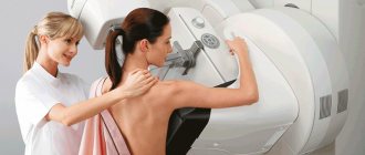
Today, the main method for diagnosing breast cancer is mammography . Regularly conducting this simple x-ray examination makes it possible to detect malignant neoplasms of the mammary glands in the early stages. Mammography using modern devices (mammographs) is a painless, but limited method of examination (a safe dose of radiation allows examination only 1-2 times a year).
To confirm the diagnosis, an ultrasound examination of the breast and puncture of the mammary gland are additionally performed. Breast puncture ( biopsy ) is the examination of tissue from a suspicious area of the breast. A small amount of tissue is taken using a thin needle (the patient feels only a slight prick) or a special device and examined in the laboratory, and a histologist or cytologist makes a conclusion.
It is also necessary to determine the prevalence of the malignant process, for which the lungs, bones and abdominal organs are examined using X-ray, tomography, ultrasound and other studies.
Practice shows that the overwhelming number of women turn to a specialist after they have independently discovered changes in the mammary gland during self-examination.
HOW TO TREAT BREAST CANCER
The complex of treatment for mammary glands includes three main components: surgical treatment, chemotherapy and radiation therapy. Surgical treatment can be limited only to the early stages of the disease. The choice of treatment depends on many factors: the type of cancer, the stage of the disease, the age and condition of the woman’s body, etc.
IS THERE A PREVENTION FOR BREAST CANCER?
It is impossible to prevent breast cancer 100%, but it is quite possible to eliminate many factors that contribute to the development of malignant tumors.
Timely pregnancy, childbirth and breastfeeding , regular visits to specialists and self-examination can significantly reduce the risk of breast cancer.
Breast cancer is a disease that is not only life-threatening, but also difficult for a woman to cope with. However, fear of this disease should not be a reason for you to avoid unpleasant thoughts, but an incentive for timely prevention of this insidious disease.
OMP doctor (polyclinic) I.S. Burlakova
Self-diagnosis
It is worth checking the mammary gland yourself for the presence of nodules or any other changes once a month after menstruation. It is more convenient to carry out home diagnostics while taking a bath or while under the shower. You should tell your doctor about any changes that are discovered as soon as possible.
Procedure for performing breast self-examination:
- Undress from the waist up and stand in front of a mirror.
- Raise your hands up and place them behind your head. Carefully examine the mammary glands. Turn right, left side.
- Feel the mammary glands while standing with your index, middle and ring fingers folded together. Start at the upper outer part of the chest and move clockwise.
- Pinch the nipple with two fingers. Check to see if anything stands out.
- Feel the mammary glands again - now in a lying position.
70% of breast cancer cases are self-diagnosed by patients through breast self-examination.
Diagnosis by an oncologist-mammologist
History taking
Diagnosing breast cancer begins with a conversation. At this stage, it is important for the doctor to evaluate the complaints and find out whether cases of breast cancer have occurred in the family, and if so, how often. This helps to suspect a hereditary form of the disease associated with mutations in the BRCA1, BRCA2, NBS1, CHECK, TP53 genes.
Breast examination
Next, the doctor examines, palpates the mammary glands, checks to see if there are any nodes or lumps in them, or if the lymph nodes in the axillary, supraclavicular and subclavian areas are enlarged.
Instrumental diagnostics
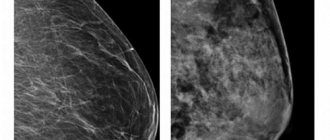
After the examination, the doctor may refer the woman for a mammogram, an X-ray of the breast. Indications for this study include lumps in the mammary gland, changes in the skin, bleeding from the nipple, as well as any other symptoms that may indicate a malignant tumor. Ultrasound examination of the mammary glands is also used. Mammography and ultrasound are complementary methods, each of them has its own advantages:
| Mammography | Ultrasound of the mammary glands |
| Allows you to detect pathological changes in the mammary gland 1.5–2 years before the onset of symptoms. If there is bloody discharge from the nipple, ductography can be performed - X-ray with contrast of the milk ducts. This helps to obtain additional useful information. High sensitivity - accurate diagnosis of up to 90% of cases of breast tumors. Ability to detect microcalcifications up to 0.5 mm. | Safety - there is no effect on the body from x-rays. Well suited for high density breast tissue in young women (up to 35–45 years). Allows you to distinguish breast cysts (cavities with fluid) from solid tumors. Allows you to assess the condition of regional lymph nodes. Well suited for monitoring needle position during biopsy. |
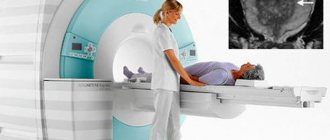
Magnetic resonance imaging is a highly informative method for diagnosing malignant breast tumors. It is used for lobular cancer, when mammography and ultrasound are uninformative, as well as for assessing the size and location of the tumor, which helps determine the tactics of surgical treatment. MRI can be used to screen women who carry abnormal genes associated with an increased risk of breast cancer and have a strong family history.
The final diagnosis is determined by the results of a biopsy. You can obtain breast tumor tissue in different ways:
- Examination of nipple discharge - tumor cells may be found in it.
- In a fine-needle biopsy, a needle is inserted into the breast tumor under ultrasound or mammography guidance.
- During a trephine biopsy (CORE biopsy), a special instrument resembling a thick hollow needle is used. It allows you to obtain more breast tumor tissue and examine it in more detail.
- In a needle-gun biopsy, a needle is inserted into the exact location using a special gun.
- A stereotactic vacuum biopsy is almost as accurate as a breast cancer biopsy during surgery, but it can be performed under local anesthesia without the need for general anesthesia. The procedure is carried out using a Bard Magnum pistol and a vacuum apparatus.
- An excisional biopsy is performed during surgery. The entire tumor along with breast tissue is sent for examination.
- Sentinel biopsy is an examination of the sentinel lymph node during surgery. It helps determine whether the tumor has spread to regional lymph nodes and whether they should be removed.
Euroonco doctor S.M. Portnoy talks about the role of biopsy in diagnosing breast cancer:
Cytological and histological examinations are carried out in the laboratory, where the structure of individual cells and tissues is assessed. Molecular genetic studies are currently available: they help to identify mutations that caused malignant degeneration and to select optimal antitumor therapy.
A biopsy can determine whether a tumor is cancerous, as well as determine its type and stage. In addition, examination of the biopsy material provides an answer to the question of whether the tumor is hormone-dependent, which also affects the treatment regimen.
Assessing the extent of cancer spread in the body
Once cancer is diagnosed, it is important to determine its stage and understand how far it has spread. The following studies are used for this:
- Ultrasound and lymph node biopsy.
- Computed tomography and MRI - they help assess the size, location of the breast tumor, and lesions in other organs.
- Metastases in the liver are diagnosed using ultrasound.
- X-rays can help identify lesions in the lungs and bones.
- PET scanning is the modern “gold standard” for diagnosing metastases of malignant tumors.

What is screening and who should undergo it?
Screening in oncology is a preventive examination that is recommended for all healthy people or those at high risk. This helps to detect cancer early and increase the chances of successful treatment.
As we have already mentioned, the risks of breast cancer increase with age. Therefore, all women over 45 years of age are recommended to undergo annual breast radiography - mammography.
At a younger age, you need to visit a mammologist once a year. During the examination, he can identify symptoms that the woman herself could not detect during self-examination of the mammary glands. If the doctor detects lumps in the breast, the first thing he will do is an ultrasound examination.
Our expert in this field:
Sergeev Pyotr Sergeevich
Oncologist, surgeon, chemotherapist, Ph.D.
Call the doctor Reviews about the doctor
What does breast cancer look like on an ultrasound? During this study, it is possible to identify a formation in the breast tissue, evaluate its shape, size, location, internal consistency, and number of foci. Ultrasound helps distinguish solid (dense) tumors from cysts - cavities with fluid inside. But the results cannot be used to judge whether the tumor is benign or malignant; this requires additional diagnostic methods.
Stages of breast cancer
Staging for breast cancer is based on the generally accepted TNM system. The T in this abbreviation indicates the size of the primary tumor:
- Tis is a “cancer in situ” that resides in the cells lining the milk ducts or lobules and does not invade adjacent tissue. This may be lobular, ductal, or Paget's carcinoma.
- T1—tumor diameter in greatest dimension is less than 2 cm.
- T2 - 2–5 cm.
- T3 - more than 5 cm.
- T4 is a tumor that has grown into the chest wall, skin, or inflammatory cancer.
The letter N indicates the presence of metastases in regional lymph nodes. N0 - there are no foci in the lymph nodes. N1, N2 and N3 - damage to different numbers of lymph nodes.
The letter M indicates the presence of distant metastases. One of two numbers can be indicated next to it: M0 - no distant metastases, M1 - distant metastases present.
Depending on the values of T, N and M, there are five main stages of breast cancer (within some of them there are substages):
- Stage 0: cancer in situ.
- Stage I: tumor in the mammary gland up to 2 cm in diameter.
- Stage II: a tumor in the mammary gland with a diameter of up to 5 cm or more, there may be metastases in the axillary lymph nodes on the affected side.
- Stage III: a tumor in the mammary gland up to 5 cm or more, can grow into the chest wall or into the skin, there are foci in the regional lymph nodes.
- Stage IV: The tumor can be of any size, it does not matter whether regional lymph nodes are affected. If distant metastases are detected, stage IV cancer is always diagnosed.
Breast cancer treatment
The treatment regimen for breast cancer should be selected individually for each patient, taking into account factors such as tumor type, stage, and sensitivity of the tumor to hormonal therapy. The general condition of the patient is also taken into account. If the tumor is detected in the early stages and the correct management tactics are chosen, then the chance of completely curing breast cancer is very high.
Surgical method
The surgical method is the dominant one in the treatment of breast cancer. If the tumor is detected early, it is possible to perform an organ-preserving operation—sectoral resection. Performing such an intervention is accompanied by an increased risk of local recurrence, and therefore it is combined with other methods, for example, radiation therapy.
In later stages, breast cancer is treated with a mastectomy—removal of the entire breast along with nearby lymph nodes. The doctor may also decide to remove the second breast if there is a high risk of developing a malignant tumor in it.
A sentinel biopsy, or sentinel lymph node biopsy, may be performed to determine whether cancer cells have spread to the lymph nodes and to determine the extent of surgery . During surgery, a radiopharmaceutical or fluorescent dye is injected into the tumor to help visualize the lymph node that first receives lymph from the breast tissue. It is removed and histological examination is performed. If no tumor cells are found in the sentinel lymph node, you can limit yourself to removing the lesion in the mammary gland. Otherwise, excision of regional lymph nodes is indicated.

Radiation therapy
In order to improve the immediate and long-term results of surgery for breast cancer, radiation therapy is used as an auxiliary method. It can be used in the preoperative period to reduce the degree of malignancy of the breast tumor, damage and regression of micrometastases. However, more often radiation therapy is used after surgery, when it is necessary to destroy cancer cells that may have remained in the body.
Chemotherapy
To treat breast cancer, chemotherapy is used before or after surgery with the aim of complete cure, prolonging life and improving its quality, or to reduce the volume of surgery. Each chemotherapy drug only affects cells in a specific phase of the cell cycle. Therefore, the most effective prescription is polychemotherapy - a combination of several drugs that have different effectiveness and mechanism of action.
Euroonko clinics use the most modern, original European and American chemotherapy drugs for the treatment of breast cancer. We have the opportunity to create a “molecular fingerprint” of the tumor and, based on its analysis, select the most effective and safe combinations of drugs.
Drug therapy: hormonal and targeted drugs
A malignant breast tumor is recognized as hormone-sensitive if more than 10% of the cells in its composition have receptors for estrogen or progesterone. In this case, hormonal therapy is used. The more the tumor has hormonal receptors, the more effective such treatment will be.
This type of therapy includes several methods that stop the production of hormones and block their flow. Today, drugs from the group of non-steroidal antihormones are increasingly used, which affect only the breast tumor and do not affect the mechanisms of hormone formation throughout the body. Therapy with hormonal drugs is prescribed both in the postoperative period to reduce the risk of recurrence, and as an independent treatment for inoperable breast tumors to control their growth.
Targeted therapy is also used to treat breast cancer—drugs that target specific target molecules in tumor cells and have minimal effects on healthy cells in the body. Targeted therapy is used alone or in combination with other treatments.
Prognosis for breast cancer
If breast cancer is diagnosed in the early stages (0-I), the five-year survival rate approaches 100%. That is, we can say that it is possible to cure almost all patients. Further, depending on the stage, the prognosis for five-year survival worsens:
- At stage II - 93%.
- At stage III - 72%.
- At stage IV - 22%.
The combination of breast cancer treatment methods available to oncologists allows in most cases to achieve remission of the disease or, at a minimum, prolong life while maintaining its quality. The effectiveness of treatment in general directly depends on whether treatment was started in a timely manner.
The maximum effectiveness of antitumor treatment is achieved only if it is carried out in accordance with international protocols and takes into account the individual characteristics of each patient. This is exactly how Euroonco oncologists prescribe treatment.
| More information about breast cancer treatment at Euroonco: | |
| Surgeons-oncologists-mammologists | from 5100 rub. |
| Removal of a breast tumor | from 92,000 rub. |
| Emergency oncology care | from 11000 rub. |
Book a consultation 24 hours a day
+7+7+78