An accessible, accurate and simple method for determining refraction is skiascopy, otherwise called shadow testing, kerato- and retinoscopy.
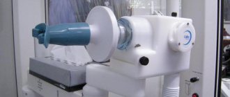
It is carried out using a special device called a skiascope.
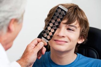
It is a round mirror with a handle with a flat and concave side. In the center of the device there is a hole for the ophthalmologist to observe the patient's eye being examined.
This technique is widely used in children who cannot talk about their symptoms and complaints, people with impaired intelligence and patients who deliberately feign refractive errors.
Indications
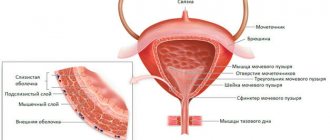
During skiascopy, the pupil is illuminated directionally and the clinical refraction is determined, i.e. the ability to focus light beams strictly on the retina. If this is observed, then they speak of normal or one hundred percent vision. Otherwise it is called emmetropia. When the focusing position of the light beams relative to the retina changes, myopia or farsightedness is diagnosed: in the first case, the intersection point is located in front of the retina, in the second - behind the retina.
Thus, the technique makes it possible to determine pathological changes in visual acuity and clinical refraction. Indications for skiascopy are:
- the need to assess refraction in children, even very young ones;
- complaints of poor vision of nearby objects (farsightedness);
- complaints of good vision of nearby objects and blurring of distant objects (myopia);
- astigmatism, or a combination of several types of refraction in one eye (plus and minus);
- the need to assess the state of visual acuity and refraction in people after surgery, in the postpartum period in a woman in labor at risk of visual impairment due to increased intraocular pressure, hemorrhage in the eye.
The technique is used when undergoing preventive examinations once a year for working people, pregnant women, for admission to educational institutions, etc. It is also an alternative way to determine the type of refraction in the absence of ophthalmological tables and special devices and for checking children.
Contraindications
Skiascopy involves cycloplegia, or artificial paralysis of the ciliary muscle of the eye using special eye drops. They can cause temporary blurred vision, pupil dilation, and increased intraocular pressure. This imposes certain restrictions on the use of this technique to determine the type and magnitude of refraction:
- glaucoma and suspicions of it;
- intolerance to drugs from the cycloplegic group (Cyclomed, Atropine, Cyclopentolate);
- the patient is drunk, under the influence of narcotic and psychotic substances;
- inappropriate behavior and aggressiveness of the patient, mental disorders;
- fear of bright light, or photophobia, which is characterized by increased tearing.
If there are contraindications or concerns, it is possible to perform skiascopy without the use of drops that lead to paralysis of the eye muscle.
Prices
| Skiaskoscopy | 200 rub. |
All prices
Skiascopy is a method for checking the functional state of the eyes, the essence of which is to study the refractoriness of the eye. Two structures are responsible for the refractoriness of the eye - the cornea and the lens. Using this procedure, it is possible to determine the degree of impairment even if a person is simulating a disease, or in a child, or in a mentally retarded patient, when it is impossible to determine visual acuity using isometry or refractometry. Synonyms for skiascopy are shadow test, keratoscopy and retinoscopy.
During the examination, the child’s answers often cannot be assessed unambiguously, since he is not able to accurately understand and determine the difference in the diopters of the glasses the doctor tries on him. Thus, the method is built on the desire to select the correct corrective agent while eliminating errors that could have been made due to objective reasons.
Preparing for the study
For correct results and accurate determination of refractive errors, the patient’s preparation for the study and the professionalism of the ophthalmologist are necessary. To do this, cycloplegia is performed through ocular instillation of special drugs, which causes temporary paralysis of the ciliary muscle of the eye. The drugs are instilled 3 days before the study twice a day and an hour before the procedure. The exact dosage and number of instillations is prescribed strictly by the doctor.
Medicines used for cycloplegia are dispensed strictly according to a doctor’s prescription due to strong side effects and the undesirability of use without a specialist’s prescription. They are prescribed in childhood before the first procedure and in difficult cases. These include Atropine eye drops.
After instillation, pupil dilation is observed, headaches, increased heart rate, increased intraocular pressure, weakness, and nausea are possible.
If the results are controversial, the use of drops is extended to 7-10 days and the skiascopy procedure is repeated.
Rapid cycloplegia is caused by drugs with Cyclopentolate (Cyclomed, Cyclopentolate-Solopharm, Cycloptik), Tropicamide (Tropicam, Midriacil). They are used three times with an interval of 10-15 minutes, then they begin to check the refraction.
Since medications can provoke an increase in intraocular pressure, patients over 40 years of age, people with a family history, or a predisposition to glaucoma first measure IOP indicators, then decide on the advisability of using these drugs.
Carrying out skiascopy
For a full examination of the central and peripheral areas of the pupil, cycloplegia is used. The patient is then seated in a chair in a darkened room with a light source or lamp placed to the side at ear level. At a distance of 67 cm for children or 1 m for adults, an ophthalmologist sits opposite the examinee with a skiascope device. It is a mirror with a hole in the center, a handle and two sides (concave and flat).
The specialist uses a mirror to direct beams of light reflected from the lamp into the patient's eye, which enters the pupil through the fundus.
The direction of gaze depends on the performance of cycloplegia or lack of preparation. In case of muscle paralysis, the patient should look at the center of the device, without instilling special drops - past the specialist’s ear.
Afterwards, the specialist moves the skiascope along the horizontal and vertical axis of the handle, the device faces the flat mirror side towards the patient. With such movements, a dark, clearly defined spot or shadow is formed. The nature of refraction is determined by the side of movement of the shadow area - farsightedness or myopia. The magnitude of deviations is determined using skiascopic rulers or corrective lenses with different optical powers (diopters).
Features of skiascopy in children
The skiascopy technique to determine refraction is performed for the first time at the age of 1 month, in the presence of complications - no later than 3 months from birth. To monitor the dynamics of refraction, a second appointment with an ophthalmologist is made at 3 and 6 months. At this time, the eye muscles are not developed, and farsightedness ranges from +1 to +3 diopters.
To conduct the examination, cycloplegia is performed by instilling short-acting drugs (Midriacyl, Tropicamide) or Atropine. Then, after the effect of the medication and the established temporary paralysis of the optic nerve, the patient is placed at a distance of 67 cm from the ophthalmologist. A specialist is responsible for moving and holding skiascopic rulers.
Skiascopy - what is it in ophthalmology?
Skiascopy or shadow test is a technique for assessing the function of the optical system of the eye, which is based on the doctor’s observation of shadows moving in the pupil when light rays reflected from mirrors hit them.
Rotations of the mirror result in the appearance of a moving shadow. Its position in the pupil depends on the refraction of the eye being examined. Skiascopy is an informative and inexpensive technique. Refractive errors can be diagnosed objectively, even if the child does not say whether he or she can see better or worse with the lenses. The approach requires experience, knowledge and time from the doctor. But in the end, a pediatric ophthalmologist, using skiascopy, can select glasses so that your child has visual acuity as close as possible to 100%.
Decoding the results
To determine and interpret the results, you need a skiascopic ruler or special lenses used in the selection of corrective glasses. If there is no shadow when reflected beams of light enter the eye, then myopia, or myopia up to 1 diopter, is diagnosed.
With greater myopia, the shadow shifts against the direction of movement. To determine the deviation, a lens with minimal optical power or a ruler with negative diopters is applied and changed to a stronger one until the shadow disappears. To determine the indicator, add 1 diopter to the optical power of the last lens and obtain the deviation value.
In case of farsightedness, a shift of the shadow in the direction of movement is noted and a ruler or lens with positive diopters is taken. The selection also begins with the minimum optical power. Further actions are identical. When the shadow disappears, to determine the amount of farsightedness or hypermetropia, one is subtracted from the optical power readings.
With complicated astigmatism, the nature of the shadow movement is multidirectional, the angle changes. Additional examinations are needed to interpret the results.
The most accurate results are obtained with extensive experience of the ophthalmologist and preparation for the examination, which consists of the use of cycloplegics.
Cylindroskiascopy
To perform cylindroskiascopy, a conventional skiascopy is first performed using spherical lenses (or rulers). The approximate position of the main meridians for an eye with astigmatism is determined, as well as the strength of the lenses that neutralize the shadow in each eye. The patient is put on a trial spectacle frame, and lenses (spherical and astigmatic) are installed in the socket opposite the eye being examined, which should simultaneously neutralize shadows in the two main meridians. Skiascopy is carried out by turning the mirror in the direction of the axis of the astigmatic lens, then in the direction of its active section. If in both cases the shadow disappears, then neutralization of ametropia has been achieved. If the shadow disappears only in the direction of the axis of the astigmatic lens, but does not disappear in the direction of its active section, the cylinder is either weakened or strengthened until the shadow disappears completely. In the case when the shadow does not disappear in both directions, first make it disappear in the direction of the axis of the astigmatic lens by selecting a sphere, then in the perpendicular direction by selecting a cylinder.
When the shadow moves outside the direction of the axis of the astigmatic lens and its active section, at an angle of 45° between them, it means that the axes of the cylinder are not aligned correctly. To eliminate this, the cylinder is rotated in the frame, ensuring that the direction of movement of the shadow coincides with the direction of the axis. Having achieved neutralization of the shadow in two sections, the spherical lens is weakened, reducing the positive or enhancing the negative lens, taking into account the distance from which skiascopy was carried out: at a distance of one meter, the correction will be 1.0D; at a distance of 67 cm - 1.5D, at a distance of 50 cm - 2.0D.
By contacting the Moscow Eye Clinic, each patient can be sure that some of the best Russian specialists will be responsible for the results of treatment. The high reputation of the clinic and thousands of grateful patients will certainly add to your confidence in the right choice. The most modern equipment for the diagnosis and treatment of eye diseases and an individual approach to the problems of each patient are a guarantee of high treatment results at the Moscow Eye Clinic. We provide diagnostics and treatment for children over 4 years of age and adults.
You can find out the cost of a particular procedure or make an appointment at the Moscow Eye Clinic by calling 8 (800) 777-38-81 (daily from 9:00 to 21:00, free for mobile phones and regions of the Russian Federation) or using the form online entries.
Useful video
The clinic doctor talks about static and dynamic skiascopy, features of its implementation in adults and children.
Author's rating
Author of the article
Alexandrova O.M.
Articles written
2100
about the author
Was the article helpful?
Rate the material on a five-point scale!
( 1 ratings, average: 4.00 out of 5)
If you have any questions or want to share your opinion or experience, write a comment below.