Raeography of the head (REG)
Posted at 23:55h in Services by doctor
Rheoencephalography of cerebral vessels is a simple but effective diagnostic method. As a result of this procedure, pathological processes such as circulatory disorders, as well as other deviations from the normal functioning of this important organ, are identified.
The method is popular with patients and doctors. This is explained not so much by the affordability of the survey, but by its high information content and the ability to quickly obtain accurate results.
A big advantage over other methods of examining cerebral vessels is its minimal invasiveness, which becomes a factor favoring the use of this diagnostic even for pediatric patients.
Prices
| Name of service (price list incomplete) | Price |
| Appointment (examination, consultation) with a neurologist, primary, therapeutic and diagnostic, outpatient | 1750 rub. |
| Consultation (interpretation) with analyzes from third parties | 2250 rub. |
| Prescription of treatment regimen (for up to 1 month) | 1800 rub. |
| Prescription of treatment regimen (for a period of 1 month) | 2700 rub. |
| Consultation with a candidate of medical sciences | 2500 rub. |
| Transcranial duplex scanning (TCDS) of cerebral vessels | 3600 rub. |
General information about the method
Rheoencephalography (REG) allows you to identify circulatory disorders in the brain even in the early stages of pathology and thereby prevent the possibility of developing complications that pose a danger to the health and life of patients.
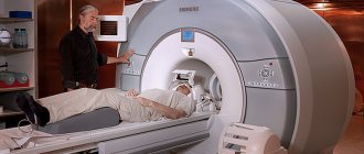
Its invaluable advantage over MRI and CT is the ability to be examined without waiting in line, which in other places takes about six months. Without detracting from the effectiveness of magnetic resonance and computed tomography, it should be noted that timely treatment is the key to victory over the disease, and in some cases, the ability to save the patient’s life.
What kind of procedure is this, who needs it, how to prepare for the examination - these are the questions that will be discussed in the article.
For what purpose is it carried out?
In addition to procedures related to the need to study pathological changes in the arteries and vessels of the brain, it is advisable to conduct REG for preventive purposes.
Operating principle of the device
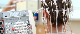
The essence of rheoencephalography is that with the help of a special device - a rheograph - an electric current of low frequency is passed through the brain, as a result of which the resistance of brain tissue is visualized on the monitor. In this way, disorders in arteries, veins and small vessels are detected .
The presence of six channels in the device makes it possible to simultaneously examine several areas of the brain.
In the projection of the studied areas, metal electrodes are installed using an elastic rubber band, which transmit the image to the monitor.
Main disadvantages of the survey
The development of medicine and related sciences is not constant - new devices and technologies appear to identify problems, including vascular ones. REG is not considered the newest of them, but all modern technologies are expensive and unaffordable for the majority of the population. Not every hospital has the funds to purchase expensive equipment and train a doctor. Consequently, queues for examinations are created in modern diagnostic centers, and sometimes the appointment lasts for many months. But what should people do in such a situation who need regular monitoring of changes in blood vessels?
In case of diseases (epilepsy), REG will not provide a complete picture and is used only as a secondary study that complements any others, for example MRI, ultrasound, EEG.
Many opponents of rheoencephalography, as an argument against this research, say that the results of the procedure cannot be accurate because the bones of the skull are unable to conduct electrical current. Deep vessels will thus remain unexplored. However, the bones of the skull have a so-called “capacitive reactance”, which does not interfere with the operation of current pulses at all.
Tags: REG
When is an REG prescribed?
- patient complaints of dizziness;
- deterioration of condition with changes in atmospheric pressure;
- osteochondrosis;
- noise in ears;
- debilitating headaches;
- suspicion of ischemic disease;
- memory losses;
- weakened vision;
- hearing loss;
- atherosclerosis;
- hypertensive crisis;
- dystonia;
- hypertension of the cerebral arteries.
For all pathologies associated with a violation of the condition of blood vessels - their blood supply, changes in blood flow speed and viscosity, it is necessary to conduct an REG.
What the study shows
- Based on rheoencephalography of the vessels of the head, specialists receive significant information about the condition of the object of examination. Among them is the possibility of studying vascular tone, their elasticity, blood circulation speed and blood inflow/outflow.
- The use of rheoencephalography makes it possible not only to identify abnormalities in the vessels of the brain, but also to monitor blood flow after complex operations or severe injuries.
- With the help of REG, various pathologies are detected, and the severity of the pathological process is established.
In this case, the high speed of obtaining results is of no small importance.
What problems are being identified?
- presence of traumatic brain injuries;
- localization of hematomas formed as a result of head trauma;
- pre-stroke condition;
- damage to blood vessels by atherosclerotic plaques (atherosclerosis);
- thrombus formation in the vessels of the brain;
- predisposition to increased blood pressure;
- diseases associated with circulatory disorders.
The procedure facilitates the task of making an accurate diagnosis, on the basis of which the doctor prescribes an adequate course of treatment. With the help of it, he subsequently monitors the effectiveness of therapy.
Due to the complete safety of such an examination for the patient’s health, it can be performed repeatedly.
One of the most significant advantages of encephalography is the ability to distinguish between pre-stroke indicators, which have certain differences for men and women.
Other features of the method
Specialists obtain even more information by conducting functional tests.
The simplest and most accessible of them is with nitroglycerin. This substance helps reduce vascular tone. This test is used to differentiate organic and functional disorders.
Cost of rheoencephalography
Equipment for conducting REG is available in many clinics, hospitals, and diagnostic centers. If you sign up for the procedure in advance, it can be done for free. Otherwise, one session will cost from 1000 to 3500 rubles, depending on the medical institution and the need for functional tests. Some centers provide the service at home, but in this case its cost will be at least 10,000 rubles.
Despite the increase in the number of specialists who are skeptical about the approach, REG of the brain is still actively used in diagnosing problems with the vessels of the central nervous system. Safe and accessible manipulation can not only identify existing problems, but also warn of potential risks. In particular, it is recommended that older people suffering from hypertension or atherosclerosis undergo it at least once a year.
How to decipher the results
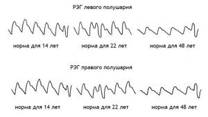
When assessing the examination results, the patient’s age must be taken into account. This is explained by the fact that the walls of blood vessels lose their elasticity over the years, become more fragile, and react differently to various stimuli.
An REG shows graphical wave fluctuations. The following indicators are taken into account:
The specialist reads the diagnostic results, taking into account the regularity of the waves, the appearance and rounding of the apex, as well as the location of the tooth and incisura.
The rate of oscillations of the wave depicted on the screen in adults differs from the manifestations of acceptable indicators in a child.
Rheoencephalographic study makes it possible to classify the condition of blood vessels according to three types of their behavior:
- Dystonic. Characterized by frequent manifestations of changes in vascular tone. Hypotonia with difficulty in venous outflow of blood and low pulse filling is more often observed.
- Angiodystonic. Its symptoms are similar to those of the previous type. The difference is that the cause of the tone disorder is a defect in the vessel wall.
- Hypertensive type according to REG. Significantly different from the species described above. Vascular tone is significantly increased. Venous outflow is impaired.
These types of behavior are not independent pathologies. They are only signs of other diseases and make it possible to identify them in the early stages of development.
You should not attempt to decipher the examination results yourself. It is better to leave this to qualified doctors who will do it professionally and establish an accurate diagnosis.
Decoding
When analyzing the rheogram, the data obtained as a result of the examination, as well as age, health status and chronic diseases are taken into account. The graph that appears on the screen or paper tape when examining a teenager will be different from the graph of an adult. In the first one, it looks like a series of uneven waves with clearly defined periodic peaks. In an adult, it looks like a straight line with evenly spaced vertices. The graph shows anacrotes, catacrotes and incisuras.
Anacrota is a wave that shoots sharply upward with a slightly rounded top. Catacrota is a wave that goes down smoothly. An incisura is a notch between a downward wave and a tooth that anticipates the next wave.
Decryption is carried out only by a specialist. During the analysis, the regularity and type of waves, features of the rounding of the top, the dicrotic tooth located after the descent of the wave, and the position of the notch are assessed. Based on the results of the analysis, the doctor determines the signs of pathologies:
- With the hypotonic type, the waves have sharp, high peaks, the tooth or additional waves are displaced, the incisura and amplitude are increased.
- The hypertensive type is characterized by prolonged anacrosis with displaced waves, additional waves are possible. The amplitude is reduced and uneven. The teeth have different shapes.
- Cerebral atherosclerosis is indicated by flat tops, soft vibrations, and the absence of additional waves on the catacrota. In severe pathology, the waves have a dome shape.
- Spasm of the walls is characterized by round peaks.
- With stagnation of venous blood and difficulties in its outflow, increased catacrota and a large number of small waves between the main ones are observed.
- Angiodystonia is manifested by floating teeth and waves on the catacrota.
How is the procedure performed?
The described diagnostic method is completely painless and safe. During the procedure, there is no impact on the patient’s skin, and no various instruments are used.
During the procedure, the patient is placed on a couch or offered to sit on a chair. To obtain more accurate information, the patient is asked to tilt his head forward, turn it to the right or to the left.
The procedure lasts 10-15 minutes. The results of the study are displayed immediately on the monitor screen and are assessed by a neurologist.
Recommendations
To avoid distorting the results, you should consider some simple tips:
- Before installing the electrodes, some areas of the head are treated with alcohol. It is advisable not to stress and take it calmly.
- Eyes should be kept closed during the procedure.
- You need to completely relax. Anxiety can cause a sharp constriction of blood vessels. This will affect the wave oscillation performance.
- It is advisable to rest for 15-20 minutes before the procedure.
- The day before the scheduled examination, you should not take medications that can affect the speed of blood flow.
- The session should not be interfered with by any objects, so you need to remove chains, earrings, hairpins and let your hair down.
If you are examining a small child, you should tell him in advance everything about the upcoming procedure. You can pick him up and sit on a chair with him. Then he will not be afraid and nervous.
How to prepare for REG of cerebral vessels
No special patient preparation is required. If the subject is taking medications that affect vascular tone, then he will have to give them up for a while - the timing is agreed with the doctor. On the day of the study, you must refrain from smoking, otherwise the narrowing of the blood channels under the influence of nicotine may lead to incorrect results. Half an hour before the session, you need to relax and calm down, so that anxiety does not cause the blood vessels to narrow and spoil the picture. At the same time, sedatives are used extremely rarely at the preparation stage; they can also reduce the information content of the method.
About contraindications
Due to the absolute absence of harm to the body, rheoencephalography has virtually no contraindications or side effects.
Such examination is contraindicated for newborn children . This is explained by the small amplitude of the reflected waves, the large size of the anacrote and the complete absence of incisura. Such readings do not provide an accurate picture of the condition of the vessels of the head.
Rheoencephalography is an effective and affordable method for examining cerebral vessels. Its widespread use is due to the presence of the device in every hospital and, of course, the absence of side effects and contraindications for use.