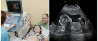
Ultrasound of the cervix is a standard examination during pregnancy, allowing to exclude the threat of premature birth. The purpose of the procedure is to determine the length of the cervix: it is this indicator that allows you to judge whether there is a risk for the mother and child.
The cervix holds the fetus inside the body. During pregnancy, it gradually shortens and opens, allowing the woman to give birth. However, if the length of this organ was less than normal from the very beginning, then this threatens not only premature birth, but also miscarriage.
Indications for ultrasound of the uterus
The content of the article
Ultrasound of the uterus
is a diagnostic examination performed to determine the anatomy, size, structure and condition of a woman's reproductive system. This procedure is usually carried out as a screening test during pregnancy and outside pregnancy to identify diseases and monitor treatment results. Ultrasound is completely painless and has no age restrictions.
Ultrasound of the uterus is prescribed for the following problems with women's health:
- irregular menstrual cycle;
- amenorrhea – complete absence of menstruation;
- prolonged menstruation;
- bleeding outside the cycle;
- copious mucous or purulent discharge with a pungent odor;
- discomfort during sexual intercourse;
- confirmation/denial of pregnancy;
- the course of pregnancy;
- inability to conceive or bear a child;
- treatment of infertility using hydrotubation;
- installation/removal of the spiral;
- pain, stinging, burning when urinating;
- suspicion of inflammatory processes;
- examination of the uterus after surgery;
- prolonged nagging pain in the lower abdomen;
- after removal of the cervix and its part;
- monitoring the postpartum condition;
- suspicion of the presence of neoplasms and clarification of their characteristics.
The procedure is also performed when the uterus has a non-standard structure and the doctor is unable to see all the features using mirrors.
Indications for use
Transvaginal ultrasound facilitates diagnosis and allows you to identify pathologies and lesions of the pelvic organs. A referral for testing can be issued by a gynecologist, reproductive specialist or surgeon.
The main indications for transvaginal ultrasound of the pelvic organs are:
- pain in the lower abdomen (any duration, frequency and character);
- suspicion of gynecological pathology;
- abnormal uterine bleeding and painful menstruation;
- disruptions in the menstrual cycle, pathological changes in the duration of menstruation;
- fertility assessment and infertility therapy;
- preparation for pregnancy and IVF procedure;
- diagnosis of early pregnancy;
- planning gynecological operations;
- monitoring the condition of the pelvic organs after gynecological manipulations (including after termination of pregnancy);
- tracking dynamics during therapy for gynecological diseases;
- suspicion of the development of neoplasms in the pelvic area;
- the presence of inflammatory and purulent-inflammatory processes;
- for choosing a contraceptive method, in some cases - its installation and control;
- premenopausal and menopausal periods;
- periodic preventive examination.
Additionally, experts advise every woman to undergo a transvaginal examination procedure at least once a year. This will allow you to identify diseases at an early stage of development and prevent the development of complications.
Types of ultrasound of the uterus: how they are performed, features, advantages
Ultrasound of the uterus and appendages can be performed in 4 ways. It is imperative that you check with your doctor which method will be used in order to take this into account when preparing for the procedure. It happens that a gynecologist recommends examination using two methods at once. Do not refuse - given the effectiveness, harmlessness and low cost of the study, this is the best option.
Transabdominal ultrasound of the uterus
The most comfortable procedure, but less informative when clarifying the nuances than other methods. Therefore, this technique is prescribed as a primary examination.
A huge advantage of the method is that with transabdominal ultrasound, a gynecologist can examine the entire genitourinary system and understand how the pathology is connected to other organs.
During a transabdominal examination of the uterus, the woman needs to lie on her back and expose her abdomen. The gynecologist applies a special gel to the area under study for better contact with the sensor and conducts the study with a special sensor. This method is ideal for pregnant women and girls who have not had sexual intercourse.
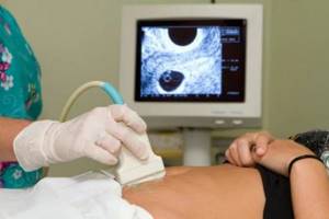
Transvaginal ultrasound of the uterus (TVU)
This technique is considered the standard for examining the uterus and appendages.
The advantage of transvaginal ultrasound of the uterus is that the examination is aimed specifically at the desired organ, and the sensor is located in close proximity to the uterus.
A woman needs to expose her lower body and take a lying position on a couch or gynecological chair. The doctor inserts a special vaginal sensor into the vagina to a depth of 5-7 cm, scanning the uterus. A condom is put on it, which completely protects the patient from infection. Contraindications to this type of ultrasound are virginity and pregnancy.
Transrectal ultrasound
This procedure is used less frequently and only when indicated. Using rectal ultrasound, it is possible to identify neoplasms associated with neighboring organs, etc.
The patient also exposes the lower part of the body, but lies on her side. During this test, a probe is inserted into the anus, scanning the uterus through the intestinal wall. The procedure does not cause discomfort, since the gynecologist uses a narrower probe than for TVU. The doctor also puts on a condom and lubricates it with gel to improve glide and reduce discomfort.
Contraindications: anal fissures, hemorrhoids, inflammation in the intestines, neoplasms of the colon.
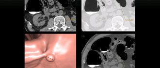
Intrauterine ultrasound
This method is the most informative, but it differs from the others and has some difficulties in execution, so it is used in special cases.
Intrauterine ultrasound is performed as follows:
- The woman undresses and lies down in the gynecological chair.
- The gynecologist opens the cervix using a speculum, then inserts a catheter for saline, which will help straighten the endometrial folds and provide a better examination. After the fluid is injected and the uterus is filled, the speculum is removed and the sensor is inserted directly into the uterine cavity.
- To assess the patency of the fallopian tubes, a saline solution with air is injected into the uterus and the movement of the bubbles is monitored.
The duration of the procedure is about 30 minutes. Intrauterine ultrasound is rarely prescribed, mainly when uterine fibroids and endometrial changes are suspected.
Methods of conducting research
The procedure can be in several options:
- Without penetration into the vagina, through the skin of the perineum. Prescribed to girls, virgins, patients with fused vaginal walls (atresia) in case of suspected abnormal development of the cervix.
- With the penetration of an ultrasound device into the vagina (transvaginally). It is prescribed to women who are sexually active, as well as at the beginning of pregnancy and in the later stages to determine the degree of maturity of the cervix.
- With penetration into the rectum (transrectal). Prescribed to virgins for a more thorough examination of the reproductive organ.
- Without penetration into the vagina, through the walls of the abdomen. This diagnostic option is applicable for virgins, pregnant women and in the presence of pathologies of vaginal development.
How to prepare for a uterine ultrasound procedure
Preparing for a uterine examination is simple and does not require much effort. By following the recommendations of the gynecologist, you can protect yourself as much as possible from discomfort and unpleasant sensations. Features of preparation for ultrasound examination depend on the method in which it will be carried out.
Preparing for a transabdominal ultrasound
The day before the procedure, you need to change your diet to reduce gas exchange as much as possible: cabbage, beans, carbonated drinks, nuts, black bread and other gas-forming foods should be excluded.
The bladder should be full. The patient is offered the choice of either drinking 1 liter 1 hour before the examination. liquids without gas, or do not urinate for 2-3 hours.
A full bladder will help make the picture clearer and provide a convenient location for the organs to be examined.
Preparing for a transvaginal ultrasound
Preparatory measures include emptying the bladder immediately before the procedure. To improve the quality of the examination, it is recommended to empty the intestines of gases the day before using special medications (Smecta, Espumisan, etc.).
Preparing for transrectal ultrasound
A woman needs to empty her rectum 6-8 hours before the examination using one of the following methods:
- small enema with cool water;
- microenema - quick-acting rectal suppository (sold in a pharmacy);
- inserting a glycerin suppository into the anus (can also be purchased at a pharmacy);
- laxative.
It is better to use modern drugs, for example, guttalax. Do not use products containing senna and other components that promote the formation of spasms.
Preparing for intrauterine ultrasound
This type of examination does not imply any preparatory procedures. Immediately before the ultrasound, you need to empty your bladder so that the organ does not put pressure on the uterus
Our advantages
By contacting our center for an ultrasound scan of the uterus and appendages, you can get:
- Accurate results To conduct ultrasound of the ovaries and uterus, modern equipment from General Electric, one of the leading manufacturers of medical equipment, is used. Thanks to this scanner, the image of the uterus and ovaries is very clear, and the professionalism of doctors allows you to accurately decipher the result;
- Quick examination. The patient undergoes the procedure strictly according to the appointment time. You do not have to wait in line and return to the clinic for the result - all data on the condition of the uterus and ovaries will be entered into the medical record within 10-15 minutes after the examination;
- Budget savings. "Alfa Health Center" is a large network of private clinics. We always offer affordable prices for ultrasound of the appendages and uterus and other procedures. Our clinic regularly holds promotions. This makes prices for ultrasound of the uterus and appendages even more profitable;
- Comprehensive services. After receiving the results of the procedure, you can immediately make an appointment and undergo examination by specialized doctors. All manipulations for the diagnosis and treatment of diseases of the uterus and ovaries can be carried out in our clinic.
What does an ultrasound of the uterus show?
Ultrasound examination of the uterus is a very effective procedure that detects dozens of diseases and pathologies. With its help, you can diagnose in a timely manner:
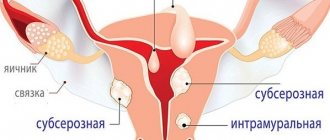
- leiomyoma – benign smooth muscle tumor;
- uterine cancer is a malignant tumor;
- adenomyosis - a benign formation (germination of the inner layer of the uterus (endometrium) into the thickness of the uterus itself);
- endometrial polyps are benign neoplasms inside the uterine cavity;
- cervical polyps - benign neoplasms in the cervical canal (warts);
- cysts - tumors with blood or other fluid inside;
- synechia - fusion of individual sections of the uterine mucosa with each other. In girls, this disease often manifests itself in the form of adhesions of the labia minora, less often - minor and major;
- separation of surgical scars;
- uterine rupture during pregnancy;
- trophoblastic disease is a generalized concept of benign and malignant pathologies of placental tissues;
- pathological abnormalities in the development of the uterus, causing infertility and miscarriage;
- pregnancy - duration, number of embryos, etc.;
- frozen pregnancy;
- ectopic pregnancy;
- pathological development of the fetus.
When is the best time to perform an ultrasound of the uterus?
Unlike ultrasound examination of any other organs, ultrasound examination of the uterus must focus on the menstrual cycle. Ideally, it should be carried out 5-7 days after the end of menstruation.
At the very beginning of the cycle, the endometrium is thinnest, which will provide the most accurate assessment of the condition. In the second half of the cycle, the growing endometrium can distort the result, hiding the tumor or the affected area. In case of acute disease, an ultrasound of the uterus is performed on the day pathological symptoms are detected.
To check the functionality of the ovaries, examinations are prescribed three times during the cycle on days 8-10, 14-16 and 23-24. If the rectal temperature does not drop for more than 2 weeks and the hCG test result is negative, then an ultrasound is performed to rule out a cyst.
During pregnancy, three mandatory ultrasounds are performed to monitor the condition of the uterus and fetus. An ultrasound examination is prescribed at the following stages of pregnancy: 10-14, 20-24, 32-34 weeks.
Diagnostic standards
The size of the cervical canal is not a constant value. Its length is determined by the duration of pregnancy, the number of previous births or primogeniture. As the period increases, the channel shortens.
During a normal pregnancy (without threat of failure), the dimensions correspond to:
- at 20 weeks ~ 40 mm;
- at 34 weeks ~ 34 mm.
If the cervical length is less than 25 mm, then it is considered short, and the pregnancy is at risk of spontaneous termination. If the size is 15 mm or less at the end of the second trimester, it is determined by a condition with a high risk of miscarriage.
You can sign up for the cervicometry procedure by calling the numbers listed on the website or by clicking the “SIGN UP” button. If necessary, you can consult a gynecologist at a special price.
Why do an ultrasound of the uterus during pregnancy?
Using an ultrasound of the uterus, the gynecologist monitors the condition of the amniotic fluid, umbilical cord, placenta and fetal development in general throughout the patient’s pregnancy. Carrying out early ultrasound diagnostics provides the specialist with the following information:
- condition of the uterus - absence or presence of deformities, tumors, polyps that can lead to miscarriage
- estimated date of conception, gestational age;
- diagnosis of multiple pregnancy.
With the help of the new device installed in our clinic, you can hear the heartbeat of the unborn child already at the first ultrasound session. The heart rate of the fetus is higher than that of a child who has already been born; the rate depends on the stage of pregnancy. For example, at 6-8 weeks the norm is 110-130 beats per minute, while at 9-12 it is already 170-190. Heart rate is one of the most important characteristics of the embryo’s condition; if the indicators exceed the norm, it allows timely diagnosis of the development of pathology and the taking of measures.
First ultrasound
10-14 weeks are the optimal period for ultrasound examination. During the first session, the gynecologist records the most important indicators (biparietal size, coccygeal-parietal size of the head), measures and controls the main characteristics of pregnancy. In addition, the thickness of the collar space is measured (allows us to determine the likelihood of the unborn child developing Down syndrome), and the size of the nasal bone is determined.
The nuchal translucency is measured in the neck area of the fetus; this is a dark stripe between the soft tissues and skin of the unborn child. The norm is the thickness of the space not exceeding 3 mm. The information is processed in the process of a comprehensive study, which allows us to determine the risk of having a baby with chromosomal abnormalities. In addition, ultrasound diagnostics allows a thorough examination of the structure of the fetal brain.
If indicated, the doctor may send the patient for an unscheduled ultrasound examination, regardless of the stage of pregnancy.
Second ultrasound
At 20-24 weeks, the expectant mother can see the arms and legs of her grown baby and examine his movements. The doctor carries out a full examination of the fetal organs, which have already formed, and carefully examines the vital organs - kidneys, heart, and so on.
During the second scheduled ultrasound diagnostic session, the embryo is measured, the amount of water and the condition of the placenta are studied. The results of the second ultrasound are certainly compared with the results of the first examination, which makes it possible to assess the intensity of development of the unborn child. And finally, the second examination allows the specialist to make sure that the fetus does not have obvious signs of chromosomal diseases. And the mother does not have any problems with the uterus and nearby organs
In most cases, a second planned ultrasound allows the doctor to find out the sex of the unborn baby. At 20-24 weeks, you can already independently see signs of gender on printouts or a monitor.
Third ultrasound
The main task of the third ultrasound during pregnancy is to check the position and condition of the unborn child before its birth. An ultrasound examination is performed at 28-32 weeks of pregnancy. The doctor determines whether the baby is breech or cephalic and makes sure there is no entanglement in the umbilical cord. This ultrasound diagnostic session makes it possible to calculate malformations, the signs of which appear only in the third trimester of pregnancy.
The last planned ultrasound for parents is no less interesting than the first, as it allows you to see the face of the unborn baby on the monitor; this is almost a newborn baby. Expert class equipment broadcasts the face of the unborn child in a three-dimensional image; parents, if desired, can film this joyful moment on video.
As a rule, the third ultrasound is combined with Doppler ultrasound; during this study, blood flow in the vessels of the embryo, uterus and umbilical cord is studied.
During pregnancy, there may be a need for additional ultrasound diagnostics, regardless of the period - an unscheduled ultrasound of the uterus is absolutely safe for the mother and child.
Three-dimensional ultrasound and 4D ultrasound
3D ultrasound of the uterus is a standard ultrasound, during which you can see an image of the unborn baby from all sides; a special program simultaneously processes the results. As a result, you can get a video recording with a three-dimensional image of the fetus in the uterus. 3D ultrasound increases the accuracy of medical diagnostics and gives parents the opportunity to “get to know” their baby before birth. 4D ultrasound differs in that the ultrasound image is broadcast in real time.
From the point of view of the emotional state, an ultrasound scan is very useful for a pregnant mother, since the study makes it possible to visually verify that the baby is okay. Often, it is the image of the baby that is displayed on the device’s monitor that becomes the moment when future parents realize the role ahead of them.
For a specialist, 3D and 4D ultrasound are important in the process of studying the fetal heart, because genetic abnormalities cannot always be identified at the prenatal stage. The heart is constantly in motion, which makes it difficult to examine. It is the combination of time study and obtaining a three-dimensional image that simplifies the diagnosis of fetal pathologies. This study also improves the quality of blood flow studies.
Why is cervicometry needed?
Ultrasound examinations of the areas of the uterine pharynx from the inside and outside, the size of the cervical canal in its closed space (the most important) are necessary to identify all existing risk factors for the threat of premature termination of pregnancy.
During cervicometry, the size of the cervix is measured and the structure of its tissues and mucous membranes is examined. The cervix is responsible for holding the unborn child in the uterine cavity; if muscle tone becomes too weak, this leads to the opening of the cervical canal, its shortening and the threat of spontaneous birth ahead of schedule.
All expectant mothers undergo an ultrasound scan during pregnancy, but for women at risk, the procedure is especially necessary. And it is very important that it be carried out methodically correctly, that is, by an experienced and qualified specialist using good equipment of the “expert” category. Ideally, this should be a color scanning device with a high screen resolution.