How is an ultrasound of the uterus and appendages performed?
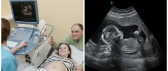
Ultrasound of the uterus and appendages does not cause pain or inconvenience to the patient, since the examination is done in most cases through the anterior abdominal wall. Before the procedure, the patient undresses to the waist or frees access to the abdominal area. During an abdominal ultrasound of the uterus and appendages, the woman lies on her back, the doctor applies a gel to her stomach and moves a sensor over it.
With other forms of examination, the position of the patient, as well as the location of the sensor, may vary. In some cases, the sensor is inserted into the rectum or vagina.
It is important to note that the decision about when and in what form is best to do an ultrasound of the uterus is made by a specialist based on a number of symptoms or pathologies of the reproductive system noticed during examination.
When is the best time to do a pelvic ultrasound?
An ultrasound performed in the first 7-10 days of the menstrual cycle will be indicative. This is relevant both for the study of the uterus and appendages, and for the diagnosis of polycystic disease, erosion and other diseases.
If uterine fibroids are suspected, the examination should be done immediately after the end of menstruation.
Endometriosis is diagnosed before menstruation. To monitor folliculogenesis (when planning pregnancy and in other cases), transvaginal ultrasound is performed on the fifth, ninth and 14-17 days of the menstrual cycle. Shifts in timing are possible in specific cases, depending on the duration of the cycle.
How to prepare for an ultrasound of the uterus?
Since the examination can be carried out in various ways, preparation for an ultrasound of the uterus will depend on the method.
Transvaginal diagnosis
On the day of the examination, it is recommended not to eat foods that cause increased gas formation. Before the procedure, you must empty your bladder.
Transrectal examination
Since the examination is performed rectally, it is necessary to prepare for an ultrasound of the uterus more thoroughly. 6-8 hours before the procedure, the patient needs to empty the rectal ampulla using an enema or laxative.
Abdominal diagnostics
Abdominal ultrasound of the uterus and appendages is performed through the anterior abdominal wall. Therefore, increased gas formation in the intestines may affect the examination results. To eliminate distortions, it is recommended to remove foods that cause increased gas formation from the diet the day before the procedure: brown bread, cabbage, legumes, carbonated drinks.
Since abdominal ultrasound of the uterus and appendages is performed with a full bladder, you need to drink a liter of water an hour before the procedure or not urinate for 2-3 hours before the examination.
Indications for ultrasound of the pelvic organs
You can have a pelvic ultrasound done at the medical center either as prescribed by your doctor or at your own discretion. Indications for ultrasound of the pelvic organs are:
- menstrual irregularities;
- sudden or prolonged bleeding;
- pain in the lower abdomen (pelvic area);
- suspicion of tumor formations;
- all variants of pregnancy (including suspected ectopic pregnancy);
- history of inflammatory diseases of the pelvic organs;
- infertility;
- preventive examination.
How is the procedure performed?
Abdominal ultrasound of the uterus and appendages is performed through the anterior abdominal wall. The patient undresses to the waist and takes a horizontal position face up. The sensor is placed on the stomach.
During a cervical examination or transvaginal ultrasound
a special sensor in a disposable condom is inserted into the vagina. Before the procedure, the patient removes clothing from the lower part of the body, including underwear. After this, the woman lies on her back and bends her knees.
For transrectal examination, the thinnest probe is used, which is inserted into the rectum. Before the procedure, the patient removes all clothing below the waist and lies on her left side. The sensor is inserted into the rectum in a special condom. A gel is used to improve the conductivity of ultrasound.
What is examined during an abdominal ultrasound?
During the study, the condition is determined:
- liver;
- gallbladder;
- kidney;
- spleen;
- pancreas;
- abdominal aorta;
- organs located in the pelvis.
An abdominal ultrasound, the price of which is quite affordable for every patient, shows the presence or absence of stones in the gall bladder or kidneys, the presence or absence of tumors, the condition of one or more organs, the presence of an inflammatory process, etc. This is an important study on which the doctor relies when making a diagnosis or determining the upcoming treatment process.
Decoding the results
On the ultrasound transcript, the uterus is pear-shaped with a smooth, clearly defined contour, the ovaries are clearly visible. Fuzzy boundaries of the uterus may indicate inflammation. An uneven contour also occurs in other pathological conditions, such as tumors.
If after an ultrasound of the uterus the interpretation shows a deviation from the norm, the specialist will prescribe additional examinations or make a diagnosis based on the available data.
Normal sizes of the uterus and ovaries on ultrasound
The size of the uterus according to ultrasound may vary, the norm depends on the age of the patient, pregnancy and the onset of menopause.
For patients aged 15-40 years, the normal sizes of the uterus and ovaries according to ultrasound data will be: uterus - 4.5-6.7 cm in length, 3-4 cm in thickness and 4.6-6.4 cm in width, each ovary – 3-4.1 cm in length, 2.0-3.1 cm in width and 14-22 mm in thickness. 20 years after menopause, the uterus becomes significantly smaller.
An increase in the size of the uterus occurs during pregnancy or due to a tumor. A deviation from the norm to a lesser extent indicates an infantile uterus.
Why do an ultrasound of the uterus during pregnancy?
Using an ultrasound of the uterus, the gynecologist monitors the condition of the amniotic fluid, umbilical cord, placenta and fetal development in general throughout the patient’s pregnancy. Carrying out early ultrasound diagnostics provides the specialist with the following information:
- condition of the uterus - absence or presence of deformities, tumors, polyps that can lead to miscarriage
- estimated date of conception, gestational age;
- diagnosis of multiple pregnancy.
With the help of the new device installed in our clinic, you can hear the heartbeat of the unborn child already at the first ultrasound session. The heart rate of the fetus is higher than that of a child who has already been born; the rate depends on the stage of pregnancy. For example, at 6-8 weeks the norm is 110-130 beats per minute, while at 9-12 it is already 170-190. Heart rate is one of the most important characteristics of the embryo’s condition; if the indicators exceed the norm, it allows timely diagnosis of the development of pathology and the taking of measures.
First ultrasound
10-14 weeks are the optimal period for ultrasound examination. During the first session, the gynecologist records the most important indicators (biparietal size, coccygeal-parietal size of the head), measures and controls the main characteristics of pregnancy. In addition, the thickness of the collar space is measured (allows us to determine the likelihood of the unborn child developing Down syndrome), and the size of the nasal bone is determined.
The nuchal translucency is measured in the neck area of the fetus; this is a dark stripe between the soft tissues and skin of the unborn child. The norm is the thickness of the space not exceeding 3 mm. The information is processed in the process of a comprehensive study, which allows us to determine the risk of having a baby with chromosomal abnormalities. In addition, ultrasound diagnostics allows a thorough examination of the structure of the fetal brain.
If indicated, the doctor may send the patient for an unscheduled ultrasound examination, regardless of the stage of pregnancy.
Second ultrasound
At 20-24 weeks, the expectant mother can see the arms and legs of her grown baby and examine his movements. The doctor carries out a full examination of the fetal organs, which have already formed, and carefully examines the vital organs - kidneys, heart, and so on.
During the second scheduled ultrasound diagnostic session, the embryo is measured, the amount of water and the condition of the placenta are studied. The results of the second ultrasound are certainly compared with the results of the first examination, which makes it possible to assess the intensity of development of the unborn child. And finally, the second examination allows the specialist to make sure that the fetus does not have obvious signs of chromosomal diseases. And the mother does not have any problems with the uterus and nearby organs
In most cases, a second planned ultrasound allows the doctor to find out the sex of the unborn baby. At 20-24 weeks, you can already independently see signs of gender on printouts or a monitor.
Third ultrasound
The main task of the third ultrasound during pregnancy is to check the position and condition of the unborn child before its birth. An ultrasound examination is performed at 28-32 weeks of pregnancy. The doctor determines whether the baby is breech or cephalic and makes sure there is no entanglement in the umbilical cord. This ultrasound diagnostic session makes it possible to calculate malformations, the signs of which appear only in the third trimester of pregnancy.
The last planned ultrasound for parents is no less interesting than the first, as it allows you to see the face of the unborn baby on the monitor; this is almost a newborn baby. Expert class equipment broadcasts the face of the unborn child in a three-dimensional image; parents, if desired, can film this joyful moment on video.
As a rule, the third ultrasound is combined with Doppler ultrasound; during this study, blood flow in the vessels of the embryo, uterus and umbilical cord is studied.
During pregnancy, there may be a need for additional ultrasound diagnostics, regardless of the period - an unscheduled ultrasound of the uterus is absolutely safe for the mother and child.
Three-dimensional ultrasound and 4D ultrasound
3D ultrasound of the uterus is a standard ultrasound, during which you can see an image of the unborn baby from all sides; a special program simultaneously processes the results. As a result, you can get a video recording with a three-dimensional image of the fetus in the uterus. 3D ultrasound increases the accuracy of medical diagnostics and gives parents the opportunity to “get to know” their baby before birth. 4D ultrasound differs in that the ultrasound image is broadcast in real time.
From the point of view of the emotional state, an ultrasound scan is very useful for a pregnant mother, since the study makes it possible to visually verify that the baby is okay. Often, it is the image of the baby that is displayed on the device’s monitor that becomes the moment when future parents realize the role ahead of them.
For a specialist, 3D and 4D ultrasound are important in the process of studying the fetal heart, because genetic abnormalities cannot always be identified at the prenatal stage. The heart is constantly in motion, which makes it difficult to examine. It is the combination of time study and obtaining a three-dimensional image that simplifies the diagnosis of fetal pathologies. This study also improves the quality of blood flow studies.
What abnormalities of the uterus and appendages will an ultrasound show?
The ultrasound method allows us to identify many pathologies of the development of the uterus, appendages and tubes and diagnose a number of diseases.
Malignant formations
The ultrasound method is considered the most informative examination of the uterus and appendages for malignant tumors. The examination visualizes the neoplasm, which allows the specialist to determine its location, the depth of germination into the muscle tissue, and the degree of damage to the lymph nodes and ovaries.
Transvaginal ultrasound of the uterus
and appendages is used as a method for diagnosing cervical cancer. The disease is widespread, and the risk of developing a tumor increases with age. Therefore, regular cervical cancer screening is recommended for all women over 30 years of age. In addition to ultrasound of the uterus and appendages, screening includes a smear for the papilloma virus and colposcopy.
Location of the fertilized egg
Atypical location of the ovum on a transvaginal ultrasound of the uterus
and appendages can be diagnosed from the third week of pregnancy.
What does an ultrasound of the pelvic organs show?
Ultrasound of the pelvis in women allows:
- determine multiple pregnancy;
- determine the location of the fertilized egg (in the uterus, in the tube, in the cervix, in the ovary);
- diagnose and assess the condition of uterine fibroids;
- determine the degree and location of a disease such as endometriosis;
- diagnose polyps of the cervix and uterine cavity, cysts, tumors, space-occupying formations of the uterus and its appendages;
- malformations of the internal genital organs (in adolescents);
- chronic inflammatory processes;
- complications after abortion and childbirth.
Our clinics in St. Petersburg
Structural subdivision of Polikarpov Alley Polikarpov 6k2 Primorsky district
- Pionerskaya
- Specific
- Commandant's
Structural subdivision of Zhukov Marshal Zhukov Ave. 28k2 Kirovsky district
- Avtovo
- Avenue of Veterans
- Leninsky Prospekt
Structural subdivision Devyatkino Okhtinskaya alley 18 Vsevolozhsk district
- Devyatkino
- Civil Prospect
- Academic
For detailed information and to make an appointment, you can call +7 (812) 640-55-25
Make an appointment
Attention!
It is possible to conduct other stock surveys at the same time by the same specialist.
Transvaginal ultrasound examination of the uterus
Transvaginal ultrasound is a procedure that shows the most accurate picture, so it can be called the most effective and efficient gynecological examination. The only disadvantage of the procedure is that, due to the peculiarities of its implementation, it can only be prescribed to women who are already sexually active.
To carry out this procedure, a woman, having first undressed as for a regular appointment with a gynecologist, sits on a gynecological chair or a special couch, then places her legs in a position favorable for examination (spread to the sides and bent at the knees). The prepared ultrasound device (with a medical condom on and a special gel applied) is inserted a minimum distance inside the vagina. Thanks to this method, an accurate picture is obtained that conveys all the features of the organ being studied. An ultrasound of the uterus should not feel painful, so if you feel discomfort or pain, be sure to point this out to the specialist performing the procedure.
In addition to the uterus, such a study will allow us to study other organs - the ovaries, fallopian tubes, bladder and more. In addition, in some cases, it is possible to conduct an exclusively transvaginal examination - for example, in case of obesity, problems with conceiving and bearing a child, excessive gas formation, and inability to keep the bladder full. Before the procedure, you should not drink water or other drinks for at least four hours before the test - your bladder must remain empty to obtain accurate data.
Transabdominal ultrasound examination of the uterus
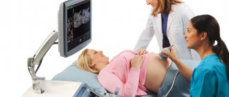
The simplest method of ultrasound of the uterus - transabdominal - is similar to most other standard ultrasound examinations. The patient lies down on the couch and removes clothes from her stomach. The doctor applies a special gel and examines the organ using an ultrasound device. Naturally, this method of research is not at all uncomfortable and does not cause any unpleasant sensations.
Unfortunately, transabdominal ultrasound of the uterus has some disadvantages. For example, a qualitative study may be hindered if the wall of the abdominal cavity is somewhat thicker than normal, or the sensor used for the procedure does not meet modern standards. In addition, the image is not very detailed, which is sometimes very important for clarifying diagnostic data.