Magnetic resonance imaging is based on exposure to a magnetic field of varying intensity. No negative consequences for the body were detected during native scanning; contraindications are individual in nature. To increase the information content, MRI with contrast is used, which allows you to visualize the slightest structural changes in tissues and organs. The number of contraindications to such an examination has been expanded, since during the scanning process a special solution is injected intravenously into the human body, which can cause a response. The procedure is considered a modern and safe technique, which helps to identify the disease at an early stage.
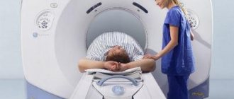
A series of MRI images of the brain for suspected Alzheimer's disease
Indications for MRI examination with contrast enhancement
1. Suspicion of a tumor
The main key to success in cancer treatment is early diagnosis. Unlike many other studies, contrast-enhanced MRI not only detects cancer in its early stages, but also determines the affected area. What may be a prerequisite for suspecting the presence of a tumor?
- independent determination of nodes, swelling, non-healing ulcers;
- pain syndrome;
- malfunction of the body;
- deviations in general analyses.
2. The need for differential diagnosis
In case of suspected cancer, differential diagnosis is an effective research method. It consists of a number of measures that exclude diagnoses that do not match the symptoms. The most comprehensive picture of the patient’s condition can be obtained with CT and MRI with contrast. With the help of the latter, examinations are carried out: the pituitary gland, brain, mammary glands, abdominal and pelvic organs, and spine.
Indications for abdominal MRI with contrast
In the vast majority of cases, MR scanning shows pathological changes in detail even without the introduction of a contrast agent. But for some pathologies, the preparation of a further treatment plan depends on the accuracy of the diagnosis, so a more thorough diagnosis is necessary.
The main indications for the use of contrast in examining the abdominal organs are as follows:
- suspicion of malignant neoplasm;
- assessment of the prevalence of the inflammatory process;
- the need for a more thorough study of the structure and functioning of the organ.
In these situations, the use of contrast agents can accurately identify pathologies and begin treatment in a timely manner.
Magnetic resonance imaging with contrast helps to identify pathologies of the following abdominal organs:
- liver;
- gallbladder and bile ducts;
- pancreas, walls of the stomach and intestines, spleen;
- the abdominal aorta and its branches, large venous trunks and their branches.
The nature and rate of accumulation of contrast in tumor tissue are the most reliable criteria for establishing the malignancy or benignity of a neoplasm. It is very important to compare the images before the contrast is administered and immediately after. Malignant neoplasms, as a rule, have uneven borders. Due to the good blood supply to the malignant tumor, the contrast quickly spreads through the bloodstream and stains the pathological tissue, which helps to obtain clear images of the tumor on MRI images.
CT will better show calcifications (pathological accumulation of calcium salts) in the kidneys, but their presence is less significant than identifying uneven distribution of contrast in the tumor. Therefore, MRI of the abdominal cavity with or without contrast has a significant advantage over CT.
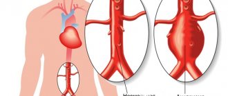
Magnetic resonance imaging with contrast can be used to diagnose serious pathologies of the abdominal aorta, such as aneurysm, thrombosis and wall tumors.
Contrast scanning of the aorta provides a unique opportunity to track all the details of blood flow (pulse wave speed, shifts of the aortic wall).
MRI is the preferred method for diagnosing aortic wall dissection with a sensitivity of 98%. Contrast-enhanced scanning is the most accurate diagnostic method to detect tumors in the aortic wall.
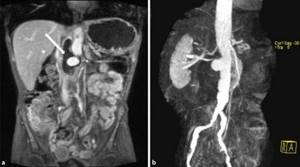
Magnetic resonance angiography of the aorta. An aneurysm and partial thrombosis are determined.
All patients with aortic disease require lifelong monitoring. In these cases, MRI scanning, as an accurate and safe method, is irreplaceable. MRI is more suitable for repeated studies in those who have already been diagnosed and require dynamic monitoring. While it is recommended to limit the number of CT scans to two times a year, magnetic resonance imaging can be done as needed, since there is no radiation exposure to the body.
Brain MRI with contrast
Due to its high information content, contrast magnetic resonance imaging is used in examining the brain. This method identifies pathologies such as:
- changes in blood vessels and tissues;
- intracranial pressure;
- tumors;
- pathologies of the central nervous system;
- infectious diseases;
- brain injuries.
Magnetic resonance imaging of the brain with contrast allows not only to make a diagnosis with great accuracy, but also to make it quickly.
What is administered during MRI with contrast?
To increase the efficiency of the magnetic resonance imaging method, special solutions are used, which are based on gadolinium metal salts. Due to their composition, contrast agents enhance the response from the tissues being studied, increasing the information content and quality of images.
The drugs used are characterized by low toxicity and hypoallergenicity, so the number of contraindications for MRI with contrast is limited.
The following solutions are most often used in modern tomography:
- Gadovist;
- Magnevist;
- Primovist;
- Dotarem;
- Omniscan.
Coloring preparations are inert, they do not take part in cellular metabolism and are excreted unchanged from the body after 24-36 hours.
MRI of the abdomen with contrast
MRI is usually used in controversial cases when diagnostics by other methods have not yielded results. Also, many procedures are quite painful and may not be reliable. Therefore, many doctors recommend immediately performing an MRI of the abdominal cavity.
with contrast.
This method helps to identify not only tumors, but also changes in pressure inside the abdominal cavity, internal bleeding and bruises, and inflammatory processes. Abdominal MRI with contrast includes an examination of the liver. This is almost the only way that allows you to determine not only the size, density and function of the organ, but also visualize the bile and pancreatic ducts. They can be changed, which leads to severe damage to the organ. Previously, the lack of such examination led to incorrect diagnoses.
What will an abdominal MRI with contrast show?
The main purposes of using contrast agents in magnetic resonance diagnostics:
- enhance the difference between the image of normal and pathologically altered tissue;
- study possible deviations in the structure and functioning of internal organs;
- distinguish a malignant tumor from a benign one.
MRI scans accurately show the soft tissues and organs of the abdomen, but are less effective at diagnosing hollow anatomical structures such as the stomach and intestines.
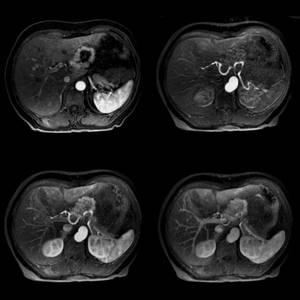
Magnetic resonance imaging of the abdomen with contrast
Contrast agent for MRI
The contrast agent is safe for humans, which allows diagnosis in the first 6 months of life. Principle of action: the drug is injected into a vein and spreads after some time. Accumulating in the affected cells, it colors them, depending on their density and structure, which increases the sensitivity of the device and the clarity of the image. The most effective contrast agents are those containing gadolinium (especially a soft and viscous silver-colored metal). Some of them are common in our country:
- Primovist
; - Magnevist;
- Omniscan;
- Gadovist;
- Let's do it.
Contrast agent for MRI - types and properties
Contrast agents can be hydrophilic complex compounds with a high concentration of gadolinium cations. Substances from the group of magnetic pharmaceuticals differ from those used in radiography. They are less toxic than contrast containing iodine salts and compounds used in CT and fluorography. Allergy to gadolinium compounds in the form of itchy eyes and hives is extremely rare. If a reaction does occur, it is milder and easily cured. Improved visualization is achieved due to the fact that gadolinium enters the soft tissues through the blood vessels and accumulates there. The peculiarity of gadolinium is its ability to enhance the magnetic signal during MRI, which improves the quality of images. There are many contrast agents. They are conventionally divided into the following groups:
- Intravascular. The dye is injected into the vein in full. The calculation is carried out using the formula 0.2 mg/kg of weight. These may be substances based on iron oxide, which has superparamagnetic properties. This also includes manganese compounds.
- Bolus contrast. The dye is also administered intravenously, but in doses through a dropper. The tomography progress and pigment supply are synchronized.
- Using oral agents, the gastrointestinal tract is scanned. In addition to manganese and gadolinium compounds, some natural products are also used, for example, blueberry and green tea for their high manganese content.
Dye tomography is more expensive than conventional tomography, but is well worth the higher cost.
Difference between MRI with and without contrast
The main difference is not only the use of contrast agent, but also the effectiveness. Contrast magnetic resonance imaging is prescribed in cases where other examinations, including conventional MRI, have not provided an absolute picture of the disease. The advantages of MRI with contrast are that it allows:
- examine in detail organs and tissues, especially the ducts: blood, bile, lymphatic;
- identify not only the neoplasms themselves, but also their number and size, even in the early stages.
Contrast magnetic resonance imaging is one of the most advanced and effective examination methods. If you have health problems, do not waste time on numerous studies. Trust the professionals of the Energo clinic in St. Petersburg. Speed up the healing process significantly.
Contraindications to MRI with contrast
There are absolute and relative contraindications to the use of contrast MRI. In some cases, restrictions depend on the individual characteristics of the patient and concomitant diseases.
Absolute contraindications to native and contrast magnetic resonance imaging are:
- the presence in the patient’s body of ferromagnetic and metal implants, prostheses (dental crowns, knitting needles, etc.);
- the presence of implanted electromagnetic devices (pacemakers, insulin pump, etc.);
- tattoos on the body using metallic inks.
These limitations are related to the nature of the scanning field: the magnet attracts metals, which can damage the equipment and aggravate the patient's condition.
The use of contrast agents based on coloring agents is not recommended in the following cases:
- first trimester of pregnancy;
- children up to 1 month;
- severe liver pathologies accompanied by severe liver failure;
- anemia;
- bronchial asthma of various etiologies;
- liver transplant operations;
- scarring and inflammatory processes in the kidney area;
- diseases of the cardiovascular system;
- lactation period.
All these contraindications are relative. If a patient needs to undergo an MRI, but has one or more limitations, then consultation with a specialist is necessary before the procedure. The doctor will premedicate and give recommendations that will help avoid negative consequences.
Lactation is not a direct contraindication, but when a contrast agent is administered, you must avoid breastfeeding for 36 hours. The drug is completely eliminated from the body after 1-2 days.
The use of beta-blockers leads to changes in the functioning of the vascular system, which negatively affects image quality. It is necessary to inform the radiologist about all prescriptions made by the attending physician.
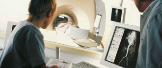
MRI angiography of the vessels of the lower extremities
An absolute contraindication to the administration of a contrast agent is an allergic reaction and individual intolerance to the components of the drug.
Contraindications for abdominal MRI with contrast agent
The coloring agent used for MRI is made from the metal gadolinium. Modern drugs have virtually no side effects, and their use does not require preliminary laboratory diagnostics to identify contraindications.
Patients with a significant medical history have an increased risk of adverse reactions to contrast agents. Information about previously observed allergic reactions to the contrast agent must be provided to the doctor before starting the study.
Bronchial asthma, pathologies of the cardiovascular system and renal failure in the acute stage require prior consultation with a specialist before conducting an MRI using contrast enhancement.
Pregnancy at any stage or constant use of certain medications are also contraindications.
In addition, the entire list of contraindications to the standard procedure without contrast remains:
- the presence of any structures, implants, foreign bodies made of ferromagnetic metals that cannot be removed;
- pacemaker, cochlear implant and other electronic devices;
- the patient’s weight is more than 130 kg, body girth at the maximum point is more than 140 cm;
- claustrophobia or the inability to maintain a stationary body position for other reasons;
- age less than 5 years.
If there are absolute contraindications to the study, the patient will be recommended other methods of diagnostic testing.
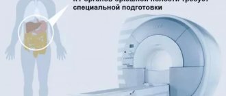
Preparing for an abdominal MRI with contrast
MRI of the abdominal organs, both with and without contrast, is performed on an empty stomach. Eating should take place no later than 6 hours before the procedure. It is also recommended to limit the consumption of foods that cause increased gas formation in the intestines 1-2 days before the test.
The decision about the possibility of intravenous contrast for children is made by the doctor based on the indications.
Immediately before the scan, the radiologist will ask the patient about possible contraindications, previous allergic reactions to contrast agents, and will also ask to remove accessories and clothing with metal fittings.